The brainstem is the posterior stalk-like part of the brain that connects the cerebrum with the spinal cord. In the human brain the brainstem is composed of the midbrain, the pons, and the medulla oblongata. The midbrain is continuous with the thalamus of the diencephalon through the tentorial notch, and sometimes the diencephalon is included in the brainstem.

The spinothalamic tract is a nerve tract in the anterolateral system in the spinal cord. This tract is an ascending sensory pathway to the thalamus. From the ventral posterolateral nucleus in the thalamus, sensory information is relayed upward to the somatosensory cortex of the postcentral gyrus.

The hypoglossal nucleus is a cranial nerve nucleus, found within the medulla. Being a motor nucleus, it is close to the midline. In the open medulla, it is visible as what is known as the hypoglossal trigone, a raised area protruding slightly into the fourth ventricle.
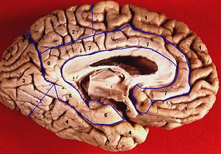
The limbic lobe is an arc-shaped cortical region of the limbic system, on the medial surface of each cerebral hemisphere of the mammalian brain, consisting of parts of the frontal, parietal and temporal lobes. The term is ambiguous, with some authors including the paraterminal gyrus, the subcallosal area, the cingulate gyrus, the parahippocampal gyrus, the dentate gyrus, the hippocampus and the subiculum; while the Terminologia Anatomica includes the cingulate sulcus, the cingulate gyrus, the isthmus of cingulate gyrus, the fasciolar gyrus, the parahippocampal gyrus, the parahippocampal sulcus, the dentate gyrus, the fimbrodentate sulcus, the fimbria of hippocampus, the collateral sulcus, and the rhinal sulcus, and omits the hippocampus.

The middle cerebral artery (MCA) is one of the three major paired cerebral arteries that supply blood to the cerebrum. The MCA arises from the internal carotid artery and continues into the lateral sulcus where it then branches and projects to many parts of the lateral cerebral cortex. It also supplies blood to the anterior temporal lobes and the insular cortices.

The dorsal root of spinal nerve is one of two "roots" which emerge from the spinal cord. It emerges directly from the spinal cord, and travels to the dorsal root ganglion. Nerve fibres with the ventral root then combine to form a spinal nerve. The dorsal root transmits sensory information, forming the afferent sensory root of a spinal nerve.

In neuroanatomy, the parieto-occipital sulcus is a deep sulcus in the cerebral cortex that marks the boundary between the cuneus and precuneus, and also between the parietal and occipital lobes. Only a small part can be seen on the lateral surface of the hemisphere, its chief part being on the medial surface.

The calcarine sulcus is an anatomical landmark located at the caudal end of the medial surface of the brain of humans and other primates. Its name comes from the Latin "calcar" meaning "spur". It is very deep, and known as a complete sulcus.

The circumflex branch of left coronary artery is a branch of the left coronary artery. It winds around the left side of the heart along the atrioventricular groove. It supplies the posterolateral portion of the left ventricle.

The most lateral of the bundles of the anterior nerve roots is generally taken as a dividing line that separates the anterolateral system into two parts. These are the anterior funiculus, between the anterior median fissure and the most lateral of the anterior nerve roots, and the lateral funiculus between the exit of these roots and the posterolateral sulcus.
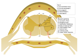
The Anterolateral sulcus of spinal cord is a landmark on the anterior side of the spinal cord. It denotes the location at which the ventral fibers leave the spinal cord.

In the human brain, the rhomboid fossa is divided into symmetrical halves by a median sulcus which reaches from the upper to the lower angles of the fossa and is deeper below than above. On either side of this sulcus is an elevation, the medial eminence, bounded laterally by a sulcus, the sulcus limitans.

In neuroanatomy, the paracentral lobule is on the medial surface of the cerebral hemisphere and is the continuation of the precentral and postcentral gyri. The paracentral lobule controls motor and sensory innervations of the contralateral lower extremity. It is also responsible for control of blushing, defecation and urination.

In neuroanatomy, the marginal sulcus is a sulcus (crevice) that may be considered the termination of the cingulate sulcus. It separates the paracentral lobule anteriorly and the precuneus posteriorly.
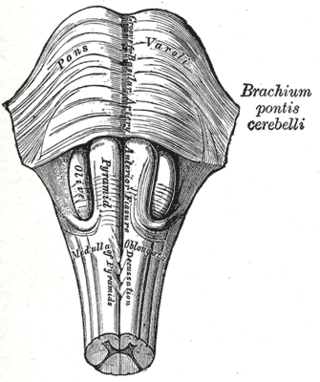
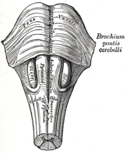
The basilar sulcus is a groove in the pons, part of the brainstem.

The inferior or orbital surface of the frontal lobe is concave, and rests on the orbital plate of the frontal bone. It is divided into four orbital gyri by a well-marked H-shaped orbital sulcus. These are named, from their position, the medial, anterior, lateral, and posterior, orbital gyri. The medial orbital gyrus presents a well-marked antero-posterior sulcus, the olfactory sulcus, for the olfactory tract; the portion medial to this is named the straight gyrus, and is continuous with the superior frontal gyrus on the medial surface.

The paracentral sulcus is a sulcus of the brain. It forms the paracentral lobule's anterior border. It is part of the cingulate sulcus.
Anterolateral sulcus may refer to:
Anterolateral may refer to:
Sulcus of spinal cord may refer to: