Arcuate nucleus (medulla)

Last updated
Arcuate nucleus (medulla)
Gray695.png
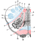
Transverse section of medulla oblongata below the middle of the olive. ("Nucleus arcuatus" visible near bottom right.)
Gray683.png
Dissection of brain-stem. Lateral view. (Labels for "External arcuate fibers" and "Dorsal external arcuate fibers" visible at lower right.)
Details
Identifiers
Latin nucleus arcuatus medullae oblongatae
NeuroNames 775
NeuroLex ID birnlex_2635
TA98 A14.1.04.256
TA2 6016
FMA 72609
Anatomical terms of neuroanatomy

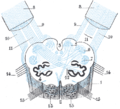
In the medulla oblongata, the arcuate nucleus is a group of neurons located on the anterior surface of the medullary pyramids. These nuclei are the extension of the pontine nuclei. [1]

Contents

They receive afferents from the corticospinal tract.[ citation needed ]

They in turn project efferents into the cerebellum through the inferior cerebellar peduncle as: [1]

Additional images

Related Research Articles

<span class="mw-page-title-main">Medulla oblongata</span> Structure of the brain stem

The medulla oblongata or simply medulla is a long stem-like structure which makes up the lower part of the brainstem. It is anterior and partially inferior to the cerebellum. It is a cone-shaped neuronal mass responsible for autonomic (involuntary) functions, ranging from vomiting to sneezing. The medulla contains the cardiac, respiratory, vomiting and vasomotor centers, and therefore deals with the autonomic functions of breathing, heart rate and blood pressure as well as the sleep–wake cycle. "Medulla" is from Latin, ‘pith or marrow’. And "oblongata" is from Latin, ‘lengthened or longish or elongated'.

<span class="mw-page-title-main">Pons</span> Part of the brainstem in humans and other bipeds

The pons is part of the brainstem that in humans and other mammals, lies inferior to the midbrain, superior to the medulla oblongata and anterior to the cerebellum.

<span class="mw-page-title-main">Brainstem</span> Posterior part of the brain, adjoining and structurally continuous

The brainstem is the stalk-like part of the brain that interconnects the cerebrum and diencephalon with the spinal cord. In the human brain, the brainstem is composed of the midbrain, the pons, and the medulla oblongata. The midbrain is continuous with the thalamus of the diencephalon through the tentorial notch.

<span class="mw-page-title-main">Glossopharyngeal nerve</span> Cranial nerve IX, for the tongue and pharynx

The glossopharyngeal nerve, also known as the ninth cranial nerve, cranial nerve IX, or simply CN IX, is a cranial nerve that exits the brainstem from the sides of the upper medulla, just anterior to the vagus nerve. Being a mixed nerve (sensorimotor), it carries afferent sensory and efferent motor information. The motor division of the glossopharyngeal nerve is derived from the basal plate of the embryonic medulla oblongata, whereas the sensory division originates from the cranial neural crest.

<span class="mw-page-title-main">Medial lemniscus</span> Ascending bundle of axons which cross in the brainstem

In neuroanatomy, the medial lemniscus, also known as Reil's band or Reil's ribbon, is a large ascending bundle of heavily myelinated axons that decussate (cross) in the brainstem, specifically in the medulla oblongata. The medial lemniscus is formed by the crossings of the internal arcuate fibers. The internal arcuate fibers are composed of axons of nucleus gracilis and nucleus cuneatus. The axons of the nucleus gracilis and nucleus cuneatus in the medial lemniscus have cell bodies that lie contralaterally.

<span class="mw-page-title-main">Nucleus ambiguus</span> Group of motor neurons in the brain stem

The nucleus ambiguus is a group of large motor neurons, situated deep in the medullary reticular formation named by Jacob Clarke. The nucleus ambiguus contains the cell bodies of neurons that innervate the muscles of the soft palate, pharynx, and larynx which are associated with speech and swallowing. As well as motor neurons, the nucleus ambiguus contains preganglionic parasympathetic neurons which innervate postganglionic parasympathetic neurons in the heart.

<span class="mw-page-title-main">Medial medullary syndrome</span> Medical condition

Medial medullary syndrome, also known as inferior alternating syndrome, hypoglossal alternating hemiplegia, lower alternating hemiplegia, or Dejerine syndrome, is a type of alternating hemiplegia characterized by a set of clinical features resulting from occlusion of the anterior spinal artery. This results in the infarction of medial part of the medulla oblongata.

<span class="mw-page-title-main">Olivary body</span>

In anatomy, the olivary bodies or simply olives are a pair of prominent oval structures in the medulla oblongata, the lower portion of the brainstem. They contain the olivary nuclei.

<span class="mw-page-title-main">Inferior cerebellar peduncle</span>

The upper part of the posterior district of the medulla oblongata is occupied by the inferior cerebellar peduncle, a thick rope-like strand situated between the lower part of the fourth ventricle and the roots of the glossopharyngeal and vagus nerves.

The dorsal longitudinal fasciculus (DLF) is a longitudinal tract interconnecting the posterior hypothalamus, and the inferior medulla oblongata. It contains both ascending tracts and descending tracts, and serves to link the forebrain, and the visceral autonomic centres of the lower brainstem. It conveys both visceral motor signals, and sensory signals.

<span class="mw-page-title-main">Vestibular nuclei</span>

The vestibular nuclei (VN) are the cranial nuclei for the vestibular nerve located in the brainstem.

<span class="mw-page-title-main">Cerebellar peduncle</span> Structure connecting the cerebellum to the brainstem

Cerebellar peduncles connect the cerebellum to the brain stem. There are six cerebellar peduncles in total, three on each side:

<span class="mw-page-title-main">Anterior external arcuate fibers</span>

The anterior external arcuate fibers vary as to their prominence: in some cases they form an almost continuous layer covering the medullary pyramids and olivary body, while in other cases they are barely visible on the surface.

<span class="mw-page-title-main">Internal arcuate fibers</span> Axons of neurons that compose the brains gracile and cuneate nuclei

In neuroanatomy, the internal arcuate fibers or internal arcuate tract are the axons of second-order sensory neurons that compose the gracile and cuneate nuclei of the medulla oblongata. These second-order neurons begin in the gracile and cuneate nuclei in the medulla. They receive input from first-order sensory neurons, which provide sensation to many areas of the body and have cell bodies in the dorsal root ganglia of the dorsal root of the spinal nerves. Upon decussation from one side of the medulla to the other, also known as the sensory decussation, they are then called the medial lemniscus.

<span class="mw-page-title-main">Dorsal column nuclei</span> Nuclei in the dorsal column of the brainstem

In neuroanatomy, the dorsal column nuclei are a pair of nuclei in the dorsal columns in the brainstem. The name refers collectively to the cuneate nucleus and gracile nucleus, which are situated at the lower end of the medulla oblongata. Both nuclei contain second-order neurons of the dorsal column–medial lemniscus pathway, which convey fine touch and proprioceptive information from the body to the brain. The dorsal column nuclei project to the thalamus.

<span class="mw-page-title-main">Medullary striae of fourth ventricle</span>

Medullary striae of fourth ventricle are a landmark of the rhomboid fossa - the floor of the fourth ventricle. They are part of the auditory system. The medullary striae are formed by crossed-over anterior internal arcuate fibers - efferents of the arcuate nucleus of medulla oblongata - as they pass laterally beneath the ependyma of the fourth ventricle to reach the contralateral cerebellum. The striae pass over the dorsal aspect of the medial vestibular nucleus.

The projection fibers consist of efferent and afferent fibers uniting the cortex with the lower parts of the brain and with the spinal cord. In human neuroanatomy, bundles of axons called tracts, within the brain, can be categorized by their function into association fibers, projection fibers, and commissural fibers.

<span class="mw-page-title-main">Medullary pyramids (brainstem)</span> White matter structures within the brainstems medulla oblongata

In neuroanatomy, the medullary pyramids are paired white matter structures of the brainstem's medulla oblongata that contain motor fibers of the corticospinal and corticobulbar tracts – known together as the pyramidal tracts. The lower limit of the pyramids is marked when the fibers cross (decussate).

<span class="mw-page-title-main">Anterior median fissure of the medulla oblongata</span>

The anterior median fissure contains a fold of pia mater, and extends along the entire length of the medulla oblongata: It ends at the lower border of the pons in a small triangular expansion, termed the foramen cecum.

References

  1. 1 2 Standring, Susan (2020). Gray's Anatomy: The Anatomical Basis of Clinical Practice (42th ed.). New York: Elsevier. p. 445.e1. ISBN   978-0-7020-7707-4. OCLC   1201341621.