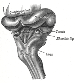
The medulla oblongata or simply medulla is a long stem-like structure which makes up the lower part of the brainstem. It is anterior and partially inferior to the cerebellum. It is a cone-shaped neuronal mass responsible for autonomic (involuntary) functions, ranging from vomiting to sneezing. The medulla contains the cardiac, respiratory, vomiting and vasomotor centers, and therefore deals with the autonomic functions of breathing, heart rate and blood pressure as well as the sleep–wake cycle. "Medulla" is from Latin, ‘pith or marrow’. And "oblongata" is from Latin, ‘lengthened or longish or elongated'.
Articles related to anatomy include:

The pons is part of the brainstem that in humans and other mammals, lies inferior to the midbrain, superior to the medulla oblongata and anterior to the cerebellum.
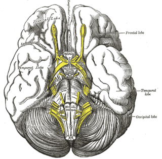
The brainstem is the stalk-like part of the brain that connects the forebrain with the spinal cord. In the human brain, the brainstem is composed of the midbrain, the pons, and the medulla oblongata. The midbrain is continuous with the thalamus of the diencephalon through the tentorial notch.

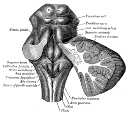
The hypoglossal nucleus is a cranial nerve nucleus, found within the medulla. Being a motor nucleus, it is close to the midline. In the open medulla, it is visible as what is known as the hypoglossal trigone, a raised area protruding slightly into the fourth ventricle.

The fourth ventricle is one of the four connected fluid-filled cavities within the human brain. These cavities, known collectively as the ventricular system, consist of the left and right lateral ventricles, the third ventricle, and the fourth ventricle. The fourth ventricle extends from the cerebral aqueduct to the obex, and is filled with cerebrospinal fluid (CSF).

The lateral ventricles are the two largest ventricles of the brain and contain cerebrospinal fluid. Each cerebral hemisphere contains a lateral ventricle, known as the left or right lateral ventricle, respectively.

In the medulla oblongata, the arcuate nucleus is a group of neurons located on the anterior surface of the medullary pyramids. These nuclei are the extension of the pontine nuclei.

The vestibular nuclei (VN) are the cranial nuclei for the vestibular nerve located in the brainstem.

Cerebellar peduncles connect the cerebellum to the brain stem. There are six cerebellar peduncles in total, three on each side:

The area postrema, a paired structure in the medulla oblongata of the brainstem, is a circumventricular organ having permeable capillaries and sensory neurons that enable its dual role to detect circulating chemical messengers in the blood and transduce them into neural signals and networks. Its position adjacent to the bilateral nuclei of the solitary tract and role as a sensory transducer allow it to integrate blood-to-brain autonomic functions. Such roles of the area postrema include its detection of circulating hormones involved in vomiting, thirst, hunger, and blood pressure control.
The medullary striae of fourth ventricle are a landmark of the rhomboid fossa - the floor of the fourth ventricle. They are part of the auditory system. The medullary striae are formed by crossed-over anterior internal arcuate fibers - efferents of the arcuate nucleus of medulla oblongata - as they pass laterally beneath the ependyma of the fourth ventricle to reach the contralateral cerebellum. The striae pass over the dorsal aspect of the medial vestibular nucleus.
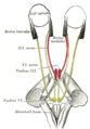
The interpeduncular fossa is a deep depression of the ventral surface of the midbrain between the two crura cerebri.

The olfactory tract is a bilateral bundle of afferent nerve fibers from the mitral and tufted cells of the olfactory bulb that connects to several target regions in the brain, including the piriform cortex, amygdala, and entorhinal cortex. It is a narrow white band, triangular on coronal section, the apex being directed upward.
The sulcus limitans is a groove on either side of the midline in the rhomboid fossa. It separates the cranial nerve motor nuclei, and the sensory nuclei. It is parallel to the median sulcus.
The vagal trigone is a triangular eminence upon the rhomboid fossa produced by the underlying dorsal nucleus of vagus nerve.
In the human brain, the rhomboid fossa is divided into symmetrical halves by a median sulcus which reaches from the upper to the lower angles of the fossa and is deeper below than above. On either side of this sulcus is an elevation, the medial eminence, bounded laterally by a sulcus, the sulcus limitans.
In the upper part of the medulla oblongata, the hypoglossal nucleus approaches the rhomboid fossa, where it lies close to the middle line, under an eminence named the hypoglossal trigone. It is a slight elevation in the floor of the inferior recess of the fourth ventricle, beneath which is the nucleus of origin of the twelfth cranial nerve.
The substantia ferruginea is an underlying patch of deeply pigmented nerve cells located in the floor of the superior part of the sulcus limitans.