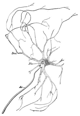
A motor nerve, or efferent nerve, is a nerve that contains exclusively efferent nerve fibers and transmits motor signals from the central nervous system (CNS) to the muscles of the body. This is different from the motor neuron, which includes a cell body and branching of dendrites, while the nerve is made up of a bundle of axons. Motor nerves act as efferent nerves which carry information out from the CNS to muscles, as opposed to afferent nerves, which transfer signals from sensory receptors in the periphery to the CNS. Efferent nerves can also connect to glands or other organs/issues instead of muscles. The vast majority of nerves contain both sensory and motor fibers and are therefore called mixed nerves.

The spinothalamic tract is a nerve tract in the anterolateral system in the spinal cord. This tract is an ascending sensory pathway to the thalamus. From the ventral posterolateral nucleus in the thalamus, sensory information is relayed upward to the somatosensory cortex of the postcentral gyrus.

The spinocerebellar tract is a nerve tract originating in the spinal cord and terminating in the same side (ipsilateral) of the cerebellum.

The dorsal root of spinal nerve is one of two "roots" which emerge from the spinal cord. It emerges directly from the spinal cord, and travels to the dorsal root ganglion. Nerve fibres with the ventral root then combine to form a spinal nerve. The dorsal root transmits sensory information, forming the afferent sensory root of a spinal nerve.

The vestibulospinal tract is a neural tract in the central nervous system. Specifically, it is a component of the extrapyramidal system and is classified as a component of the medial pathway. Like other descending motor pathways, the vestibulospinal fibers of the tract relay information from nuclei to motor neurons. The vestibular nuclei receive information through the vestibulocochlear nerve about changes in the orientation of the head. The nuclei relay motor commands through the vestibulospinal tract. The function of these motor commands is to alter muscle tone, extend, and change the position of the limbs and head with the goal of supporting posture and maintaining balance of the body and head.

In the spinal cord, the most lateral of the bundles of the ventral nerve roots is generally taken as a dividing line that separates the antero-lateral region into two parts: an anterior funiculus , between the anterior median fissure and the most lateral of the ventral nerve roots; and a lateral funiculus, between the exit of these roots and the posterolateral sulcus.
The most lateral of the bundles of the anterior nerve roots is generally taken as a dividing line that separates the anterolateral system into two parts. These are the anterior funiculus, between the anterior median fissure and the most lateral of the anterior nerve roots, and the lateral funiculus between the exit of these roots and the posterolateral sulcus.
The anterior grey column is the front column of grey matter in the spinal cord. It is one of the three grey columns. The anterior grey column contains alpha motor neurons that affect the skeletal muscles while the posterior grey column receives information regarding touch and sensation.

The posterior thoracic nucleus, is a group of interneurons found in the medial part of lamina VII, also known as the intermediate zone, of the spinal cord. It is located from the cervical segment C8 to lumbar segment L3 of the spinal cord and is an important structure for proprioception of the lower limb.

The lateral vestibular nucleus is the continuation upward and lateralward of the principal nucleus, and in it terminate many of the ascending branches of the vestibular nerve.

The rhomboid fossa is a rhombus-shaped depression that is the anterior part of the fourth ventricle. Its anterior wall, formed by the back of the pons and the medulla oblongata, constitutes the floor of the fourth ventricle.
The projection fibers consist of efferent and afferent fibers uniting the cortex with the lower parts of the brain and with the spinal cord. In human neuroanatomy, bundles of axons called tracts, within the brain, can be categorized by their function into association fibers, projection fibers, and commissural fibers.

In neuroanatomy, the medullary pyramids are paired white matter structures of the brainstem's medulla oblongata that contain motor fibers of the corticospinal and corticobulbar tracts – known together as the pyramidal tracts. The lower limit of the pyramids is marked when the fibers cross (decussate).

A nerve fascicle is a bundle of nerve fibers belonging to a nerve in the peripheral nervous system. A nerve fascicle is also called a fasciculus, as is a nerve tract in the central nervous system.
Funiculus is any cord-like structure in anatomy or biology, and may refer to:

The medial vestibular nucleus is one of the vestibular nuclei. It is located in the medulla oblongata.

The spinal cord is a long, thin, tubular structure made up of nervous tissue that extends from the medulla oblongata in the brainstem to the lumbar region of the vertebral column (backbone) of vertebrate animals. The center of the spinal cord is hollow and contains a structure called the central canal, which contains cerebrospinal fluid. The spinal cord is also covered by meninges and enclosed by the neural arches. Together, the brain and spinal cord make up the central nervous system.

A nerve tract is a bundle of nerve fibers (axons) connecting nuclei of the central nervous system. In the peripheral nervous system, this is known as a nerve fascicle, and has associated connective tissue. The main nerve tracts in the central nervous system are of three types: association fibers, commissural fibers, and projection fibers. A nerve tract may also be referred to as a commissure, decussation, or neural pathway. A commissure connects the two cerebral hemispheres at the same levels, while a decussation connects at different levels.
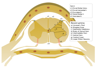
On either side of the posterior median sulcus of the spinal cord, and at a short distance from it, the posterior nerve roots are attached along a vertical furrow named the posterolateral sulcus. The portion of the medulla spinalis which lies between this and the posterior median sulcus is named the posterior funiculus.
The solitariospinal tract is a descending nerve tract that controls breathing by promoting the action of inspiratory muscles. It consists of a small group of axons originating in the nucleus solitarius of the medulla oblongata, and projects to the motor neurons of the phrenic nerve and of motor neurons of the thoracic nerves. In the spinal cord, it descends in the anterior funiculus and lateral funiculus. Section of the anterior and lateral funiculi appears to abolish rhythmic breathing.
