Angiotensin is a peptide hormone that causes vasoconstriction and an increase in blood pressure. It is part of the renin–angiotensin system, which regulates blood pressure. Angiotensin also stimulates the release of aldosterone from the adrenal cortex to promote sodium retention by the kidneys.

The juxtaglomerular apparatus is a structure in the kidney that regulates the function of each nephron, the functional units of the kidney. The juxtaglomerular apparatus is named because it is next to (juxta-) the glomerulus.
The control of ventilation is the physiological mechanisms involved in the control of breathing, which is the movement of air into and out of the lungs. Ventilation facilitates respiration. Respiration refers to the utilization of oxygen and balancing of carbon dioxide by the body as a whole, or by individual cells in cellular respiration.
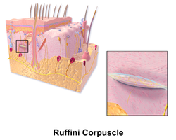
A cutaneous receptor is the type of sensory receptor found in the skin. They are a part of the somatosensory system. Cutaneous receptors include mechanoreceptors, nociceptors (pain), and thermoreceptors (temperature).
A mechanoreceptor, also called mechanoceptor, is a sensory receptor that responds to mechanical pressure or distortion. Mechanoreceptors are innervated by sensory neurons that convert mechanical pressure into electrical signals that, in animals, are sent to the central nervous system.

The dorsal column–medial lemniscus pathway (DCML) is a sensory pathway of the central nervous system that conveys sensations of fine touch, vibration, two-point discrimination, and proprioception from the skin and joints. It transmits information from the body to the primary somatosensory cortex in the postcentral gyrus of the parietal lobe of the brain. The pathway receives information from sensory receptors throughout the body, and carries this in nerve tracts in the white matter of the dorsal column of the spinal cord to the medulla, where it is continued in the medial lemniscus, on to the thalamus and relayed from there through the internal capsule and transmitted to the somatosensory cortex. The name dorsal-column medial lemniscus comes from the two structures that carry the sensory information: the dorsal columns of the spinal cord, and the medial lemniscus in the brainstem.

Tactile corpuscles or Meissner's corpuscles are a type of mechanoreceptor discovered by anatomist Georg Meissner (1829–1905) and Rudolf Wagner. This corpuscle is a type of nerve ending in the skin that is responsible for sensitivity to pressure. In particular, they have their highest sensitivity when sensing vibrations between 10 and 50 hertz. They are rapidly adaptive receptors. They are most concentrated in thick hairless skin, especially at the finger pads.

Sensory neurons, also known as afferent neurons, are neurons in the nervous system, that convert a specific type of stimulus, via their receptors, into action potentials or graded receptor potentials. This process is called sensory transduction. The cell bodies of the sensory neurons are located in the dorsal ganglia of the spinal cord.

The subcutaneous tissue, also called the hypodermis, hypoderm, subcutis, or superficial fascia, is the lowermost layer of the integumentary system in vertebrates. The types of cells found in the layer are fibroblasts, adipose cells, and macrophages. The subcutaneous tissue is derived from the mesoderm, but unlike the dermis, it is not derived from the mesoderm's dermatome region. It consists primarily of loose connective tissue, and contains larger blood vessels and nerves than those found in the dermis. It is a major site of fat storage in the body.
The Pacinian corpuscle, lamellar corpuscle or Vater-Pacini corpuscle is one of the four major types of mechanoreceptors for mechanical sensation) found in mammalian skin. This type of mechanoreceptor is found in both hairy, and hairless skin, viscera, joints, and attached to the periosteum of bone, primarily responsible for sensitivity to vibration. A few are also sensitive to quasi-static or low frequency pressure stimuli. Most of them respond only to sudden disturbances and are especially sensitive to vibration of a few hundreds hertz. The vibrational role may be used for detecting surface texture, such as rough and smooth. Most of the Pacinian corpuscles act as rapidly adapting mechanoreceptors. Groups of corpuscles respond to pressure changes, such as on grasping or releasing an object.
Merkel nerve endings are mechanoreceptors, a type of sensory receptor, that are found in the basal epidermis and hair follicles. They are nerve endings and provide information on mechanical pressure, position, and deep static touch features, such as shapes and edges.
In the physiology of the kidney, renal blood flow (RBF) is the volume of blood delivered to the kidneys per unit time. In humans, the kidneys together receive roughly 25% of cardiac output, amounting to 1.2 - 1.3 L/min in a 70-kg adult male. It passes about 94% to the cortex. RBF is closely related to renal plasma flow (RPF), which is the volume of blood plasma delivered to the kidneys per unit time.
The cough reflex occurs when stimulation of cough receptors in the respiratory tract by dust or other foreign particles produces a cough, which causes rapidly moving air which usually remove the foreign material before it reaches the lungs. This typically clears particles from the bronchi and trachea, the tubes that feed air to lung tissue from the nose and mouth. The larynx and carina are especially sensitive. Cough receptors in the surface cells (epithelium) of the respiratory tract are also sensitive to chemicals. Terminal bronchioles and even the alveoli are sensitive to chemicals such as sulfur dioxide gas or chlorine gas.
In endocrinology, permissiveness is a biochemical phenomenon in which the presence of one hormone is required in order for another hormone to exert its full effects on a target cell. Hormones can interact in permissive, synergistic, or antagonistic ways. The chemical classes of hormones include amines, polypeptides, glycoproteins and steroids. Permissive hormones act as precursors to active hormones and may be classified as either prohormones or prehormones. It stimulate the formation of receptors of that hormone.
Tonic in physiology refers to a physiological response which is slow and may be graded. This term is typically used in opposition to a fast response. For instance, tonic muscles are contrasted by the more typical and much faster twitch muscles, while tonic sensory nerve endings are contrasted to the much faster phasic sensory nerve endings.
William Francis Ganong Jr. was an American physiologist at the University of California, San Francisco (UCSF), and was one of the first scientists to trace how the brain controls important internal functions of the body.
Juxtacapillary receptors, J-receptors, or pulmonary C-fiber receptors are sensory nerve endings located within the alveolar walls in juxtaposition to the pulmonary capillaries of the lung, and are innervated by fibers of the vagus nerve. Although their functional role is unclear, J-receptors respond to events such as pulmonary edema, pulmonary emboli, pneumonia, congestive heart failure and barotrauma, which cause a decrease in oxygenation and thus lead to an increase in respiration. They may be also stimulated by hyperinflation of the lung as well as intravenous or intracardiac administration of chemicals such as capsaicin. The stimulation of the J-receptors causes a reflex increase in breathing rate, and is also thought to be involved in the sensation of dyspnea, the subjective sensation of difficulty breathing. The reflex response that is produced is apnea followed by rapid breathing, bradycardia, and hypotension. The physiologic role of this reflex is uncertain, but it probably occurs in pathologic states such as pulmonary congestion or embolization. These receptors were discovered by Autar Paintal.
The Golgi tendon reflex (also called inverse stretch reflex, autogenic inhibition, tendon reflex) is an inhibitory effect on the muscle resulting from the muscle tension stimulating Golgi tendon organs (GTO) of the muscle, and hence it is self-induced. The reflex arc is a negative feedback mechanism preventing too much tension on the muscle and tendon. When the tension is extreme, the inhibition can be so great it overcomes the excitatory effects on the muscle's alpha motoneurons causing the muscle to suddenly relax. This reflex is also called the inverse myotatic reflex, because it is the inverse of the stretch reflex.

Touch is perceiving the environment using skin. Specialized receptors in the skin send signals to the brain indicating light and soft pressure, hot and cold, body position and pain. It is a subset of the sensory nervous system, which also includes the visual, auditory, olfactory, gustatory and vestibular senses.

The Golgi tendon organ (GTO) is a proprioceptor – a type of sensory receptor that senses changes in muscle tension. It lies at the interface between a muscle and its tendon known as the musculotendinous junction also known as the myotendinous junction. It provides the sensory component of the Golgi tendon reflex.