Myelin incisure

Last updated
Myelin incisure
Gray631.png
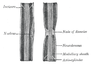
Diagram of longitudinal sections of medullated nerve fibers. (Incisure labeled at upper left.)
Details
System Nervous system
Identifiers
TH H2.00.06.2.03015
Anatomical terms of microanatomy

Myelin incisures (a.k.a. Schmidt-Lanterman clefts, Schmidt-Lanterman incisures, clefts of Schmidt-Lanterman, segments of Lanterman, medullary segments), are small pockets of cytoplasm left behind during the Schwann cell myelination process. They are histological evidence of the small amount of cytoplasm that remains in the inner layer of the myelin sheath created by Schwann cells wrapping tightly around an axon (nerve fiber).

Cytoplasm all of the contents of a cell excluding the plasma membrane and nucleus, but including other subcellular structures

In cell biology, the cytoplasm is all of the material within a cell, enclosed by the cell membrane, except for the cell nucleus. The material inside the nucleus and contained within the nuclear membrane is termed the nucleoplasm. The main components of the cytoplasm are cytosol – a gel-like substance, the organelles – the cell's internal sub-structures, and various cytoplasmic inclusions. The cytoplasm is about 80% water and usually colorless.

Schwann cell

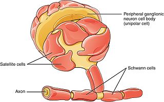
Schwann cells or neurolemmocytes are the principal glia of the peripheral nervous system (PNS). Glial cells function to support neurons and in the PNS, also include satellite cells, olfactory ensheathing cells, enteric glia and glia that reside at sensory nerve endings, such as the Pacinian corpuscle. The two types of Schwann cells are myelinating and nonmyelinating. Myelinating Schwann cells wrap around axons of motor and sensory neurons to form the myelin sheath. The Schwann cell promoter is present in the downstream region of the human dystrophin gene that gives shortened transcript that are again synthesized in a tissue-specific manner.

Histology study of the microscopic anatomy of cells and tissues of plants and animals

Histology, also microanatomy, is the branch of biology which studies the tissues of animals and plants using microscopy. It is commonly studied using a light microscope or electron microscope, the specimen having been sectioned, stained, and mounted on a microscope slide. Histological studies may be conducted using tissue culture, where live animal cells are isolated and maintained in an artificial environment for various research projects. The ability to visualize or differentially identify microscopic structures is frequently enhanced through the use of staining. Histology is one of the major preclinical subjects in medical school. Medical students are expected to be familiar with the morphological features and function of all cells and tissues of the human body from an early stage of their studies, so histology often stretches over several semesters.

In the peripheral nervous system (PNS) axons can be either myelinated or unmyelinated. Myelination refers to the insulation of an axon with concentric surrounding layers of lipid membrane (myelin) produced by Schwann cells. These layers are generally uniform and continuous, but due to imperfect nature of the process by which Schwann cells wrap the nerve axon, this wrapping process can sometimes leave behind small pockets of residual cytoplasm displaced to the periphery during the formation of the myelin sheath. These pockets, or "incisures", can subdivide the myelinated axon into irregular portions. These staggered clefts also provide communication channels between layers by connecting the outer collar of cytoplasm of the Schwann cell to the deepest layer of myelin sheath. Primary incisures which appear ab initio in myelination and always extend across the whole radial thickness of the myelin sheath but initially around only part of its circumference. Secondary incisures appear later, in regions of a compact myelin sheath, initially traversing only part of its radial thickness but commonly occupying its whole circumference. [1]

Peripheral nervous system part of the nervous system

The peripheral nervous system (PNS) is one of two components that make up the nervous system of bilateral animals, with the other part being the central nervous system (CNS). The PNS consists of the nerves and ganglia outside the brain and spinal cord. The main function of the PNS is to connect the CNS to the limbs and organs, essentially serving as a relay between the brain and spinal cord and the rest of the body. Unlike the CNS, the PNS is not protected by the vertebral column and skull, or by the blood–brain barrier, which leaves it exposed to toxins and mechanical injuries.

Lipid A substance of biological origin that is soluble in nonpolar solvents

In biology and biochemistry, a lipid is a biomolecule that is soluble in nonpolar solvents. Non-polar solvents are typically hydrocarbons used to dissolve other naturally occurring hydrocarbon lipid molecules that do not dissolve in water, including fatty acids, waxes, sterols, fat-soluble vitamins, monoglycerides, diglycerides, triglycerides, and phospholipids.

Myelin fatty white substance, multi-layered membrane, unique to the nervous system, that functions as an insulator to greatly increase the velocity of axonal impulse conduction; it is an outgrowth of a type of glial cell

Myelin is a lipid-rich (fatty) substance formed in the central nervous system (CNS) by glial cells called oligodendrocytes, and in the peripheral nervous system (PNS) by Schwann cells. Myelin insulates nerve cell axons to increase the speed at which information travels from one nerve cell body to another or, for example, from a nerve cell body to a muscle. The myelinated axon can be likened to an electrical wire with insulating material (myelin) around it. However, unlike the plastic covering on an electrical wire, myelin does not form a single long sheath over the entire length of the axon. Rather, each myelin sheath insulates the axon over a single section and, in general, each axon comprises multiple long myelinated sections separated from each other by short gaps called Nodes of Ranvier. Each myelin sheath is formed by the concentric wrapping of an oligodendrocyte or Schwann cell process around the axon.

Related Research Articles

Axon The long process of a neuron that conducts nerve impulses, usually away from the cell body to the terminals and varicosities, which are sites of storage and release of neurotransmitter.

An axon, or nerve fiber, is a long, slender projection of a nerve cell, or neuron, in vertebrates, that typically conducts electrical impulses known as action potentials away from the nerve cell body. The function of the axon is to transmit information to different neurons, muscles, and glands. In certain sensory neurons, such as those for touch and warmth, the axons are called afferent nerve fibers and the electrical impulse travels along these from the periphery to the cell body, and from the cell body to the spinal cord along another branch of the same axon. Axon dysfunction has caused many inherited and acquired neurological disorders which can affect both the peripheral and central neurons. Nerve fibers are classed into three types – group A nerve fibers, group B nerve fibers, and group C nerve fibers. Groups A and B are myelinated, and group C are unmyelinated. These groups include both sensory fibers and motor fibers. Another classification groups only the sensory fibers as Type I, Type II, Type III, and Type IV.

Central nervous system part of the nervous system consisting of the brain and spinal cord

The central nervous system (CNS) is the part of the nervous system consisting of the brain and spinal cord. The CNS is so named because it integrates the received information and coordinates and influences the activity of all parts of the bodies of bilaterally symmetric animals—that is, all multicellular animals except sponges and radially symmetric animals such as jellyfish—and it contains the majority of the nervous system. Many consider the retina and the optic nerve, as well as the olfactory nerves and olfactory epithelium as parts of the CNS, synapsing directly on brain tissue without intermediate ganglia. As such, the olfactory epithelium is the only central nervous tissue in direct contact with the environment, which opens up for therapeutic treatments. The CNS is contained within the dorsal body cavity, with the brain housed in the cranial cavity and the spinal cord in the spinal canal. In vertebrates, the brain is protected by the skull, while the spinal cord is protected by the vertebrae. The brain and spinal cord are both enclosed in the meninges. Within the CNS, the interneuronal space is filled with a large amount of supporting non-nervous cells called neuroglial cells.

Nerve enclosed, cable-like bundle of axons in the peripheral nervous system

A nerve is an enclosed, cable-like bundle of nerve fibres called axons, in the peripheral nervous system. A nerve provides a common pathway for the electrochemical nerve impulses called action potentials that are transmitted along each of the axons to peripheral organs or, in the case of sensory nerves, from the periphery back to the central nervous system. Each axon within the nerve is an extension of an individual neuron, along with other supportive cells such as Schwann cells that coat the axons in myelin.

Neurilemma

Neurilemma is the outermost nucleated cytoplasmic layer of Schwann cells that surrounds the axon of the neuron. It forms the outermost layer of the nerve fiber in the peripheral nervous system.

Saltatory conduction propagation of action potentials along a nerve cell

Saltatory conduction is the propagation of action potentials along myelinated axons from one node of Ranvier to the next node, increasing the conduction velocity of action potentials. The uninsulated nodes of Ranvier are the only places along the axon where ions are exchanged across the axon membrane, regenerating the action potential between regions of the axon that are insulated by myelin, unlike electrical conduction in a simple circuit.

Nervous tissue main component of the nervous system

Nervous tissue, also called neural tissue or nerve tissue, is the main tissue component of the nervous system. The nervous system regulates and controls bodily functions and activity and consists of two parts: the central nervous system (CNS) comprising the brain and spinal cord, and the peripheral nervous system (PNS) comprising the branching peripheral nerves. It is composed of neurons, or nerve cells, which receive and transmit impulses, and neuroglia, also known as glial cells or glia, which assist the propagation of the nerve impulse as well as provide nutrients to the neurons.

Oligodendrocyte neural cell type

Oligodendrocytes, or oligodendroglia, are a type of neuroglia whose main functions are to provide support and insulation to axons in the central nervous system of some vertebrates, equivalent to the function performed by Schwann cells in the peripheral nervous system. Oligodendrocytes do this by creating the myelin sheath, which is 80% lipid and 20% protein. A single oligodendrocyte can extend its processes to 50 axons, wrapping approximately 1 μm of myelin sheath around each axon; Schwann cells, on the other hand, can wrap around only one axon. Each oligodendrocyte forms one segment of myelin for several adjacent axons.

Node of Ranvier An axon part that is a gap in the myelin where voltage-gated sodium channels cluster and saltatory conduction is executed.

Nodes of Ranvier, also known as myelin-sheath gaps, occur along a myelinated axon where the axolemma is exposed to the extracellular space. Nodes of Ranvier are uninsulated and highly enriched in ion channels, allowing them to participate in the exchange of ions required to regenerate the action potential. Nerve conduction in myelinated axons is referred to as saltatory conduction due to the manner in which the action potential seems to "jump" from one node to the next along the axon. This results in faster conduction of the action potential.

Wallerian degeneration

Wallerian degeneration is an active process of degeneration that results when a nerve fiber is cut or crushed and the part of the axon distal to the injury degenerates. A related process of dying back or retrograde degeneration known as 'Wallerian-like degeneration' occurs in many neurodegenerative diseases, especially those where axonal transport is impaired. Primary culture studies suggest that a failure to deliver sufficient quantities of the essential axonal protein NMNAT2 is a key initiating event.

The axolemma is the cell membrane surrounding an axon. It is responsible for maintaining the membrane potential of the neuron, and it contains ion channels through which ions can flow rapidly. When this occurs, the voltage inside the axon changes, and depolarization or hyperpolarization of the membrane can occur. Adequate depolarization can lead to an action potential, which travels down the axon in a self-propagating manner as more ion channels open due to stimulation by the influx of positive ions. An unmyelinated axolemma has a high capacitance which imposes a restraint on the conduction speed.. The constricted axon segment is one of the few locations in which there is ten times more schwann cell membrane than axolemma, while other portions they have equal distributions.

Posterolateral tract

The posterolateral tract is a small strand situated in relation to the tip of the posterior column close to the entrance of the posterior nerve roots. It is present throughout the spinal cord, and is most developed in the upper cervical regions.

Perineurium sheath around nerve bundles

In the peripheral nervous system, the myelin sheath of each axon in a nerve is wrapped in a delicate protective sheath known as the endoneurium. Within the nerve, axons targeting the same anatomical location are bundled together into groups known as fascicles, each surrounded by another protective sheath known as the perineurium. Several fascicles may be in turn bundled together with a blood supply and fatty tissue within yet another sheath, the epineurium. This grouping structure is analogous to the muscular organization system of epimysium, perimysium and endomysium.

Endoneurium

The endoneurium is a layer of delicate connective tissue around the myelin sheath of each myelinated nerve fiber. Its component cells are called endoneurial cells. The endoneuria with their enclosed nerve fibers are bundled into groups called nerve fascicles, each fascicle within its own protective sheath called a perineurium. In sufficiently large nerves multiple fascicles, each with its blood supply and fatty tissue, may be bundled within yet another sheath, the epineurium.

Remyelination is the process of propagating oligodendrocyte precursor cells to form oligodendrocytes to create new myelin sheaths on demyelinated axons in the CNS. This is a process naturally regulated in the body and tends to be very efficient in a healthy CNS. The process creates a thinner myelin sheath than normal, but it helps to protect the axon from further damage, from overall degeneration, and proves to increase conductance once again. The processes underlying remyelination are under investigation in the hope of finding treatments for Demyelinating diseases, such as multiple sclerosis.

Nerve injury damage to a nerve

Nerve injury is injury to nervous tissue. There is no single classification system that can describe all the many variations of nerve injury. In 1941, Seddon introduced a classification of nerve injuries based on three main types of nerve fiber injury and whether there is continuity of the nerve. Usually, however, (peripheral) nerve injury is classified in five stages, based on the extent of damage to both the nerve and the surrounding connective tissue, since supporting glial cells may be involved. Unlike in the central nervous system, neuroregeneration in the peripheral nervous system is possible. The processes that occur in peripheral regeneration can be divided into the following major events: Wallerian degeneration, axon regeneration/growth, and nerve reinnervation. The events that occur in peripheral regeneration occur with respect to the axis of the nerve injury. The proximal stump refers to the end of the injured neuron that is still attached to the neuron cell body; it is the part that regenerates. The distal stump refers to the end of the injured neuron that is still attached to the end of the axon; it is the part of the neuron that will degenerate but that remains in the area toward which the regenerating axon grows. The study of peripheral nerve injury began during the American Civil War and has greatly expanded to the point of using growth-promoting molecules.

Myelinogenesis is generally the proliferation of myelin sheaths in the nervous system, and specifically the progressive myelination of nerve axon fibers in the central nervous system. This is a non-simultaneous process that occurs primarily postnatally in mammalian species, beginning in the embryo during the midst of early development and finishing after birth.

In neurobiology, a mesaxon is a pair of parallel plasma membranes of a Schwann cell, marking the point of edge-to-edge contact by the Schwann cell encircling the axon. A single Schwann cell of the peripheral nervous system will wrap around and support only one individual axon, while the oligodendrocytes found in the central nervous system can wrap around and support 5-8 axons. Thin unmyelinated axons are often bundled, with several unmyelinated axons to a single mesaxon.

Anti-MAG Peripheral Neuropathy is a specific type of peripheral neuropathy in which the person’s own immune system attacks cells that are specific in maintaining a healthy nervous system. As these cells are destroyed by antibodies, the nerve cells in the surrounding region begin to lose function and create many problems in both sensory and motor function. Specifically, antibodies against myelin-associated glycoprotein (MAG) damage Schwann cells. While the disorder occurs in only 10% of those afflicted with peripheral neuropathy, people afflicted have symptoms such as muscle weakness, sensory problems, and other motor deficits usually starting in the form of a tremor of the hands or trouble walking. There are, however, multiple treatments that range from simple exercises in order to build strength to targeted drug treatments that have been shown to improve function in people with this type of peripheral neuropathy.

References

  1. Small, J. R., Ghabriel, M. N., & Allt, G. (1987). The development of Schmidt-Lanterman incisures: an electron microscope study. Journal of Anatomy, 150, 277–286.

This article incorporates text in the public domain from page 727 of the 20th edition of Gray's Anatomy (1918)

The public domain consists of all the creative works to which no exclusive intellectual property rights apply. Those rights may have expired, been forfeited, expressly waived, or may be inapplicable.

<i>Grays Anatomy</i> English-language textbook of human anatomy

Gray's Anatomy is an English language textbook of human anatomy originally written by Henry Gray and illustrated by Henry Vandyke Carter. Earlier editions were called Anatomy: Descriptive and Surgical, Anatomy of the Human Body and Gray's Anatomy: Descriptive and Applied, but the book's name is commonly shortened to, and later editions are titled, Gray's Anatomy. The book is widely regarded as an extremely influential work on the subject, and has continued to be revised and republished from its initial publication in 1858 to the present day. The latest edition of the book, the 41st, was published in September 2015.