Satellite glial cell

Last updated
Satellite glial cell
1318b DRG.jpg
Satellite glial cells surround the cell bodies of sensory neurons
Details
LocationSurface of neuron bodies in sensory, sympathetic and parasympathetic ganglia
Identifiers
Latin gliocytus ganglionicus
NeuroLex ID sao792373294
TH H2.00.06.2.02002
Anatomical terms of microanatomy

Satellite glial cells, formerly called amphicytes, [1] are glial cells that cover the surface of neuron cell bodies in ganglia of the peripheral nervous system. Thus, they are found in sensory, sympathetic, and parasympathetic ganglia. [2] [3] Both satellite glial cells (SGCs) and Schwann cells (the cells that ensheathe some nerve fibers in the PNS) are derived from the neural crest of the embryo during development. [4] SGCs have been found to play a variety of roles, including control over the microenvironment of sympathetic ganglia. [3] They are thought to have a similar role to astrocytes in the central nervous system (CNS). [3] They supply nutrients to the surrounding neurons and also have some structural function. Satellite cells also act as protective, cushioning cells. Additionally, they express a variety of receptors that allow for a range of interactions with neuroactive chemicals. [5] Many of these receptors and other ion channels have recently been implicated in health issues including chronic pain [6] and herpes simplex. [7] There is much more to be learned about these cells, and research surrounding additional properties and roles of the SGCs is ongoing. [8]

Contents

Structure

Satellite glial cells are expressed throughout the sympathetic and parasympathetic ganglia in their respective nervous system divisions. Gray839.png
Satellite glial cells are expressed throughout the sympathetic and parasympathetic ganglia in their respective nervous system divisions.

Satellite glial cells are a type of glia found in the peripheral nervous system, specifically in sensory, [2] sympathetic, and parasympathetic ganglia. [3] They compose the thin cellular sheaths that surround the individual neurons in these ganglia.

In a SGC, the cell body is denoted by the region containing the single, relatively large nucleus. Each side of the cell body extends outward, forming perineuronal processes. The region containing the nucleus has the largest volume of cytoplasm, making this region of the SGC sheath thicker. [3] The sheath can be even thicker if multiple SGCs are layered on top of one another, each measuring 0.1 micrometres (3.9×10⁻⁶ in). [9]

Despite their flattened shape, satellite glial cells contain all common organelles necessary to make cellular products and to maintain the homeostatic environment of the cell. The plasma membrane of SGCs is thin and not very dense, [10] and it is associated with adhesion molecules, [11] receptors for neurotransmitters and other molecules, [10] and ion channels, specifically potassium ion channels. [12] Within individual SGCs, there is both rough endoplasmic reticulum [13] and smooth endoplasmic reticulum, but the latter is much less abundant. [10] Most often the Golgi apparatus and the centrioles in an SGC are found in a region very close to the cell's nucleus. On the other hand, mitochondria are found throughout the cytoplasm [10] along with the organelles involved in autophagy and other forms of catabolic degradation, such as lysosomes, lipofuscin granules, and peroxisomes. [14] Both microtubules and intermediate filaments can be seen throughout the cytoplasm, and most often they lie parallel to the SGC sheath. These filaments are found in greater concentrations at the axon hillock and at the beginning portion of an axon in an SGC of the sympathetic ganglia. [10] In some SGCs of the sensory ganglia researchers have seen a single cilium that extends outward from the cell surface near the nucleus and into the extracellular space of a deep indentation in the plasma membrane. [15] The cilium, however, only has the nine pairs of peripheral microtubules while it lacks the axial pair of microtubules, making its structure very similar to the cilia of neurons, Schwann cells, and astrocytes of the CNS. [10]

In sensory ganglia

Satellite glial cells in sensory ganglia are laminar cells that wrap around sensory neurons. [2] An envelope of multiple SGCs completely surrounds each sensory neuron. [2] The number of SGCs that make up the sheath increases proportionately with the volume of the neuron which it surrounds. Additionally, the volume of the sheath itself increases proportionately with the volume and surface area of the neuron's somata. The distance of extracellular space between the sheath and the neuronal plasma membrane measures 20 nanometres (7.9×10⁻⁷ in), allowing the neuron and its SGC sheath to form a single anatomical and functional unit. [16] These individual units are separated by areas of connective tissue. However, there are some sensory neurons that occupy the same space within connective tissue and are therefore grouped together in a “cluster” of two or three neurons. Most often each individual neuron in a cluster is still surrounded by its own SGC sheath, but in some cases it is missing. [17] Some sensory neurons have small projections called microvilli that extend outward from their cell surfaces. Due to their close proximity to the SGC sheath, these microvilli of the neuronal plasma membrane reach into the grooves of the sheath, allowing for possible exchange of materials between the cells. [18]

In sympathetic ganglia

In the sympathetic ganglia, satellite glial cells are one of three main types of cells, the other two being the sympathetic ganglion neurons and small intensely fluorescent (SIF) cells. [3] SIF cells of sympathetic ganglia are separated into groups, each of which is surrounded by an SGC sheath. [19] The SGCs of the sympathetic ganglia come from the neural crest and do not proliferate during embryonic development until the neurons are present and mature, indicating that the neurons signal the division and maturation of the SGCs. [4] The SGCs of sympathetic ganglia follow the same basic structure as the SGCs of sensory ganglia, except that sympathetic ganglia also receive synapses. Therefore, the SGC sheath of sympathetic neurons must extend even further to cover the axon hillock near the somata. [20] Like the regions of the sheath near the glial nucleus, the regions of the sheath at the axon hillocks are thicker than those surrounding the rest of the neuron. This indicates that the SGCs play a role in the synaptic environment, thereby influencing synaptic transmission.

Differences from other glial cells

Many people liken SGCs to the astrocytes of the CNS because they share certain anatomical and physiological properties, such as the presence of neurotransmitter transporters and the expression of glutamine synthetase. [3] However, there are distinguishing factors that put SGCs in their own distinct category of glial cells. SGCs most often surround individual sensory and parasympathetic neurons with a complete, unbroken sheath while most neurons of sympathetic ganglia lack a completely continuous SGC sheath, allowing for limited direct exchange of materials between the extracellular space of the neuron and the space within the connective tissue where the SGCs are situated. [9] Furthermore, gap junctions exist between SGCs in the sheaths of adjacent neurons as well as between SGCs in the same sheath (reflexive gap junctions). [2] These gap junctions have been identified through the use of electron microscopy and weight tracer markers, such as Lucifer yellow or neurobiotin. The degree to which SGCs are coupled to SGCs of another sheath or to SGCs of the same sheath is dependent on the pH of the cellular environment. [2]

From studies on rats and mice, researchers have found that satellite glial cells express many neurotransmitter receptors, such as muscarinic acetylcholine and erythropoietin receptors. [2] In order to differentiate between SGCs and other glial cells researchers have used markers to identify which proteins are found in different cells. Although SGCs express glial fibrillary acidic protein (GFAP) [21] and different S-100 proteins, [22] the most useful marker available today for SGC identification is glutamine synthetase (GS). The levels of GS are relatively low at rest, but they greatly increase if the neuron undergoes axonal damage. [2] Furthermore, SGCs also possess mechanisms to release cytokines, adenosine triphosphate (ATP), and other chemical messengers. [3]

Function

Research is currently ongoing in determining the physiological role of satellite glial cells. Current theories suggest that SGCs have a significant role in controlling the microenvironment of the sympathetic ganglia. This is based on the observation that SGCs almost completely envelop the neuron and can regulate the diffusion of molecules across the cell membrane. [3] It has been previously shown that when fluorescent protein tracers are injected into the cervical ganglion in order to bypass the circulatory system, they are not found on the neuron surface. This suggests that the SGCs can regulate the extracellular space of individual neurons. [23] Some speculate that SGCs in the autonomic ganglia have a similar role to the blood–brain barrier as a functional barrier to large molecules. [24]

SGCs role as a regulator of neuronal microenvironment is further characterized by its electrical properties which are very similar to those of astrocytes. [25] Astrocytes have a well studied and defined role in controlling the microenvironment within the brain, therefore researchers are investigating any homologous role of SGCs within the sympathetic ganglia. An established mode of controlling the microenvironment in sensory ganglia is the uptake of substances by specialized transporters which carry neurotransmitters into cells when coupled with Na+ and Cl. [26] Transporters for glutamate and gamma-Aminobutyric acid (GABA) [27] have been found in SGCs. They appear to be actively engaged in the control of the composition of the extracellular space of the ganglia. The enzyme glutamine synthetase, which catalyzes the conversion of glutamate into glutamine, is found in large amounts in SGCs. [28] Additionally, SGCs contain the glutamate related enzymes glutamate dehydrogenase and pyruvate carboxylase, and thus can supply the neurons not only with glutamine, but also with malate and lactate. [28]

Molecular properties

Unlike their adjacent neurons, SGCs do not have synapses but are equipped with receptors for a variety of neuroactive substances that are analogous to those found in neurons. [5] Axon terminals as well as other parts of the neuron carry receptors to substances such as acetylcholine (ACh), GABA, glutamate, ATP, noradrenaline, substance P, and capsaicin that directly affect the physiology of these cells. [29] Current research is revealing that SGCs are also able to respond to some of the same chemical stimuli as neurons. The research is ongoing and SGCs role in injury repair mechanisms is not yet fully understood.

Molecular characteristics of SGCs

Molecule [2] Type of GangliaMethod of DetectionComments
Glutamine synthetaseMouse TG IHC Catalyzes the condensation of glutamate and ammonia to form glutamine
GFAPRat DRG, TGIHCUpregulated by nerve damage
S100 Rat DRGIHCUpregulated by nerve damage
Endothelin ETB receptor Rat, rabbit DRGIHC, autoradiography Blockers of ETs are shown to alleviate pain in animal models
Bradykinin B2 receptor Rat DRG Electrophysiology Involved in the inflammatory process
P2Y receptor Mouse TG Ca2+ imaging, IHCContributes to nociception
ACh muscarinic receptor Rat DRGIHC, mRNA (ISH)Role not well defined in sensory ganglia
NGF trkA receptor Rat DRG Immuno-EM May play a role in response to neuronal injury
TGFα Rat DRGmRNA (ISH), IHCStimulates neural proliferation after injury
Erythropoietin receptor Rat DRGIHC
TNF-αMouse DRG, TGIHCInflammatory mediator increased by nerve crush, herpes simplex activation
IL-6 Mouse TGIHCCytokine released during inflammation, increased by UV irradiation
ERK Rat DRGIHCInvolved in functions including the regulation of meiosis, and mitosis
JAK2 Rat DRGIHCSignaling protein a part of the type II cytokine receptor family
Somatostatin sst1 receptor Rat DRGIHCSomatostatin inhibits the release of many hormones and other secretory proteins
GABA transporterRat DRGAutoradiography
Glutamate transporter Rat DRGmRNA (ISH), IHC, AutoradiographyTerminates the excitatory neurotransmitter signal by removal (uptake) of glutamate
Guanylate cyclase Rat DRG, TGIHC for cGMP Second messenger that internalizes the message carried by intercellular messengers such as peptide hormones and NO
PGD synthase Chick DRGIHCKnown to function as a neuromodulator as well as a trophic factor in the central nervous system

Clinical significance

Chronic pain

Glial cells, including SGCs, have long been recognized for their roles in response to neuronal damage and injury. SCGs have specifically been implicated in a new role involving the creation and persistence of chronic pain, which may involve hyperalgesia and other forms of spontaneous pain. [30]

Secretion of bioactive molecules

SGCs have the ability to release cytokines and other bioactive molecules that transmit pain neuronally. [6] Neurotrophins and tumor necrosis factor α (TNFα) are other cellular factors that work to sensitize neurons to pain. [30] SGCs are present in the PNS in fewer numbers than other more well-known types of glial cells, like astrocytes, but have been determined to affect nociception because of some of their physiological and pharmacological properties. [21] In fact, just like astrocytes, SGCs have the ability to sense and regulate neighboring neuronal activity. [30] First, after a period of nerve cell injury, SGCs are known to up-regulate GFAP and to undergo cell division. They have the ability to release chemoattractants, which are analogous to those released by Schwann cells and contribute to the recruitment and proliferation of macrophages. Additionally, several research groups have found that SGC coupling increases after nerve damage, which has an effect on the perception of pain, likely for several reasons. Normally, the gap junctions between SGCs are used in order to redistribute potassium ions between adjacent cells. However, in coupling of SGCs, the number of gap junctions greatly increases. This may possibly be to deal with larger amounts of ATP and glutamate, which eventually leads to increased recycling of the glutamate. The increased levels of glutamate lead to over excitation and an increase in nociception. [21]

Expression of receptors and ion channels

Representation of a typical P2X receptor subunit associated with the plasma membrane. SchematicP2XRsubunitJPG.jpg
Representation of a typical P2X receptor subunit associated with the plasma membrane.

Various neuronal receptors present on SGCs have been named as participants in ATP-evoked pain signals, particularly the homomultimer P2X3 and the heteromultimer P2X2/3 purinoceptors. In general, the P2X family of receptors responds to neuronally released ATP. Each of the P2X subtypes are found in sensory neurons with the exception of the P2X7 receptor, which is selectively expressed by glial cells, including SGCs. The receptor has been implicated in the release of interleukin IL-1β from macrophages or microglia and astrocytes. The receptor likely has a part in the cascade of events that end with inflammation and neuropathic pain. It has been discovered that this receptor has an antagonist in the form of A-317491, which, when present, has the ability to reduce both the evoked and unprompted firing of various classes of spinal neurons, as well as to inhibit release of IL-1β. However, the outside influences of receptors P2X3 and P2Y1 are believed to complicate the interactions between P2X7 and its antagonist, making it a non-ideal target when using pharmacological strategy. [6]

P2Y receptors are also found on both neurons and glial cells. Their role is less clear than that of the P2X receptors, but it has been noted they have several conflicting functions. In some cases, these receptors act as analgesics, as P2Y1 has the ability to inhibit the action of P2X3. In other cases, the receptors contribute to nociception through the modulation of the extracellular concentration of calcitonin gene related peptide (CGRP). These conflicting roles are being researched further so that they may serve as potential targets for the development of a variety of therapeutic drugs. [6]

SGCs also express a specific type of channel, the Kir4.1 channel, which works to maintain the desired low extracellular K+ concentration in order to control hyperexcitability, which is known to cause migraines. Additionally, extracellular K+ concentration has been found to be controlled by guanine nucleoside guanosine (Guo). Guo, which may be involved in neuron-to-SGC communication and interaction in sensory ganglia, is also a potential target that could control the alterations of extracellular K+ concentration associated with chronic pain. [6]

Herpes simplex

Herpes simplex virions. Herpes simplex virions, TEM.jpg
Herpes simplex virions.

Sensory ganglia have been associated with infections from viruses like herpes simplex, which can exist in a dormant state within the ganglia for decades after the primary infection. [31] When the virus becomes reactivated, blisters on the skin and mucous membranes appear. During the latent stage of the virus, the viruses are rarely located in the SGCs within the sensory ganglia, but the SGCs may still play an important role within the disease. [7] It has been proposed that SGCs act to create walls to prevent the spread of the virus from infected to uninfected neurons. [32] [33] If this wall of protection was to break down, then the infection could become more widespread. [34] This property may be explained by looking at the location and arrangement of the SGCs, as they are centered on the neurons, allowing them to protect the neurons. It has also been proposed that SGCs may have a job in ridding the ganglia of the virus and in protecting and repairing the nervous system after the virus has left the dormant stage. [2]

Research directions

The majority of the information available on the subject of SGCs comes from research which was focused on the sensory neurons that the SGCs surround rather than the SGCs themselves. In the future, researchers plan to give more time and attention to the SGCs, which have many supportive and protective functions essential for life. [2] Neurotransmitter and hormone receptors on SGCs in situ rather than in culture will likely be explored and definitively characterized. [2] Changes in the receptors caused by various mutations and diseases will also be explored in order to determine the effect of these conditions. [2] Additionally, the mechanisms behind neuronal-SGC communication is essentially unidentified, though it is likely that the various receptors both the neurons and SGCs have are used for chemical signaling, perhaps with P2Y. [35] Ca2+ and NO and their effects must also be observed to gain further understanding of interactions between the two types of cells. [2] Finally, the possibility of an influence of SGCs on synaptic transmission within autonomic ganglia provides another direction for future research. [8]

See also

List of distinct cell types in the adult human body

Related Research Articles

<span class="mw-page-title-main">Neuron</span> Electrically excitable cell found in the nervous system of animals

Within a nervous system, a neuron, neurone, or nerve cell is an electrically excitable cell that fires electric signals called action potentials across a neural network. Neurons communicate with other cells via synapses, which are specialized connections that commonly use minute amounts of chemical neurotransmitters to pass the electric signal from the presynaptic neuron to the target cell through the synaptic gap.

<span class="mw-page-title-main">Nervous system</span> Part of an animal that coordinates actions and senses

In biology, the nervous system is the highly complex part of an animal that coordinates its actions and sensory information by transmitting signals to and from different parts of its body. The nervous system detects environmental changes that impact the body, then works in tandem with the endocrine system to respond to such events. Nervous tissue first arose in wormlike organisms about 550 to 600 million years ago. In vertebrates it consists of two main parts, the central nervous system (CNS) and the peripheral nervous system (PNS). The CNS consists of the brain and spinal cord. The PNS consists mainly of nerves, which are enclosed bundles of the long fibers or axons, that connect the CNS to every other part of the body. Nerves that transmit signals from the brain are called motor nerves or efferent nerves, while those nerves that transmit information from the body to the CNS are called sensory nerves or afferent. Spinal nerves are mixed nerves that serve both functions. The PNS is divided into three separate subsystems, the somatic, autonomic, and enteric nervous systems. Somatic nerves mediate voluntary movement. The autonomic nervous system is further subdivided into the sympathetic and the parasympathetic nervous systems. The sympathetic nervous system is activated in cases of emergencies to mobilize energy, while the parasympathetic nervous system is activated when organisms are in a relaxed state. The enteric nervous system functions to control the gastrointestinal system. Both autonomic and enteric nervous systems function involuntarily. Nerves that exit from the cranium are called cranial nerves while those exiting from the spinal cord are called spinal nerves.

<span class="mw-page-title-main">Schwann cell</span> Glial cell type

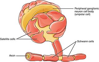
Schwann cells or neurolemmocytes are the principal glia of the peripheral nervous system (PNS). Glial cells function to support neurons and in the PNS, also include satellite cells, olfactory ensheathing cells, enteric glia and glia that reside at sensory nerve endings, such as the Pacinian corpuscle. The two types of Schwann cells are myelinating and nonmyelinating. Myelinating Schwann cells wrap around axons of motor and sensory neurons to form the myelin sheath. The Schwann cell promoter is present in the downstream region of the human dystrophin gene that gives shortened transcript that are again synthesized in a tissue-specific manner.

The development of the nervous system, or neural development (neurodevelopment), refers to the processes that generate, shape, and reshape the nervous system of animals, from the earliest stages of embryonic development to adulthood. The field of neural development draws on both neuroscience and developmental biology to describe and provide insight into the cellular and molecular mechanisms by which complex nervous systems develop, from nematodes and fruit flies to mammals.

<span class="mw-page-title-main">Sympathetic nervous system</span> Part of the autonomic nervous system which stimulates fight-or-flight responses

The sympathetic nervous system (SNS) is one of the three divisions of the autonomic nervous system, the others being the parasympathetic nervous system and the enteric nervous system. The enteric nervous system is sometimes considered part of the autonomic nervous system, and sometimes considered an independent system.

<span class="mw-page-title-main">Nervous tissue</span> Main component of the nervous system

Nervous tissue, also called neural tissue, is the main tissue component of the nervous system. The nervous system regulates and controls body functions and activity. It consists of two parts: the central nervous system (CNS) comprising the brain and spinal cord, and the peripheral nervous system (PNS) comprising the branching peripheral nerves. It is composed of neurons, also known as nerve cells, which receive and transmit impulses, and neuroglia, also known as glial cells or glia, which assist the propagation of the nerve impulse as well as provide nutrients to the neurons.

<span class="mw-page-title-main">Glia</span> Support cells in the nervous system

Glia, also called glial cells(gliocytes) or neuroglia, are non-neuronal cells in the central nervous system (brain and spinal cord) and the peripheral nervous system that do not produce electrical impulses. The neuroglia make up more than one half the volume of neural tissue in our body. They maintain homeostasis, form myelin in the peripheral nervous system, and provide support and protection for neurons. In the central nervous system, glial cells include oligodendrocytes, astrocytes, ependymal cells and microglia, and in the peripheral nervous system they include Schwann cells and satellite cells.

<span class="mw-page-title-main">Astrocyte</span> Type of brain cell

Astrocytes, also known collectively as astroglia, are characteristic star-shaped glial cells in the brain and spinal cord. They perform many functions, including biochemical control of endothelial cells that form the blood–brain barrier, provision of nutrients to the nervous tissue, maintenance of extracellular ion balance, regulation of cerebral blood flow, and a role in the repair and scarring process of the brain and spinal cord following infection and traumatic injuries. The proportion of astrocytes in the brain is not well defined; depending on the counting technique used, studies have found that the astrocyte proportion varies by region and ranges from 20% to around 40% of all glia. Another study reports that astrocytes are the most numerous cell type in the brain. Astrocytes are the major source of cholesterol in the central nervous system. Apolipoprotein E transports cholesterol from astrocytes to neurons and other glial cells, regulating cell signaling in the brain. Astrocytes in humans are more than twenty times larger than in rodent brains, and make contact with more than ten times the number of synapses.

<span class="mw-page-title-main">Astrogliosis</span> Increase in astrocytes in response to brain injury

Astrogliosis is an abnormal increase in the number of astrocytes due to the destruction of nearby neurons from central nervous system (CNS) trauma, infection, ischemia, stroke, autoimmune responses or neurodegenerative disease. In healthy neural tissue, astrocytes play critical roles in energy provision, regulation of blood flow, homeostasis of extracellular fluid, homeostasis of ions and transmitters, regulation of synapse function and synaptic remodeling. Astrogliosis changes the molecular expression and morphology of astrocytes, in response to infection for example, in severe cases causing glial scar formation that may inhibit axon regeneration.

Oligodendrocyte progenitor cells (OPCs), also known as oligodendrocyte precursor cells, NG2-glia, O2A cells, or polydendrocytes, are a subtype of glia in the central nervous system named for their essential role as precursors to oligodendrocytes. They are typically identified in the human by co-expression of PDGFRA and CSPG4.

<span class="mw-page-title-main">Glutamate receptor</span> Cell-surface proteins that bind glutamate and trigger changes which influence the behavior of cells

Glutamate receptors are synaptic and non synaptic receptors located primarily on the membranes of neuronal and glial cells. Glutamate is abundant in the human body, but particularly in the nervous system and especially prominent in the human brain where it is the body's most prominent neurotransmitter, the brain's main excitatory neurotransmitter, and also the precursor for GABA, the brain's main inhibitory neurotransmitter. Glutamate receptors are responsible for the glutamate-mediated postsynaptic excitation of neural cells, and are important for neural communication, memory formation, learning, and regulation.

<span class="mw-page-title-main">Neuroimmune system</span>

The neuroimmune system is a system of structures and processes involving the biochemical and electrophysiological interactions between the nervous system and immune system which protect neurons from pathogens. It serves to protect neurons against disease by maintaining selectively permeable barriers, mediating neuroinflammation and wound healing in damaged neurons, and mobilizing host defenses against pathogens.

Glutamate transporters are a family of neurotransmitter transporter proteins that move glutamate – the principal excitatory neurotransmitter – across a membrane. The family of glutamate transporters is composed of two primary subclasses: the excitatory amino acid transporter (EAAT) family and vesicular glutamate transporter (VGLUT) family. In the brain, EAATs remove glutamate from the synaptic cleft and extrasynaptic sites via glutamate reuptake into glial cells and neurons, while VGLUTs move glutamate from the cell cytoplasm into synaptic vesicles. Glutamate transporters also transport aspartate and are present in virtually all peripheral tissues, including the heart, liver, testes, and bone. They exhibit stereoselectivity for L-glutamate but transport both L-aspartate and D-aspartate.

Gliosis is a nonspecific reactive change of glial cells in response to damage to the central nervous system (CNS). In most cases, gliosis involves the proliferation or hypertrophy of several different types of glial cells, including astrocytes, microglia, and oligodendrocytes. In its most extreme form, the proliferation associated with gliosis leads to the formation of a glial scar.

<span class="mw-page-title-main">Radial glial cell</span> Bipolar-shaped progenitor cells of all neurons in the cerebral cortex and some glia

Radial glial cells, or radial glial progenitor cells (RGPs), are bipolar-shaped progenitor cells that are responsible for producing all of the neurons in the cerebral cortex. RGPs also produce certain lineages of glia, including astrocytes and oligodendrocytes. Their cell bodies (somata) reside in the embryonic ventricular zone, which lies next to the developing ventricular system.

<span class="mw-page-title-main">Glia limitans</span> Thin astrocyte membrane surrounding the brain and spinal cord

The glia limitans, or the glial limiting membrane, is a thin barrier of astrocyte foot processes associated with the parenchymal basal lamina surrounding the brain and spinal cord. It is the outermost layer of neural tissue, and among its responsibilities is the prevention of the over-migration of neurons and neuroglia, the supporting cells of the nervous system, into the meninges. The glia limitans also plays an important role in regulating the movement of small molecules and cells into the brain tissue by working in concert with other components of the central nervous system (CNS) such as the blood–brain barrier (BBB).

Gliotransmitters are chemicals released from glial cells that facilitate neuronal communication between neurons and other glial cells. They are usually induced from Ca2+ signaling, although recent research has questioned the role of Ca2+ in gliotransmitters and may require a revision of the relevance of gliotransmitters in neuronal signalling in general.

In biochemistry, the glutamate–glutamine cycle is a cyclic metabolic pathway which maintains an adequate supply of the neurotransmitter glutamate in the central nervous system. Neurons are unable to synthesize either the excitatory neurotransmitter glutamate, or the inhibitory GABA from glucose. Discoveries of glutamate and glutamine pools within intercellular compartments led to suggestions of the glutamate–glutamine cycle working between neurons and astrocytes. The glutamate/GABA–glutamine cycle is a metabolic pathway that describes the release of either glutamate or GABA from neurons which is then taken up into astrocytes. In return, astrocytes release glutamine to be taken up into neurons for use as a precursor to the synthesis of either glutamate or GABA.

<span class="mw-page-title-main">Outline of the human nervous system</span> Overview of and topical guide to the human nervous system

The following diagram is provided as an overview of and topical guide to the human nervous system:

<span class="mw-page-title-main">Alexei Verkhratsky</span> Ukrainian professor and researcher

Alexei Verkhratsky, sometimes spelled Alexej, is a professor of neurophysiology at the University of Manchester best known for his research on the physiology and pathophysiology of neuroglia, calcium signalling, and brain ageing. He is an elected member and vice-president of Academia Europaea, of the German National Academy of Sciences Leopoldina, of the Real Academia Nacional de Farmacia (Spain), of the Slovenian Academy of Sciences and Arts, of Polish Academy of Sciences, and Dana Alliance for Brain Initiatives, among others. Since 2010, he is a Ikerbasque Research Professor and from 2012 he is deputy director of the Achucarro Basque Center for Neuroscience in Bilbao. He is a distinguished professor at Jinan University, China Medical University of Shenyang, and Chengdu University of Traditional Chinese Medicine and is an editor-in-chief of Cell Calcium, receiving editor for Cell Death and Disease, and Acta Physiologica and member of editorial board of many academic journals.

References

  1. Krstić, Radivoj V. (1985). "Glia of the Central Nervous System. Ependymal Cells". General Histology of the Mammal. Springer. pp. 302–303. doi:10.1007/978-3-642-70420-8_147. ISBN   978-3-642-70420-8.
  2. 1 2 3 4 5 6 7 8 9 10 11 12 13 14 15 Hanani M (June 2005). "Satellite glial cells in sensory ganglia: from form to function" (PDF). Brain Res. Brain Res. Rev. 48 (3): 457–76. doi:10.1016/j.brainresrev.2004.09.001. PMID   15914252. S2CID   5316025.
  3. 1 2 3 4 5 6 7 8 9 Hanani M (September 2010). "Satellite glial cells in sympathetic and parasympathetic ganglia: in search of function". Brain Res Rev. 64 (2): 304–27. doi:10.1016/j.brainresrev.2010.04.009. PMID   20441777. S2CID   11833205.
  4. 1 2 Hall AK; Landis SC (September 1992). "Division and migration of satellite glia in the embryonic rat superior cervical ganglion". J. Neurocytol. 21 (9): 635–47. doi:10.1007/bf01191725. PMID   1403009. S2CID   42893326.
  5. 1 2 Shinder V; Devor M (September 1994). "Structural basis of neuron-to-neuron cross-excitation in dorsal root ganglia". J. Neurocytol. 23 (9): 515–31. doi:10.1007/bf01262054. PMID   7815085. S2CID   37909973.
  6. 1 2 3 4 5 Villa G; Fumagalli M; Verderio C; Abbracchio MP; Ceruti S (February 2010). "Expression and contribution of satellite glial cells purinoceptors to pain transmission in sensory ganglia: an update". Neuron Glia Biol. 6 (1): 31–42. doi:10.1017/S1740925X10000086. PMID   20604978.
  7. 1 2 Levin MJ; Cai GY; Manchak MD; Pizer LI (June 2003). "Varicella-zoster virus DNA in cells isolated from human trigeminal ganglia". J. Virol. 77 (12): 6979–87. doi:10.1128/jvi.77.12.6979-6987.2003. PMC   156183 . PMID   12768016.
  8. 1 2 Hanani M (February 2010). "Satellite glial cells: more than just 'rings around the neuron'". Neuron Glia Biol. 6 (1): 1–2. doi: 10.1017/S1740925X10000104 . PMID   20604976.
  9. 1 2 Dixon JS (January 1969). "Changes in the fine structure of satellite cells surrounding chromatolytic neurons". Anat. Rec. 163 (1): 101–9. doi:10.1002/ar.1091630112. PMID   5763130. S2CID   36355664.
  10. 1 2 3 4 5 6 Pannese E (February 2010). "The structure of the perineuronal sheath of satellite glial cells (SGCs) in sensory ganglia". Neuron Glia Biol. 6 (1): 3–10. doi:10.1017/S1740925X10000037. PMID   20604977.
  11. Mirsky R; Jessen KR; Schachner M; Goridis C (December 1986). "Distribution of the adhesion molecules N-CAM and L1 on peripheral neurons and glia in adult rats". J. Neurocytol. 15 (6): 799–815. doi:10.1007/bf01625196. PMID   3819781. S2CID   12144849.
  12. Hibino H, Horio Y, Fujita A, et al. (October 1999). "Expression of an inwardly rectifying K(+) channel, Kir4.1, in satellite cells of rat cochlear ganglia". Am. J. Physiol. 277 (4 Pt 1): C638–44. doi:10.1152/ajpcell.1999.277.4.C638. PMID   10516093. S2CID   44872893.
  13. HESS A (December 1955). "The fine structure of young and old spinal ganglia". Anat. Rec. 123 (4): 399–423. doi:10.1002/ar.1091230403. PMID   13292772. S2CID   40915651.
  14. Citkowitz E; Holtzman E (January 1973). "Peroxisomes in dorsal root ganglia". J. Histochem. Cytochem. 21 (1): 34–41. doi: 10.1177/21.1.34 . PMID   4694538.
  15. Pannese E (July 1964). "Number And Structure Of Perisomatic Satellite Cells Of Spinal Ganglia Under Normal Conditions Or During Axon Regeneration And Neuronal Hypertrophy". Z Zellforsch Mikrosk Anat. 63 (4): 568–92. doi:10.1007/bf00339491. PMID   14254752. S2CID   6191814.
  16. Pannese E (1981). "The satellite cells of the sensory ganglia". Adv Anat Embryol Cell Biol. 65: 1–111. doi:10.1007/978-3-642-67750-2_1. PMID   7013430.
  17. Pannese E; Ledda M; Arcidiacono G; Rigamonti L (May 1991). "Clusters of nerve cell bodies enclosed within a common connective tissue envelope in the spinal ganglia of the lizard and rat". Cell Tissue Res. 264 (2): 209–14. doi:10.1007/BF00313957. PMID   1878941. S2CID   617044.
  18. Pannese E (2002). Perikaryal surface specializations of neurons in sensory ganglia. International Review of Cytology. Vol. 220. pp. 1–34. doi:10.1016/S0074-7696(02)20002-9. ISBN   9780123646248. PMID   12224547.
  19. Elfvin LG (January 1968). "A new granule-containing nerve cell in the inferior mesenteric ganglion of the rabbit". J. Ultrastruct. Res. 22 (1): 37–44. doi:10.1016/s0022-5320(68)90048-8. PMID   5653898.
  20. Elfvin LG (November 1971). "Ultrastructural studies on the synaptology of the inferior mesenteric ganglion of the cat. I. Observations on the cell surface of the postganglionic perikarya". J. Ultrastruct. Res. 37 (3): 411–25. doi:10.1016/s0022-5320(71)80135-1. PMID   4331152.
  21. 1 2 3 Jasmin L; Vit JP; Bhargava A; Ohara PT (February 2010). "Can satellite glial cells be therapeutic targets for pain control?". Neuron Glia Biol. 6 (1): 63–71. doi:10.1017/S1740925X10000098. PMC   3139431 . PMID   20566001.
  22. Ichikawa H; Jacobowitz DM; Sugimoto T (February 1997). "S100 protein-immunoreactive primary sensory neurons in the trigeminal and dorsal root ganglia of the rat". Brain Res. 748 (1–2): 253–7. doi:10.1016/S0006-8993(96)01364-9. PMID   9067472. S2CID   31518343.
  23. Allen DT; Kiernan JA (April 1994). "Permeation of proteins from the blood into peripheral nerves and ganglia". Neuroscience. 59 (3): 755–64. doi:10.1016/0306-4522(94)90192-9. PMID   8008217. S2CID   2516314.
  24. Ten Tusscher MP; Klooster J; Vrensen GF (June 1989). "Satellite cells as blood-ganglion cell barrier in autonomic ganglia". Brain Res. 490 (1): 95–102. doi:10.1016/0006-8993(89)90434-4. PMID   2474362. S2CID   22719441.
  25. Bowery NG; Brown DA; Marsh S (August 1979). "gamma-Aminobutyric acid efflux from sympathetic glial cells: effect of 'depolarizing' agents". J. Physiol. 293: 75–101. doi:10.1113/jphysiol.1979.sp012879. PMC   1280703 . PMID   501652.
  26. Alvarez-Leefmans FJ; León-Olea M; Mendoza-Sotelo J; Alvarez FJ; Antón B; Garduño R (2001). "Immunolocalization of the Na(+)-K(+)-2Cl(-) cotransporter in peripheral nervous tissue of vertebrates". Neuroscience. 104 (2): 569–82. doi:10.1016/S0306-4522(01)00091-4. PMID   11377856. S2CID   36201377.
  27. Berger UV; Hediger MA (June 2000). "Distribution of the glutamate transporters GLAST and GLT-1 in rat circumventricular organs, meninges, and dorsal root ganglia". J. Comp. Neurol. 421 (3): 385–99. doi:10.1002/(SICI)1096-9861(20000605)421:3<385::AID-CNE7>3.0.CO;2-S. PMID   10813794. S2CID   38551356.
  28. 1 2 Miller KE; Richards BA; Kriebel RM (August 2002). "Glutamine-, glutamine synthetase-, glutamate dehydrogenase- and pyruvate carboxylase-immunoreactivities in the rat dorsal root ganglion and peripheral nerve". Brain Res. 945 (2): 202–11. doi:10.1016/S0006-8993(02)02802-0. PMID   12126882. S2CID   2140577.
  29. Julius D; Basbaum AI (September 2001). "Molecular mechanisms of nociception". Nature. 413 (6852): 203–10. Bibcode:2001Natur.413..203J. doi:10.1038/35093019. PMID   11557989. S2CID   13263269.
  30. 1 2 3 Gosselin RD; Suter MR; Ji RR; Decosterd I (October 2010). "Glial cells and chronic pain". Neuroscientist. 16 (5): 519–31. doi:10.1177/1073858409360822. PMC   3017463 . PMID   20581331.
  31. Steiner I (August 1996). "Human herpes viruses latent infection in the nervous system". Immunol. Rev. 152: 157–73. doi:10.1111/j.1600-065X.1996.tb00915.x. PMID   8930672. S2CID   31268611.
  32. LaVail JH; Topp KS; Giblin PA; Garner JA (August 1997). "Factors that contribute to the transneuronal spread of herpes simplex virus". J. Neurosci. Res. 49 (4): 485–96. doi:10.1002/(SICI)1097-4547(19970815)49:4<485::AID-JNR9>3.0.CO;2-4. PMID   9285524. S2CID   22825338.
  33. Wilkinson R; Leaver C; Simmons A; Pereira RA (August 1999). "Restricted replication of herpes simplex virus in satellite glial cell cultures clonally derived from adult mice". J. Neurovirol. 5 (4): 384–91. doi:10.3109/13550289909029479. PMID   10463860.
  34. Elson K; Speck P; Simmons A (May 2003). "Herpes simplex virus infection of murine sensory ganglia induces proliferation of neuronal satellite cells". J. Gen. Virol. 84 (Pt 5): 1079–84. doi: 10.1099/vir.0.19035-0 . PMID   12692271.
  35. Weick M, Cherkas PS, Härtig W, et al. (2003). "P2 receptors in satellite glial cells in trigeminal ganglia of mice". Neuroscience. 120 (4): 969–77. doi:10.1016/S0306-4522(03)00388-9. PMID   12927203. S2CID   25702841.