P2Y receptor

Last updated

P2Y receptors are a family of purinergic G protein-coupled receptors, stimulated by nucleotides such as adenosine triphosphate, adenosine diphosphate, uridine triphosphate, uridine diphosphate and UDP-glucose.To date, 8 P2Y receptors have been cloned in humans: P2Y1, P2Y2, P2Y4, P2Y6, P2Y11, P2Y12, P2Y13 and P2Y14. [1]

Contents

P2Y receptors are present in almost all human tissues where they exert various biological functions based on their G-protein coupling. P2Y receptors mediate responses including vasodilation, [2] blood clotting, [3] and immune response. [4] Due to their ubiquity and variety in function, they are a common biological target in pharmacological development. [3]

Structure

P2Y12 structure as generated by PYMOL with color-coded helices P2Y12 cartoon structure (generated by pymol).png
P2Y12 structure as generated by PYMOL with color-coded helices

P2Y receptors are membrane proteins belonging to the class A family of G protein-coupled receptors (GPCRs). [5] [6] P2Y receptor proteins display large-scale structural domains typical of GPCRs, consisting of seven hydrophobic transmembrane helices connected by three short extracellular loops and three variably sized intracellular loops; an extracellular N-terminus; and an intracellular C-terminus. [7] The extracellular regions interact with the receptor ligands, while the intracellular regions activate the G protein, control receptor internalization, and mediate dimerization. [6] Similar to other GPCRs, P2Y receptors can form both homodimers and heterodimers. These dimeric forms may vary significantly in their biochemical and pharmacological properties from the monomeric receptor.

In addition to the structural domains typical of all GPCRs, some structural elements are common across P2Y receptor subtypes. All P2Y receptors contain four extracellular cysteine residues which can form two disulfide bridges, one between the N-terminus domain and the proximal extracellular loop and another between the two remaining extracellular loops. [6] These disulfide bonds have been shown to be involved in ligand binding and signal transduction. [8] In addition, several polar residues found within the transmembrane helices are highly conserved across both species and receptor subtypes. Mutational analysis has suggested that these residues are integral to the ligand-binding mechanism of P2Y receptors. Outside of these conserved regions, the P2Y receptor family exhibits unusually high diversity in primary structure, with P2Y1 sharing only 19% of its primary structure with P2Y12. [6] Despite this, the individual P2Y subtypes are highly conserved across species, with human and mouse P2Y receptors sharing 95% of amino acids.

The ligand-binding mechanisms of P2Y receptors are not currently well established. [8] The binding complex of P2Y receptors with ATP is of significant interest, as no P2Y receptor contains amino acids sequences similar to any of the many established ATP-binding sites. [7] Recent x-ray crystallography of the human P2Y12 receptor has shown several structural irregularities in regions that are typically highly conserved across GPCRs. [8]

In contrast to the unusual structure and behavior of the extracellular ligand binding domains, P2Y intracellular domains appear to be structurally and mechanistically similar to other GPCRs. [6]

Signal transduction

P2Y receptors respond either positively or negatively to the presence of nucleotides in extracellular solution. [9] Nucleotides may be divided into two categories: purines and pyrimidines. Individual P2Y receptor species may respond to only purines, only pyrimidines, or both; the activation profiles of the eight known P2Y receptors are listed below. [9]

P2Y speciesReceptivity
P2Y1Activation by purines [9]
P2Y2Activation by purines and pyrimidines triphosphates [9]
P2Y4Activation by pyrimidines [9]
P2Y6Activation by pyrimidines [9]
P2Y11Activation by purines [9]
P2Y12Inactivation by ADP through G1 protein [9]
P2Y13Inactivation by ADP through G1 protein [9]
P2Y14Activation by UDP-Glucose [9]

The activity of P2Y receptors is linked to a signal cascade originating in regulation of the flow of Ca2+ and K+ ions by the receptor's interactions with G proteins, modulating access to Ca2+ and K+ channels, though the exact behavior is dependent upon individual receptor species. [10] Voltage-independent Ca2+ channels allow for the free flow of Ca2+ ions from the cell activated by P2Y receptors. [10] Oscillation of Ca2+ concentration is directly affected by the signal-transduction activity of P2Y1; specifically, through protein kinase C phosphorylation of Thr339 in the carboxy terminus of the P2Y1 receptor. [10]

Changes in the concentration of Ca2+ have many important ramifications for the cell, including regulation of cell metabolism (e.g. autophagy initiation / regulation), ATP production (through Ca2+ entering the mitochondrial outer mitochondrial membrane and stimulation of mitochondrial dehydrogenases leading to the production of ATP), and the possibility of triggering apoptosis. [11] [12] Both autophagy and apoptosis are cell stress responses that play significant roles in cells' overall life cycles, though autophagy seeks to preserve the viability of the cell by recycling unit parts of organelles, while apoptosis acts in the interest of the whole organism at the expense of the cell undergoing apoptosis. [12]

Pharmacology

Clopidogrel (Plavix), an inhibitor of the P2Y12 receptor, was formerly the second best-selling drug in the world Plavix 2007-04-19.jpg
Clopidogrel (Plavix), an inhibitor of the P2Y12 receptor, was formerly the second best-selling drug in the world

Many commonly prescribed medications target P2Y receptors, and active research is being conducted into developing new drugs targeting these receptors. [3] The most commonly prescribed drug targeting P2Y receptors is clopidogrel, an antiplatelet medication which acts on the P2Y12 receptor in a manner shared with other thienopyridines. [14] Other pharmaceutical applications include denufosol, which targets P2Y2 and is being investigated for the treatment of cystic fibrosis, and diquafosol, a P2Y2 agonist used in the treatment of dry eye disease. [15] [16] [17]

P2Y6 receptors have been shown to play a role in cerebral vasodilation. UDP-analogs which bind to this receptor have been investigated as possible treatments for migraines. [18] [17]

P2Y11 is a regulator of immune response, and a common polymorphism carried by almost 20% of North European caucasians give increased risk of myocardial infarction, making P2Y11 an interesting drug target candidate for treatment of myocardial infarction. [4] [16] [17]

In addition to established uses, pharmaceutical research has been conducted into the role of P2Y receptors in osteoporosis, [2] diabetes, [19] and cardio-protection. [20] [17]

Coupling

The biological effects of P2Y receptor activation depends on how they couple to downstream signalling pathways, either via Gi, Gq/11 or Gs G proteins. Human P2Y receptors have the following G protein coupling: [21]

ProteinGeneCoupling Nucleotide
P2RY1 P2RY1 Gq/11 ADP
P2RY2 P2RY2 Gq/11 (and Gi) ATP, UTP
P2RY4 P2RY4 Gq/11 (and Gi) UTP
P2RY5 / LPA6 LPAR6 Lysophosphatidic acid [22]
P2RY6 P2RY6 Gq/11 UDP
P2RY8 P2RY8 orphan receptor
P2RY9 / LPAR4 / GPR23 LPAR4 Lysophosphatidic acid
P2RY10 P2RY10 orphan receptor
P2RY11 P2RY11 Gq/11 and Gs ATP
P2RY12 P2RY12 Gi ADP
P2RY13 P2RY13 Gi ADP
P2RY14 P2RY14 Gi UDP-glucose

The gaps in P2Y receptor numbering is due to that several receptors (P2Y3, P2Y5, P2Y7, P2Y8, P2Y9, P2Y10) were thought to be P2Y receptors when they were cloned, when in fact they are not.

See also

Related Research Articles

<span class="mw-page-title-main">G protein-coupled receptor</span> Class of cell surface receptors coupled to G-protein-associated intracellular signaling

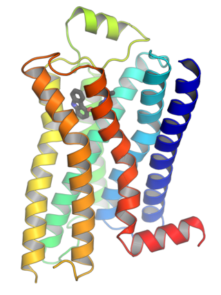
G protein-coupled receptors (GPCRs), also known as seven-(pass)-transmembrane domain receptors, 7TM receptors, heptahelical receptors, serpentine receptors, and G protein-linked receptors (GPLR), form a large group of evolutionarily related proteins that are cell surface receptors that detect molecules outside the cell and activate cellular responses. They are coupled with G proteins. They pass through the cell membrane seven times in form of six loops of amino acid residues, which is why they are sometimes referred to as seven-transmembrane receptors. Ligands can bind either to the extracellular N-terminus and loops or to the binding site within transmembrane helices. They are all activated by agonists, although a spontaneous auto-activation of an empty receptor has also been observed.

<span class="mw-page-title-main">Receptor (biochemistry)</span> Protein molecule receiving signals for a cell

In biochemistry and pharmacology, receptors are chemical structures, composed of protein, that receive and transduce signals that may be integrated into biological systems. These signals are typically chemical messengers which bind to a receptor and produce physiological responses such as change in the electrical activity of a cell. For example, GABA, an inhibitory neurotransmitter inhibits electrical activity of neurons by binding to GABAA receptors. There are three main ways the action of the receptor can be classified: relay of signal, amplification, or integration. Relaying sends the signal onward, amplification increases the effect of a single ligand, and integration allows the signal to be incorporated into another biochemical pathway.

Second messengers are intracellular signaling molecules released by the cell in response to exposure to extracellular signaling molecules—the first messengers. Second messengers trigger physiological changes at cellular level such as proliferation, differentiation, migration, survival, apoptosis and depolarization.

In biology, cell signaling or cell communication is the ability of a cell to receive, process, and transmit signals with its environment and with itself. Cell signaling is a fundamental property of all cellular life in prokaryotes and eukaryotes. Signals that originate from outside a cell can be physical agents like mechanical pressure, voltage, temperature, light, or chemical signals. Cell signaling can occur over short or long distances, and as a result can be classified as autocrine, juxtacrine, intracrine, paracrine, or endocrine. Signaling molecules can be synthesized from various biosynthetic pathways and released through passive or active transports, or even from cell damage.

P2Y<sub>12</sub> Protein-coding gene in the species Homo sapiens

P2Y12 is a chemoreceptor for adenosine diphosphate (ADP) that belongs to the Gi class of a group of G protein-coupled (GPCR) purinergic receptors. This P2Y receptor family has several receptor subtypes with different pharmacological selectivity, which overlaps in some cases, for various adenosine and uridine nucleotides. The P2Y12 receptor is involved in platelet aggregation and is thus a biological target for the treatment of thromboembolisms and other clotting disorders. Two transcript variants encoding the same isoform have been identified for this gene.

<span class="mw-page-title-main">Purinergic receptor</span> Family of cell membrane receptors in almost all tissues

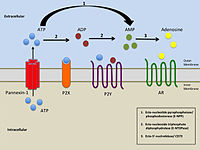
Purinergic receptors, also known as purinoceptors, are a family of plasma membrane molecules that are found in almost all mammalian tissues. Within the field of purinergic signalling, these receptors have been implicated in learning and memory, locomotor and feeding behavior, and sleep. More specifically, they are involved in several cellular functions, including proliferation and migration of neural stem cells, vascular reactivity, apoptosis and cytokine secretion. These functions have not been well characterized and the effect of the extracellular microenvironment on their function is also poorly understood.

<span class="mw-page-title-main">P2X purinoreceptor</span>

The P2X receptors, also ATP-gated P2X receptor cation channel family, is a protein family that consists of cation-permeable ligand-gated ion channels that open in response to the binding of extracellular adenosine 5'-triphosphate (ATP). They belong to a larger family of receptors known as the ENaC/P2X superfamily. ENaC and P2X receptors have similar 3-D structures and are homologous. P2X receptors are present in a diverse array of organisms including humans, mouse, rat, rabbit, chicken, zebrafish, bullfrog, fluke, and amoeba.

<span class="mw-page-title-main">Ectonucleotidase</span>

Ectonucleotidases consist of families of nucleotide metabolizing enzymes that are expressed on the plasma membrane and have externally oriented active sites. These enzymes metabolize nucleotides to nucleosides. The contribution of ectonucleotidases in the modulation of purinergic signaling depends on the availability and preference of substrates and on cell and tissue distribution.

<span class="mw-page-title-main">P2RY1</span> Protein-coding gene in the species Homo sapiens

P2Y purinoceptor 1 is a protein that in humans is encoded by the P2RY1 gene.

<span class="mw-page-title-main">P2RY2</span> Protein-coding gene in the species Homo sapiens

P2Y purinoceptor 2 is a protein that in humans is encoded by the P2RY2 gene.

<span class="mw-page-title-main">GPR17</span> Protein-coding gene in the species Homo sapiens

Uracil nucleotide/cysteinyl leukotriene receptor is a G protein-coupled receptor that in humans is encoded by the GPR17 gene located on chromosome 2 at position q21. The actual activating ligands for and some functions of this receptor are disputed.

<span class="mw-page-title-main">P2RY6</span> Protein-coding gene in the species Homo sapiens

P2Y purinoceptor 6 is a protein that in humans is encoded by the P2RY6 gene.

<span class="mw-page-title-main">P2RY11</span> Protein-coding gene in the species Homo sapiens

P2Y purinoceptor 11 is a protein that in humans is encoded by the P2RY11 gene.

<span class="mw-page-title-main">P2RY14</span> Protein-coding gene in the species Homo sapiens

P2Y purinoceptor 14 is a protein that in humans is encoded by the P2RY14 gene.

<span class="mw-page-title-main">LPAR6</span> Protein-coding gene in the species Homo sapiens

Lysophosphatidic acid receptor 6, also known as LPA6, P2RY5 and GPR87, is a protein that in humans is encoded by the LPAR6 gene. LPA6 is a G protein-coupled receptor that binds the lipid signaling molecule lysophosphatidic acid (LPA).

<span class="mw-page-title-main">OXGR1</span> Protein-coding gene in the species Homo sapiens

2-Oxoglutarate receptor 1 (OXGR1), also known as cysteinyl leukotriene receptor E (CysLTE) and GPR99, is a protein that in humans is encoded by the OXGR1 gene. The Gene has recently been nominated as a receptor not only for 2-oxogluterate but also for the three cysteinyl leukotrienes (CysLTs), particularly leukotriene E4 (LTE4) and to far lesser extents LTC4 and LTE4. Recent studies implicate GPR99 as a cellular receptor which is activated by LTE4 thereby causing these cells to contribute to mediating various allergic and hypersensitivity responses.

<span class="mw-page-title-main">P2RY10</span> Protein-coding gene in the species Homo sapiens

Putative P2Y purinoceptor 10 is a protein that, in humans, is encoded by the P2RY10 gene.

<span class="mw-page-title-main">P2RY4</span> Protein-coding gene in the species Homo sapiens

P2Y purinoceptor 4 is a protein that in humans is encoded by the P2RY4 gene.

P2 receptor may refer to:

<span class="mw-page-title-main">Purinergic signalling</span> Signalling complex involving purine nucleosides and their receptors

Purinergic signalling is a form of extracellular signalling mediated by purine nucleotides and nucleosides such as adenosine and ATP. It involves the activation of purinergic receptors in the cell and/or in nearby cells, thereby regulating cellular functions.

References

  1. Abbracchio MP, Burnstock G, Boeynaems JM, Barnard EA, Boyer JL, Kennedy C, Knight GE, Fumagalli M, Gachet C, Jacobson KA, Weisman GA (September 2006). "International Union of Pharmacology LVIII: update on the P2Y G protein-coupled nucleotide receptors: from molecular mechanisms and pathophysiology to therapy". Pharmacological Reviews. 58 (3): 281–341. doi:10.1124/pr.58.3.3. PMC   3471216 . PMID   16968944.
  2. 1 2 Romanello M, Bivi N, Pines A, Deganuto M, Quadrifoglio F, Moro L, Tell G (October 2006). "Bisphosphonates activate nucleotide receptors signaling and induce the expression of Hsp90 in osteoblast-like cell lines". Bone. 39 (4): 739–53. doi:10.1016/j.bone.2006.03.011. PMID   16697713.
  3. 1 2 3 Erlinge D (2011-01-01). "P2Y receptors in health and disease". Pharmacology of Purine and Pyrimidine Receptors. Advances in Pharmacology. Vol. 61. pp. 417–39. doi:10.1016/B978-0-12-385526-8.00013-8. ISBN   9780123855268. PMID   21586366.
  4. 1 2 Amisten S, Melander O, Wihlborg AK, Berglund G, Erlinge D (January 2007). "Increased risk of acute myocardial infarction and elevated levels of C-reactive protein in carriers of the Thr-87 variant of the ATP receptor P2Y11". European Heart Journal. 28 (1): 13–8. doi: 10.1093/eurheartj/ehl410 . PMID   17135283.
  5. Jacobson KA, Jayasekara MP, Costanzi S (September 2012). "Molecular Structure of P2Y Receptors: Mutagenesis, Modeling, and Chemical Probes". Wiley Interdisciplinary Reviews: Membrane Transport and Signaling. 1 (6): 815–827. doi:10.1002/wmts.68. PMC   3547624 . PMID   23336097.
  6. 1 2 3 4 5 von Kügelgen I, Harden TK (2011). "Chapter 12: Molecular Pharmacology, Physiology, and Structure of the P2Y Receptors". In Jacobson KA, Linden J (eds.). Pharmacology of Purine and Pyrimidine Receptors. Elsevier. pp. 373–399. ISBN   978-0-12-385526-8 . Retrieved 8 November 2018.
  7. 1 2 Dubyak, George R. (2013). "P2Y Receptors". Encyclopedia of Biological Chemistry (2nd. ed.). Cleveland, OH: Elsevier Inc. pp. 375–378. doi:10.1016/B978-0-12-378630-2.00350-9. ISBN   978-0-12-378631-9.
  8. 1 2 3 Zhang K, Zhang J, Gao ZG, Zhang D, Zhu L, Han GW, et al. (May 2014). "Structure of the human P2Y12 receptor in complex with an antithrombotic drug". Nature. 509 (7498): 115–8. Bibcode:2014Natur.509..115Z. doi:10.1038/nature13083. PMC   4174307 . PMID   24670650.
  9. 1 2 3 4 5 6 7 8 9 10 Tulapurkar ME, Schäfer R, Hanck T, Flores RV, Weisman GA, González FA, Reiser G (June 2005). "Endocytosis mechanism of P2Y2 nucleotide receptor tagged with green fluorescent protein: clathrin and actin cytoskeleton dependence". Cellular and Molecular Life Sciences. 62 (12): 1388–99. doi:10.1007/s00018-005-5052-0. PMID   15924261.
  10. 1 2 3 Van Kolen K, Slegers H (September 2006). "Integration of P2Y receptor-activated signal transduction pathways in G protein-dependent signalling networks". Purinergic Signalling. 2 (3): 451–69. doi:10.1007/s11302-006-9008-0. PMC   2254474 . PMID   18404483.
  11. Hajnóczky G, Csordás G, Das S, Garcia-Perez C, Saotome M, Sinha Roy S, Yi M (2006-11-01). "Mitochondrial calcium signalling and cell death: approaches for assessing the role of mitochondrial Ca2+ uptake in apoptosis". Cell Calcium. 40 (5–6): 553–60. doi:10.1016/j.ceca.2006.08.016. PMC   2692319 . PMID   17074387.
  12. 1 2 Decuypere JP, Bultynck G, Parys JB (September 2011). "A dual role for Ca(2+) in autophagy regulation". Cell Calcium. 50 (3): 242–50. doi:10.1016/j.ceca.2011.04.001. PMID   21571367.
  13. Doll J, Zeitler E, Becker R (July 2013). "Generic clopidogrel: time to substitute?". JAMA. 310 (2): 145–6. doi:10.1001/jama.2013.7155. PMID   23839745.
  14. von Kügelgen I (2017). 12 Receptor. Advances in Experimental Medicine and Biology. Vol. 1051. pp. 123–138. doi:10.1007/5584_2017_98. ISBN   978-981-10-7610-7. PMID   28921447.
  15. Peral A, Domínguez-Godínez CO, Carracedo G, Pintor J (April 2008). "Therapeutic targets in dry eye syndrome". Drug News & Perspectives. 21 (3): 166–76. PMID   18560615.
  16. 1 2 Wan HX, Hu JH, Xie R, Yang SM, Dong H (May 2016). "Important roles of P2Y receptors in the inflammation and cancer of digestive system". Oncotarget. 7 (19): 28736–47. doi:10.18632/oncotarget.7518. PMC   5053759 . PMID   26908460.
  17. 1 2 3 4 von Kügelgen I, Hoffmann K (May 2016). "Pharmacology and structure of P2Y receptors". Neuropharmacology. 104: 50–61. doi:10.1016/j.neuropharm.2015.10.030. PMID   26519900.
  18. Malmsjö M, Hou M, Pendergast W, Erlinge D, Edvinsson L (May 2003). "Potent P2Y6 receptor mediated contractions in human cerebral arteries". BMC Pharmacology. 3 (1): 4. doi: 10.1186/1471-2210-3-4 . PMC   156657 . PMID   12737633.
  19. Amisten S, Meidute-Abaraviciene S, Tan C, Olde B, Lundquist I, Salehi A, Erlinge D (September 2010). "ADP mediates inhibition of insulin secretion by activation of P2Y13 receptors in mice". Diabetologia. 53 (9): 1927–34. doi: 10.1007/s00125-010-1807-8 . PMID   20526761.
  20. Yitzhaki S, Shainberg A, Cheporko Y, Vidne BA, Sagie A, Jacobson KA, Hochhauser E (October 2006). "Uridine-5'-triphosphate (UTP) reduces infarct size and improves rat heart function after myocardial infarct". Biochemical Pharmacology. 72 (8): 949–55. doi:10.1016/j.bcp.2006.07.019. PMC   4429760 . PMID   16939682.
  21. HUGO Gene Nomenclature Committee, Gene Family: Purinergic receptors P2Y (P2RY) , retrieved 2017-01-26.
  22. Pasternack SM, von Kügelgen I, Aboud KA, Lee YA, Rüschendorf F, Voss K, Hillmer AM, Molderings GJ, Franz T, Ramirez A, Nürnberg P, Nöthen MM, Betz RC (March 2008). "G protein-coupled receptor P2Y5 and its ligand LPA are involved in maintenance of human hair growth". Nature Genetics. 40 (3): 329–34. doi:10.1038/ng.84. PMID   18297070.