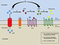
Part of a series on |
Purinergic signalling |
---|
Concepts |
Membrane transporters |
Purinergic receptors, also known as purinoceptors, are a family of plasma membrane molecules that are found in almost all mammalian tissues. [1] Within the field of purinergic signalling, these receptors have been implicated in learning and memory, locomotor and feeding behavior, and sleep. [2] More specifically, they are involved in several cellular functions, including proliferation and migration of neural stem cells, vascular reactivity, apoptosis and cytokine secretion. [2] [3] These functions have not been well characterized and the effect of the extracellular microenvironment on their function is also poorly understood.
The term purinergic receptor was originally introduced to illustrate specific classes of membrane receptors that mediate relaxation of gut smooth muscle as a response to the release of ATP (P2 receptors) or adenosine (P1 receptors). P2 receptors have further been divided into five subclasses: P2X, P2Y, P2Z, P2U, and P2T. To distinguish P2 receptors further, the subclasses have been divided into families of metabotropic (P2Y, P2U, and P2T) and ionotropic receptors (P2X and P2Z). [4]
In 2014, the first purinergic receptor in plants, DORN1, was discovered. [5]
Name | Activation | Class |
P1 receptors | adenosine | G protein-coupled receptors |
P2Y receptors | nucleotides | G protein-coupled receptors |
P2X receptors | ATP | ligand-gated ion channel |
There are three known distinct classes of purinergic receptors, known as P1, P2X, and P2Y receptors. [What about P2Z,U,T?]
P2X receptors are ligand-gated ion channels, whereas the P1 and P2Y receptors are G protein-coupled receptors. These ligand-gated ion channels are nonselective cation channels responsible for mediating excitatory postsynaptic responses, similar to nicotinic and ionotropic glutamate receptors. [6] P2X receptors are distinct from the rest of the widely known ligand-gated ion channels, as the genetic encoding of these particular channels indicates the presence of only two transmembrane domains within the channels. [1] These receptors are greatly distributed in neurons and glial cells throughout the central and peripheral nervous systems. [1] P2X receptors mediate a large variety of responses including fast transmission at central synapses, contraction of smooth muscle cells, platelet aggregation, macrophage activation, and apoptosis. [2] [7] Moreover, these receptors have been implicated in integrating functional activity between neurons, glial, and vascular cells in the central nervous system, thereby mediating the effects of neural activity during development, neurodegeneration, inflammation, and cancer. [2] The physiological modulator Zn2+ allosterically enhances ATP-induced inward cation currents in the P2X4 receptor by binding to cysteine 132 and cystine 149 residues on the extracellular domain of the P2X4 protein. [8] [9]
Both of these metabotropic receptors are distinguished by their reactivity to specific activators. P1 receptors are preferentially activated by adenosine and P2Y receptors are preferentially more activated by ATP. P1 and P2Y receptors are known to be widely distributed in the brain, heart, kidneys, and adipose tissue. Xanthines (e.g. caffeine) specifically block adenosine receptors, and are known to induce a stimulating effect to one's behavior. [10]
Inhibitors of purinergic receptors include clopidogrel, prasugrel and ticlopidine, as well as ticagrelor. All of these are antiplatelet agents that block P2Y12 receptors.
Data obtained from using P2 receptor-selective antagonists has produced evidence supporting ATP's ability to initiate and maintain chronic pain states after exposure to noxious stimuli. It is believed that ATP functions as a pronociceptive neurotransmitter, acting at specific P2X and P2Y receptors in a systemized manner, which ultimately (as a response to noxious stimuli) serve to initiate and sustain heightened states of neuronal excitability. This recent knowledge of purinergic receptors' effects on chronic pain provide promise in discovering a drug that specifically targets individual P2 receptor subtypes. While some P2 receptor-selective compounds have proven useful in preclinical trials, more research is required to understand the potential viability of P2 receptor antagonists for pain. [11]
Recent research has identified a role for microglial P2X receptors in neuropathic pain and inflammatory pain, especially the P2X4 and P2X7 receptors. [12] [13] [14] [15] [16]
Purinergic receptors have been suggested to play a role in the treatment of cytotoxic edema and brain infarctions. It was found that with treatment of the purinergic ligand 2-methylthioladenosine 5' diphosphate (2-MeSADP), which is an agonist and has a high preference for the purinergic receptor type 1 isoform (P2Y1R), significantly contributes to the reduction of an ischemic lesions caused by cytotoxic edema. Further pharmacological evidence has suggested that 2MeSADP protection is controlled by enhanced astrocyte mitochondrial metabolism through increased inositol triphosphate-dependent calcium release. There is evidence suggesting a relationship between the levels of ATP and cytotoxic edema, where low ATP levels are associated with an increased prevalence of cytotoxic edema. It is believed that mitochondria play an essential role in the metabolism of astrocyte energy within the penumbra of ischemic lesions. By enhancing the source of ATP provided by mitochondria, there could be a similar 'protective' effect for brain injuries in general. [17]
Purinergic receptors have been implicated in the vascular complications associated with diabetes due to the effect of high-glucose concentration on ATP-mediated responses in human fibroblasts. [18]
Astrocytes, also known collectively as astroglia, are characteristic star-shaped glial cells in the brain and spinal cord. They perform many functions, including biochemical control of endothelial cells that form the blood–brain barrier, provision of nutrients to the nervous tissue, maintenance of extracellular ion balance, regulation of cerebral blood flow, and a role in the repair and scarring process of the brain and spinal cord following infection and traumatic injuries. The proportion of astrocytes in the brain is not well defined; depending on the counting technique used, studies have found that the astrocyte proportion varies by region and ranges from 20% to around 40% of all glia. Another study reports that astrocytes are the most numerous cell type in the brain. Astrocytes are the major source of cholesterol in the central nervous system. Apolipoprotein E transports cholesterol from astrocytes to neurons and other glial cells, regulating cell signaling in the brain. Astrocytes in humans are more than twenty times larger than in rodent brains, and make contact with more than ten times the number of synapses.
Astrogliosis is an abnormal increase in the number of astrocytes due to the destruction of nearby neurons from central nervous system (CNS) trauma, infection, ischemia, stroke, autoimmune responses or neurodegenerative disease. In healthy neural tissue, astrocytes play critical roles in energy provision, regulation of blood flow, homeostasis of extracellular fluid, homeostasis of ions and transmitters, regulation of synapse function and synaptic remodeling. Astrogliosis changes the molecular expression and morphology of astrocytes, in response to infection for example, in severe cases causing glial scar formation that may inhibit axon regeneration.
The neuroimmune system is a system of structures and processes involving the biochemical and electrophysiological interactions between the nervous system and immune system which protect neurons from pathogens. It serves to protect neurons against disease by maintaining selectively permeable barriers, mediating neuroinflammation and wound healing in damaged neurons, and mobilizing host defenses against pathogens.
In the physiology of the kidney, tubuloglomerular feedback (TGF) is a feedback system inside the kidneys. Within each nephron, information from the renal tubules is signaled to the glomerulus. Tubuloglomerular feedback is one of several mechanisms the kidney uses to regulate glomerular filtration rate (GFR). It involves the concept of purinergic signaling, in which an increased distal tubular sodium chloride concentration causes a basolateral release of adenosine from the macula densa cells. This initiates a cascade of events that ultimately brings GFR to an appropriate level.
The ATP-gated P2X receptor cation channel family, or P2X receptor family, is a protein family that consists of cation-permeable ligand-gated ion channels that open in response to the binding of extracellular adenosine 5'-triphosphate (ATP). They belong to a larger family of receptors known as the ENaC/P2X superfamily. ENaC and P2X receptors have similar 3-D structures and are homologous. P2X receptors are present in a diverse array of organisms including humans, mouse, rat, rabbit, chicken, zebrafish, bullfrog, fluke, and amoeba.
Satellite glial cells, formerly called amphicytes, are glial cells that cover the surface of neuron cell bodies in ganglia of the peripheral nervous system. Thus, they are found in sensory, sympathetic, and parasympathetic ganglia. Both satellite glial cells (SGCs) and Schwann cells are derived from the neural crest of the embryo during development. SGCs have been found to play a variety of roles, including control over the microenvironment of sympathetic ganglia. They are thought to have a similar role to astrocytes in the central nervous system (CNS). They supply nutrients to the surrounding neurons and also have some structural function. Satellite cells also act as protective, cushioning cells. Additionally, they express a variety of receptors that allow for a range of interactions with neuroactive chemicals. Many of these receptors and other ion channels have recently been implicated in health issues including chronic pain and herpes simplex. There is much more to be learned about these cells, and research surrounding additional properties and roles of the SGCs is ongoing.
P2Y receptors are a family of purinergic G protein-coupled receptors, stimulated by nucleotides such as adenosine triphosphate, adenosine diphosphate, uridine triphosphate, uridine diphosphate and UDP-glucose.To date, 8 P2Y receptors have been cloned in humans: P2Y1, P2Y2, P2Y4, P2Y6, P2Y11, P2Y12, P2Y13 and P2Y14.
Ectonucleotidases consist of families of nucleotide metabolizing enzymes that are expressed on the plasma membrane and have externally oriented active sites. These enzymes metabolize nucleotides to nucleosides. The contribution of ectonucleotidases in the modulation of purinergic signaling depends on the availability and preference of substrates and on cell and tissue distribution.
Gliotransmitters are chemicals released from glial cells that facilitate neuronal communication between neurons and other glial cells. They are usually induced from Ca2+ signaling, although recent research has questioned the role of Ca2+ in gliotransmitters and may require a revision of the relevance of gliotransmitters in neuronal signalling in general.
P2X purinoceptor 7 is a protein that in humans is encoded by the P2RX7 gene.
P2Y purinoceptor 1 is a protein that in humans is encoded by the P2RY1 gene.
P2Y purinoceptor 2 is a protein that in humans is encoded by the P2RY2 gene.
P2 receptor may refer to:
P2X purinoceptor 4 is a protein that in humans is encoded by the P2RX4 gene. The product of this gene belongs to the family of purinoceptors for ATP. Multiple alternatively spliced transcript variants have been identified for this gene although their full-length natures have not been determined.
P2X purinoceptor 5 is a protein that in humans is encoded by the P2RX5 gene.
P2X purinoceptor 3 is a protein that in humans is encoded by the P2RX3 gene.
The rostral ventromedial medulla (RVM), or ventromedial nucleus of the spinal cord, is a group of neurons located close to the midline on the floor of the medulla oblongata. The rostral ventromedial medulla sends descending inhibitory and excitatory fibers to the dorsal horn spinal cord neurons. There are 3 categories of neurons in the RVM: on-cells, off-cells, and neutral cells. They are characterized by their response to nociceptive input. Off-cells show a transitory decrease in firing rate right before a nociceptive reflex, and are theorized to be inhibitory. Activation of off-cells, either by morphine or by any other means, results in antinociception. On-cells show a burst of activity immediately preceding nociceptive input, and are theorized to be contributing to the excitatory drive. Neutral cells show no response to nociceptive input.
Purinergic signalling is a form of extracellular signalling mediated by purine nucleotides and nucleosides such as adenosine and ATP. It involves the activation of purinergic receptors in the cell and/or in nearby cells, thereby regulating cellular functions.
Microglia are the primary immune cells of the central nervous system, similar to peripheral macrophages. They respond to pathogens and injury by changing morphology and migrating to the site of infection/injury, where they destroy pathogens and remove damaged cells.
Ream Al-Hasani is a British neuroscientist and pharmacologist as well as an assistant professor of anesthesiology at Washington University in St. Louis. Al-Hasani studies the endogenous opioid system to understand how to target it therapeutically to treat addiction, affective disorders, and chronic pain.