TNP refers to the chemical compound 2,4,6-trinitrophenol, also known as Picric acid.[2] It is a primary constituent of many unexploded landmines, and is a cousin to TNT, but less stable.[2] It is recognized as an environmental contaminant and is toxic to many organisms.[2] It is still commonly used in the manufacturing of fireworks, explosives, and rocket fuels, as well as in leather, pharmaceutical, and dye industries.[2]
ATP is an essential mediator of life.[1] It is used to overcome unfavorable energy barriers to initiate and fuel chemical reactions.[1] It is also used to drive biological machinery and regulate a number of processes via protein-phosphorylation.[1] However, the proteins that bind ATP for both regulation and enzymatic reactions are very diverse—many yet undiscovered—and for many proteins their relationship to ATP in terms of number of binding sites, binding constants, and dissociation constants remain unclear.[1]
TNP-ATP
Conjugating TNP to ATP renders this nucleotide triphosphate fluorescent and colored whilst allowing it to retain its biological activity.[1] TNP-ATP is thus a fluorescentanalog of ATP.[3] This conjugation is very useful in providing information about interactions between ATP and an ATP-binding protein because TNP-ATP interacts with proteins and enzymes as a substitute for its parent nucleotide, and has a strong binding affinity for most systems that require ATP.[1]
TNP is excited at a wavelength of 408 and 470nm, and fluoresces in the 530–560nm range.[1][2][4][5] This is a very useful range of excitation because it is far from where proteins or nucleotides absorb.[1] When TNP-ATP is in water or other aqueous solutions, this emission is very weak.[1][6] However, once TNP-ATP binds to a protein, there is a dramatic increase in fluorescent intensity.[1][3][5][6] This property enables researchers to study various proteins’ binding interaction with ATP. Thus, with enhanced fluorescence, it can be seen whether a protein binds to ATP.[1]
When TNP-ATP in water is excited at 410nm, TNP-ATP shows a single fluorescence maximum at 561nm.[6] This maximum shifts as the fluid's viscosity changes. For example, in N,N-dimethylformamide, instead of having its maxima at 561nm as in water, the maxima is instead at 533nm.[6]
Binding to a protein will also change the wavelength of maximal emission, as well as a change in fluorescent intensity.[6] For example, binding to the chemotaxis protein CheA indicates a severalfold enhancement of fluorescence intensity and a blue-shift in wavelength of the maximal emission.[6]
Using this TNP nucleotide analog has been shown in many instances to be superior to traditional radionucleotide-labelling based techniques.[1] The health concerns and the cost associated with the use of radioactive isotopes makes TNP-ATP an attractive alternative.[1]
The first fluorescent ribose-modified ATP is 2’,3’-O-(2,4,7-trinitrocyclohexadienylidene) adenosine 5’triphosphate (TNP-ATP), and was introduced in 1973 by Hiratsuka and Uchida.[1][4] TNP-ATP was originally synthesized to investigate the ATP binding site of myosin ATPase.[1][3] Reports of TNP-ATP’s success in the investigation of this motor protein extended TNP-ATP’s use to other proteins and enzymes.[1] TNP-ATP has now been used as a spectroscopic probe for numerous proteins suspected to have ATP interactions.[1] These include several protein kinases, ATPases, myosin, and other nucleotide binding proteins.[1] Over the past twenty years, there have been hundreds of papers describing TNP-ATP’s use and applications.[1] Many applications involving this fluorescently labeled nucleotide have helped to clarify structure-function relationships of many ATP-requiring proteins and enzymes.[1][3][4][5][6] There have also been a growing number of papers that display TNP-ATP use as a means of assessing the ATP-binding capacity of various mutant proteins.[1][6]
Preparation
Preparing TNP-ATP is a one-step synthesis that is relatively safe and easy.[1] Adenosine’s ribose moiety can be trinitrophenylated by 2,4,6-trinitrobenzene-1-sulfonate (TNBS).[4] The resulting compound assumes a bright orange color and has visible absorption characteristics, as is characteristic of a Meiseinheimer spiro complex compound linking.[1][4]
To see the exact method of preparion, please refer to T. Hiratsuka's and K. Uchida's paper "Preparation and Properties of 2'(r 3')-O(2,4,6-trinitrophenyl) Adenosine 5'-triphosphate, an Analog of Adenosine Triphosphate," found in the reference section.
To revert TNP-ATP back to its constituent parts, or in other words to hydrolyze TNP-ATP to give equilmolar amounts of picric acid (TNP) and ATP, TNP-ATP should be treated with 1 M HCl at 100 degrees Celsius for 1.5 hours.[4] This is because if TNP-ATP is acidified under mild conditions, it results in the opening of the dioxolane ring attached to the 2’-oxygen, leaving a 3’O-TNP derivative as the only product.[1]
Storage
TNP-ATP should be stored at −20 degrees Celsius, in the dark, and used under minimal lighting conditions.[6] When in solution, TNP-ATP has a shelf-life of about 30 days.
pKa and Isosbestic Point
When absorption was measured against wavelength at various pH values, the changes at wavelength 408nm and 470nm yielded a sigmoidal line with a midpoint at 5.1.[4] This indicated that the absorbance at these two wavelengths depends upon the ionization of the chromophoric portion of TNP-ATP and is unaffected by ionization of ATP.[4] Although this ionization constant of 5.1 is not in physiological range, it has been shown that the absorbance of TNP-ATP is sensitive enough to detect changes due to slight shifts in neutral pH.[4]Spectroscopic superposition indicated TNP-ATP’s isosbestic point to be 339nm.[4]
Constants and calculations
At low concentrations of TNP-ATP (≤1μM), fluorescent intensity is proportional to the concentration of TNP added.[6] However, at concentrations exceeding 1μM, inner filter effects cause this relationship to no longer be linear.[6] To correct this, researchers must determine the ratio of the predicted theoretical fluorescence intensity (assuming linearity) to the observed fluorescence intensity and then apply this correction factor.[6] However, in most cases, researchers will try to keep the concentration of TNP to lower than 1μM.[1][2][3][5][6]
To determine binding affinities, TNP-ATP is added to a solution and then titrated with protein.[5][6] This produces a saturation curve from which the binding affinity can be determined.[5][6] The number of binding sites may also be determined through this saturation curve by looking to see if there are sudden changes in slope.[5] One can also titrate a fixed amount of protein with increasing additions of TNP-ATP to obtain a saturation curve.[6] To do so, however, may get complicated due to the inner filter effects that will need to be corrected for.[6]
To determine dissociation constants, TNP-ATP can be competed off of a protein with ATP.[5][6] The value of the dissociation constant Kd for a single-site binding can then be obtained by applying the Langmuir equation for a curve fit:
where RFU is relative fluorescent units, RFUobs is the fluorescence observed, RFUfree is the fluorescence of free TNP-ATP, and RFUbound is the fluorescence of TNP-ATP when completely bound to a protein.[5]
To measure an ATP competitor, one can add competitor to pre-incubated samples of protein:TNP-ATP. The fraction of TNP-ATP bound to the protein can be calculated via:
where θ is that fraction, and RFUmax is the value of fluorescence intensity at saturation, meaning when 100% of TNP-ATP is bound.[5]
The dissociation constants for TNP and competitor can then be calculated through the equation:[5]
For reasons not yet fully understood, TNP-ATP typically binds the ATP binding sites of proteins and enzymes anywhere from one to three times tighter than regular ATP.[1][6] The dissociation constants are usually around 0.3–50μM.[1]
Other uses
In addition to using TNP-ATP to determine whether a protein binds ATP, its binding affinity and dissociation constants, and number of binding sites, TNP-ATP can also be used in ligand binding studies.[1] To do this, titrations of the protein are added to TNP-ATP. Then, ligand is added to displace the bound analog.[1] This is measured by decreases in fluorescence.[1] One can also do this by titrating protein with TNP-ATP in the presence and absence of varying concentrations of the ligand of interest.[1] Using either experiment will allow the binding affinity of the ligand to protein to be measured.
TNP-ATP is also valuable fluorescence acceptor.[1][2] This is because, as with any good acceptor, TNP-ATP absorbs over a wide wavelength range that corresponds to the range of emission of common FRET donors.[2] Thus, TNP-ATP can be used to look at the conformational changes that proteins undergo.[2] For example, Na+/K+ ATPase, the distance between the active site and Cys457 was shown to change from 25 Angstroms to 28 Angstroms in changing from the Na+ conformation to the K+ conformation.[1]
In addition to fluorescent spectroscopy, TNP-ATP is very useful in fluorescent microscopy.[1] This is because it greatly increases the sensitivity of the observations when bound to proteins—the enhanced fluorescence greatly reduces the problem of background fluorescence.[1] This is especially true under epifluorescent illumation (illumination and light are both on the same side of the specimen).[1]
TNP-ATP has also been used in X-ray crystallography because it can be used to determine binding constants of crystallized substrates. This technique also demonstrates the structure of proteins in the presence or absence of TNP-ATP, which may or may not correspond to the structure of proteins when they bind ATP.[1][6]
Related Research Articles
Adenosine triphosphate (ATP) is an organic compound and hydrotrope that provides energy to drive many processes in living cells, e.g. muscle contraction, nerve impulse propagation, condensate dissolution, and chemical synthesis. Found in all known forms of life, ATP is often referred to as the "molecular unit of currency" of intracellular energy transfer. When consumed in metabolic processes, it converts either to adenosine diphosphate (ADP) or to adenosine monophosphate (AMP). Other processes regenerate ATP so that the human body recycles its own body weight equivalent in ATP each day. It is also a precursor to DNA and RNA, and is used as a coenzyme.
Cyclic adenosine monophosphate is a second messenger important in many biological processes. cAMP is a derivative of adenosine triphosphate (ATP) and used for intracellular signal transduction in many different organisms, conveying the cAMP-dependent pathway. It should not be confused with 5'-AMP-activated protein kinase.
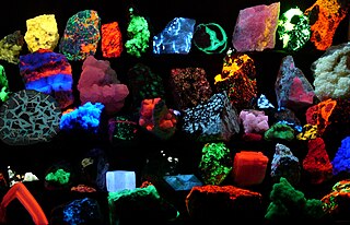
Fluorescence is the emission of light by a substance that has absorbed light or other electromagnetic radiation. It is a form of luminescence. In most cases, the emitted light has a longer wavelength, and therefore lower energy, than the absorbed radiation. The most striking example of fluorescence occurs when the absorbed radiation is in the ultraviolet region of the spectrum, and thus invisible to the human eye, while the emitted light is in the visible region, which gives the fluorescent substance a distinct color that can be seen only when exposed to UV light. Fluorescent materials cease to glow nearly immediately when the radiation source stops, unlike phosphorescent materials, which continue to emit light for some time after.
Nucleotides are organic molecules consisting of a nucleoside and a phosphate. They serve as monomeric units of the nucleic acid polymers deoxyribonucleic acid (DNA) and ribonucleic acid (RNA), both of which are essential biomolecules within all life-forms on Earth.
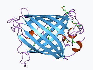
The green fluorescent protein (GFP) is a protein composed of 238 amino acid residues that exhibits bright green fluorescence when exposed to light in the blue to ultraviolet range. Similar proteins that also fluoresce green are found in many marine organisms, but the label GFP traditionally refers to this particular protein, which was first isolated from the jellyfish Aequorea victoria and is sometimes called—when such precision is required—avGFP.
Luciferase is a generic term for the class of oxidative enzymes that produce bioluminescence, and is usually distinguished from a photoprotein. The name was first used by Raphaël Dubois who invented the words luciferin and luciferase, for the substrate and enzyme, respectively. Both words are derived from the Latin word lucifer – meaning lightbearer.
Fluorescence spectroscopy is a type of electromagnetic spectroscopy that analyzes fluorescence from a sample. It involves using a beam of light, usually ultraviolet light, that excites the electrons in molecules of certain compounds and causes them to emit light; typically, but not necessarily, visible light. A complementary technique is absorption spectroscopy. In the special case of single molecule fluorescence spectroscopy, intensity fluctuations from the emitted light are measured from either single fluorophores, or pairs of fluorophores.
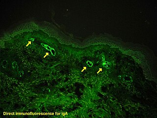
Immunofluorescence is a technique used for light microscopy with a fluorescence microscope and is used primarily on microbiological samples. This technique uses the specificity of antibodies to their antigen to target fluorescent dyes to specific biomolecule targets within a cell, and therefore allows visualization of the distribution of the target molecule through the sample. The specific region an antibody recognizes on an antigen is called an epitope. There have been efforts in epitope mapping since many antibodies can bind the same epitope and levels of binding between antibodies that recognize the same epitope can vary. Additionally, the binding of the fluorophore to the antibody itself cannot interfere with the immunological specificity of the antibody or the binding capacity of its antigen. Immunofluorescence is a widely used example of immunostaining and is a specific example of immunohistochemistry. This technique primarily makes use of fluorophores to visualise the location of the antibodies.
Fluorescence recovery after photobleaching (FRAP) is a method for determining the kinetics of diffusion through tissue or cells. It is capable of quantifying the two dimensional lateral diffusion of a molecularly thin film containing fluorescently labeled probes, or to examine single cells. This technique is very useful in biological studies of cell membrane diffusion and protein binding. In addition, surface deposition of a fluorescing phospholipid bilayer allows the characterization of hydrophilic surfaces in terms of surface structure and free energy.
Plate readers, also known as microplate readers or microplate photometers, are instruments which are used to detect biological, chemical or physical events of samples in microtiter plates. They are widely used in research, drug discovery, bioassay validation, quality control and manufacturing processes in the pharmaceutical and biotechnological industry and academic organizations. Sample reactions can be assayed in 1-1536 well format microtiter plates. The most common microplate format used in academic research laboratories or clinical diagnostic laboratories is 96-well with a typical reaction volume between 100 and 200 µL per well. Higher density microplates are typically used for screening applications, when throughput and assay cost per sample become critical parameters, with a typical assay volume between 5 and 50 µL per well. Common detection modes for microplate assays are absorbance, fluorescence intensity, luminescence, time-resolved fluorescence, and fluorescence polarization.
Glutamate dehydrogenase is an enzyme, present in most microbes and the mitochondria of eukaryotes, as are some of the other enzymes required for urea synthesis, that converts glutamate to α-ketoglutarate, and vice versa. In animals, the produced ammonia is usually used as a substrate in the urea cycle. Typically, the α-ketoglutarate to glutamate reaction does not occur in mammals, as glutamate dehydrogenase equilibrium favours the production of ammonia and α-ketoglutarate. Glutamate dehydrogenase also has a very low affinity for ammonia, and therefore toxic levels of ammonia would have to be present in the body for the reverse reaction to proceed. However, in brain, the NAD+/NADH ratio in brain mitochondria encourages oxidative deamination. In bacteria, the ammonia is assimilated to amino acids via glutamate and aminotransferases. In plants, the enzyme can work in either direction depending on environment and stress. Transgenic plants expressing microbial GLDHs are improved in tolerance to herbicide, water deficit, and pathogen infections. They are more nutritionally valuable.
Nucleoside-diphosphate kinases are enzymes that catalyze the exchange of terminal phosphate between different nucleoside diphosphates (NDP) and triphosphates (NTP) in a reversible manner to produce nucleotide triphosphates. Many NDP serve as acceptor while NTP are donors of phosphate group. The general reaction via ping-pong mechanism is as follows: XDP + YTP ←→ XTP + YDP. NDPK activities maintain an equilibrium between the concentrations of different nucleoside triphosphates such as, for example, when guanosine triphosphate (GTP) produced in the citric acid (Krebs) cycle is converted to adenosine triphosphate (ATP). Other activities include cell proliferation, differentiation and development, signal transduction, G protein-coupled receptor, endocytosis, and gene expression.
Stimulated emission depletion (STED) microscopy is one of the techniques that make up super-resolution microscopy. It creates super-resolution images by the selective deactivation of fluorophores, minimising the area of illumination at the focal point, and thus enhancing the achievable resolution for a given system. It was developed by Stefan W. Hell and Jan Wichmann in 1994, and was first experimentally demonstrated by Hell and Thomas Klar in 1999. Hell was awarded the Nobel Prize in Chemistry in 2014 for its development. In 1986, V.A. Okhonin had patented the STED idea. This patent was, perhaps, unknown to Hell and Wichmann in 1994.
Systematic evolution of ligands by exponential enrichment (SELEX), also referred to as in vitro selection or in vitro evolution, is a combinatorial chemistry technique in molecular biology for producing oligonucleotides of either single-stranded DNA or RNA that specifically bind to a target ligand or ligands. These single-stranded DNA or RNA are commonly referred to as aptamers. Although SELEX has emerged as the most commonly used name for the procedure, some researchers have referred to it as SAAB and CASTing SELEX was first introduced in 1990. In 2015 a special issue was published in the Journal of Molecular Evolution in the honor of quarter century of the SELEX discovery.
The Qubit fluorometer is a lab instrument developed and distributed by Invitrogen that, among other applications, is used for the quantification of DNA, RNA, and protein.
Fluorescent glucose biosensors are devices that measure the concentration of glucose in diabetic patients by means of sensitive protein that relays the concentration by means of fluorescence, an alternative to amperometric sension of glucose. Due to the prevalence of diabetes, it is the prime drive in the construction of fluorescent biosensors. A recent development has been approved by the FDA allowing a new continuous glucose monitoring system called EverSense, which is a 90 day glucose monitor using fluorescent biosensors.
Fluorescent chloride sensors are used for chemical analysis. The discoveries of chloride (Cl−) participations in physiological processes stimulates the measurements of intracellular Cl− in live cells and the development of fluorescent tools referred below.
Calcium imaging is a microscopy technique to optically measure the calcium (Ca2+) status of an isolated cell, tissue or medium. Calcium imaging takes advantage of calcium indicators, fluorescent molecules that respond to the binding of Ca2+ ions by changing their fluorescence properties. Two main classes of calcium indicators exist: chemical indicators and genetically encoded calcium indicators (GECI). This technique has allowed studies of calcium signalling in a wide variety of cell types. In neurons, electrical activity is always accompanied by an influx of Ca2+ ions. Thus, calcium imaging can be used to monitor the electrical activity in hundreds of neurons in cell culture or in living animals, which has made it possible to dissect the function of neuronal circuits.
Fluorescence imaging is a type of non-invasive imaging technique that can help visualize biological processes taking place in a living organism. Images can be produced from a variety of methods including: microscopy, imaging probes, and spectroscopy.
GrpE is a bacterial nucleotide exchange factor that is important for regulation of protein folding machinery, as well as the heat shock response. It is a heat-inducible protein and during stress it prevents unfolded proteins from accumulating in the cytoplasm. Accumulation of unfolded proteins in the cytoplasm can lead to cell death.
1 2 3 4 5 6 7 8 9 10 11 12 13 14 15 16 17 18 19 20 Stewart, Richard C.; VanBruggen, Ricaele; Ellefson, Dolph D.; Wolfe, Alan J. (September 1998). "TNP-ATP and TNP-ADP as Probes of the Nucleotide Binding Site of CheA, the Histidine Protein Kinase in the Chemotaxis Signal Transduction Pathway of Escherichia Coli". Biochemistry. 37 (35): 12269–12279. doi:10.1021/bi980970n. PMID9724541.
This page is based on this Wikipedia article Text is available under the CC BY-SA 4.0 license; additional terms may apply. Images, videos and audio are available under their respective licenses.