Related Research Articles
G protein-coupled receptors (GPCRs), also known as seven-(pass)-transmembrane domain receptors, 7TM receptors, heptahelical receptors, serpentine receptors, and G protein-linked receptors (GPLR), form a large group of evolutionarily related proteins that are cell surface receptors that detect molecules outside the cell and activate cellular responses. They are coupled with G proteins. They pass through the cell membrane seven times in form of six loops of amino acid residues, which is why they are sometimes referred to as seven-transmembrane receptors. Ligands can bind either to the extracellular N-terminus and loops or to the binding site within transmembrane helices. They are all activated by agonists, although a spontaneous auto-activation of an empty receptor has also been observed.
A nucleoside triphosphate is a nucleoside containing a nitrogenous base bound to a 5-carbon sugar, with three phosphate groups bound to the sugar. They are the molecular precursors of both DNA and RNA, which are chains of nucleotides made through the processes of DNA replication and transcription. Nucleoside triphosphates also serve as a source of energy for cellular reactions and are involved in signalling pathways.
In biology, cell signaling or cell communication is the ability of a cell to receive, process, and transmit signals with its environment and with itself. Cell signaling is a fundamental property of all cellular life in prokaryotes and eukaryotes. Signals that originate from outside a cell can be physical agents like mechanical pressure, voltage, temperature, light, or chemical signals. Cell signaling can occur over short or long distances, and as a result can be classified as autocrine, juxtacrine, intracrine, paracrine, or endocrine. Signaling molecules can be synthesized from various biosynthetic pathways and released through passive or active transports, or even from cell damage.

Purinergic receptors, also known as purinoceptors, are a family of plasma membrane molecules that are found in almost all mammalian tissues. Within the field of purinergic signalling, these receptors have been implicated in learning and memory, locomotor and feeding behavior, and sleep. More specifically, they are involved in several cellular functions, including proliferation and migration of neural stem cells, vascular reactivity, apoptosis and cytokine secretion. These functions have not been well characterized and the effect of the extracellular microenvironment on their function is also poorly understood.

The P2X receptors, also ATP-gated P2X receptor cation channel family, is a protein family that consists of cation-permeable ligand-gated ion channels that open in response to the binding of extracellular adenosine 5'-triphosphate (ATP). They belong to a larger family of receptors known as the ENaC/P2X superfamily. ENaC and P2X receptors have similar 3-D structures and are homologous. P2X receptors are present in a diverse array of organisms including humans, mouse, rat, rabbit, chicken, zebrafish, bullfrog, fluke, and amoeba.
A nucleotidase is a hydrolytic enzyme that catalyzes the hydrolysis of a nucleotide into a nucleoside and a phosphate.
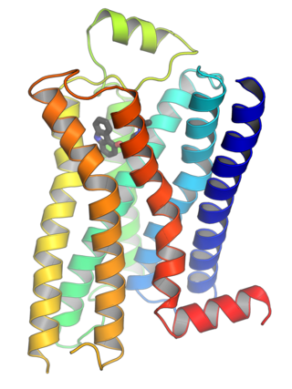
P2Y receptors are a family of purinergic G protein-coupled receptors, stimulated by nucleotides such as adenosine triphosphate, adenosine diphosphate, uridine triphosphate, uridine diphosphate and UDP-glucose.To date, 8 P2Y receptors have been cloned in humans: P2Y1, P2Y2, P2Y4, P2Y6, P2Y11, P2Y12, P2Y13 and P2Y14.
Ectonucleotidases consist of families of nucleotide metabolizing enzymes that are expressed on the plasma membrane and have externally oriented active sites. These enzymes metabolize nucleotides to nucleosides. The contribution of ectonucleotidases in the modulation of purinergic signaling depends on the availability and preference of substrates and on cell and tissue distribution.

P2X purinoceptor 7 is a protein that in humans is encoded by the P2RX7 gene.

P2Y purinoceptor 1 is a protein that in humans is encoded by the P2RY1 gene.

P2X purinoceptor 1, also ATP receptor, is a protein that in humans is encoded by the P2RX1 gene.

P2Y purinoceptor 2 is a protein that in humans is encoded by the P2RY2 gene.

Ectonucleoside triphosphate diphosphohydrolase-1 also known as CD39, is a typical cell surface enzyme with a catalytic site on the extracellular face.

P2Y purinoceptor 11 is a protein that in humans is encoded by the P2RY11 gene.
P2Y purinoceptor 14 is a protein that in humans is encoded by the P2RY14 gene.

Putative P2Y purinoceptor 10 is a protein that, in humans, is encoded by the P2RY10 gene.
P2X purinoceptor 4 is a protein that in humans is encoded by the P2RX4 gene. The product of this gene belongs to the family of purinoceptors for ATP. Multiple alternatively spliced transcript variants have been identified for this gene although their full-length natures have not been determined.
Pseudoapoptosis can be defined from multiple viewpoints, with an underlying premise of the differences in cellular processes and states relating to apoptosis. Pseudoapoptosis can be referred to as an apoptotic-like cellular state that can be readily reversed, or as a process that induces rapid apoptosis through the introduction of drugs such as bleomycin.

The rostral ventromedial medulla (RVM), or ventromedial nucleus of the spinal cord, is a group of neurons located close to the midline on the floor of the medulla oblongata. The rostral ventromedial medulla sends descending inhibitory and excitatory fibers to the dorsal horn spinal cord neurons. There are 3 categories of neurons in the RVM: on-cells, off-cells, and neutral cells. They are characterized by their response to nociceptive input. Off-cells show a transitory decrease in firing rate right before a nociceptive reflex, and are theorized to be inhibitory. Activation of off-cells, either by morphine or by any other means, results in antinociception. On-cells show a burst of activity immediately preceding nociceptive input, and are theorized to be contributing to the excitatory drive. Neutral cells show no response to nociceptive input.
Purinergic signalling is a form of extracellular signalling mediated by purine nucleotides and nucleosides such as adenosine and ATP. It involves the activation of purinergic receptors in the cell and/or in nearby cells, thereby regulating cellular functions.
References
- 1 2 Jacob, Fenila; Novo, Claudina Pérez; Bachert, Claus; Van Crombruggen, Koen (13 February 2013). "Purinergic signaling in inflammatory cells: P2 receptor expression, functional effects, and modulation of inflammatory responses". Purinergic Signalling. 9 (3): 285–306. doi:10.1007/s11302-013-9357-4. PMC 3757148 . PMID 23404828.
- ↑ Boeynaems, J. M.; Communi, D.; Gonzalez, N. S.; Robaye, B. (2005). "Overview of the P2 receptors". Seminars in Thrombosis and Hemostasis. 31 (2): 139–49. doi:10.1055/s-2005-869519. PMID 15852217. S2CID 260318518.
| | This biochemistry article is a stub. You can help Wikipedia by expanding it. |