![]() | |||
| |||
Names | |||
---|---|---|---|
Preferred IUPAC name Pyrimidine-2,4(1H,3H)-dione | |||
Other names
| |||
Identifiers | |||
3D model (JSmol) |
| ||
3DMet | |||
606623 | |||
ChEBI | |||
ChEMBL | |||
ChemSpider | |||
DrugBank | |||
ECHA InfoCard | 100.000.565 | ||
EC Number |
| ||
2896 | |||
KEGG | |||
PubChem CID | |||
RTECS number |
| ||
UNII | |||
CompTox Dashboard (EPA) | |||
| |||
Properties | |||
C4H4N2O2 | |||
Molar mass | 112.08676 g/mol | ||
Appearance | Solid | ||
Density | 1.32 g/cm3 | ||
Melting point | 335 °C (635 °F; 608 K) [1] | ||
Boiling point | N/A – decomposes | ||
Soluble | |||
Hazards | |||
Occupational safety and health (OHS/OSH): | |||
Main hazards | carcinogen and teratogen with chronic exposure | ||
GHS labelling: | |||
![]() ![]() | |||
Warning | |||
H315, H319, H335, H361 | |||
P201, P202, P261, P264, P271, P280, P281, P302+P352, P304+P340, P305+P351+P338, P308+P313, P312, P321, P332+P313, P337+P313, P362, P403+P233, P405, P501 | |||
NFPA 704 (fire diamond) | |||
Flash point | Non-flammable | ||
Related compounds | |||
Related compounds | Thymine Cytosine | ||
Except where otherwise noted, data are given for materials in their standard state (at 25 °C [77 °F], 100 kPa). |
Uracil ( /ˈjʊərəsɪl/ ) (symbol U or Ura) is one of the four nucleotide bases in the nucleic acid RNA. The others are adenine (A), cytosine (C), and guanine (G). In RNA, uracil binds to adenine via two hydrogen bonds. In DNA, the uracil nucleobase is replaced by thymine (T). Uracil is a demethylated form of thymine.
Uracil is a common and naturally occurring pyrimidine derivative. [2] The name "uracil" was coined in 1885 by the German chemist Robert Behrend, who was attempting to synthesize derivatives of uric acid. [3] Originally discovered in 1900 by Alberto Ascoli, it was isolated by hydrolysis of yeast nuclein; [4] it was also found in bovine thymus and spleen, herring sperm, and wheat germ. [5] It is a planar, unsaturated compound that has the ability to absorb light. [6]
Uracil that was formed extraterrestrially has been detected in the Murchison meteorite, [7] in a near-Earth asteroid, [8] and possibly on the surface of the moon Titan. [9] It has been synthesized under cold laboratory conditions similar to outer space, from pyrimidine embedded in water ice and exposed to ultraviolet light. [10]
In RNA, uracil base-pairs with adenine and replaces thymine during DNA transcription. Methylation of uracil produces thymine. [11] In DNA, the evolutionary substitution of thymine for uracil may have increased DNA stability and improved the efficiency of DNA replication (discussed below). Uracil pairs with adenine through hydrogen bonding. When base pairing with adenine, uracil acts as both a hydrogen bond acceptor and a hydrogen bond donor. In RNA, uracil binds with a ribose sugar to form the ribonucleoside uridine. When a phosphate attaches to uridine, uridine 5′-monophosphate is produced. [6]
Uracil undergoes amide-imidic acid tautomeric shifts because any nuclear instability the molecule may have from the lack of formal aromaticity is compensated by the cyclic-amidic stability. [5] The amide tautomer is referred to as the lactam structure, while the imidic acid tautomer is referred to as the lactim structure. These tautomeric forms are predominant at pH 7. The lactam structure is the most common form of uracil.
Uracil also recycles itself to form nucleotides by undergoing a series of phosphoribosyltransferase reactions. [2] Degradation of uracil produces the substrates β-alanine, carbon dioxide, and ammonia. [2]
Oxidative degradation of uracil produces urea and maleic acid in the presence of H2O2 and Fe 2+ or in the presence of diatomic oxygen and Fe2+.
Uracil is a weak acid. The first site of ionization of uracil is not known. [12] The negative charge is placed on the oxygen anion and produces a pKa of less than or equal to 12. The basic pKa = −3.4, while the acidic pKa = 9.389. In the gas phase, uracil has four sites that are more acidic than water. [13]
Uracil is rarely found in DNA, and this may have been an evolutionary change to increase genetic stability. This is because cytosine can deaminate spontaneously to produce uracil through hydrolytic deamination. Therefore, if there were an organism that used uracil in its DNA, the deamination of cytosine (which undergoes base pairing with guanine) would lead to formation of uracil (which would base pair with adenine) during DNA synthesis. Uracil-DNA glycosylase excises uracil bases from double-stranded DNA. This enzyme would therefore recognize and cut out both types of uracil – the one incorporated naturally, and the one formed due to cytosine deamination, which would trigger unnecessary and inappropriate repair processes. [14]
This problem is believed to have been solved in terms of evolution, that is by "tagging" (methylating) uracil. Methylated uracil is identical to thymine. Hence the hypothesis that, over time, thymine became standard in DNA instead of uracil. So cells continue to use uracil in RNA, and not in DNA, because RNA is shorter-lived than DNA, and any potential uracil-related errors do not lead to lasting damage. Apparently, either there was no evolutionary pressure to replace uracil in RNA with the more complex thymine, or uracil has some chemical property that is useful in RNA, which thymine lacks. Uracil-containing DNA still exists, for example in
Organisms synthesize uracil, in the form of uridine monophosphate (UMP), by decarboxylating orotidine 5'-monophosphate (orotidylic acid). In humans this decarboxylation is achieved by the enzyme UMP synthase. In contrast to the purine nucleotides, the pyrimidine ring (orotidylic acid) that leads uracil is synthesized first and then linked to ribose phosphate, forming UMP. [16]
There are many laboratory synthesis of uracil available. The first reaction is the simplest of the syntheses, by adding water to cytosine to produce uracil and ammonia: [2]
The most common way to synthesize uracil is by the condensation of malic acid with urea in fuming sulfuric acid: [5]
Uracil can also be synthesized by a double decomposition of thiouracil in aqueous chloroacetic acid. [5]
Photodehydrogenation of 5,6-diuracil, which is synthesized by beta-alanine reacting with urea, produces uracil. [17]
In 2009, NASA scientists reported having produced uracil from pyrimidine and water ice by exposing it to ultraviolet light under space-like conditions. [10] This suggests a possible natural original source for uracil. [18] In 2014, NASA scientists reported that additional complex DNA and RNA organic compounds of life, including uracil, cytosine and thymine, have been formed in the laboratory under outer space conditions, starting with ice, pyrimidine, ammonia, and methanol, which are compounds found in astrophysical environments. [19] Pyrimidine, like polycyclic aromatic hydrocarbons (PAHs), a carbon-rich chemical found in the Universe, may have been formed in red giants or in interstellar dust and gas clouds. [20]
Based on 12C/13C isotopic ratios of organic compounds found in the Murchison meteorite, it is believed that uracil, xanthine, and related molecules can also be formed extraterrestrially. [7] Data from the Cassini mission, orbiting in the Saturn system, suggests that uracil is present in the surface of the moon Titan. [9] In 2023, uracil was observed in a sample from 162173 Ryugu, a near-Earth asteroid, with no exposure to Earth's biosphere, giving further evidence for synthesis in space. [8]
Uracil readily undergoes regular reactions including oxidation, nitration, and alkylation. While in the presence of phenol (PhOH) and sodium hypochlorite (NaOCl), uracil can be visualized in ultraviolet light. [5] Uracil also has the capability to react with elemental halogens because of the presence of more than one strongly electron donating group. [5]
Uracil readily undergoes addition to ribose sugars and phosphates to partake in synthesis and further reactions in the body. Uracil becomes uridine, uridine monophosphate (UMP), uridine diphosphate (UDP), uridine triphosphate (UTP), and uridine diphosphate glucose (UDP-glucose). Each one of these molecules is synthesized in the body and has specific functions.
When uracil reacts with anhydrous hydrazine, a first-order kinetic reaction occurs and the uracil ring opens up. [21] If the pH of the reaction increases to > 10.5, the uracil anion forms, making the reaction go much more slowly. The same slowing of the reaction occurs if the pH decreases, because of the protonation of the hydrazine. [21] The reactivity of uracil remains unchanged, even if the temperature changes. [21]
Uracil's use in the body is to help carry out the synthesis of many enzymes necessary for cell function through bonding with riboses and phosphates. [2] Uracil serves as allosteric regulator and coenzyme for reactions in animals and in plants. [22] UMP controls the activity of carbamoyl phosphate synthetase and aspartate transcarbamoylase in plants, while UDP and UTP regulate CPSase II activity in animals. UDP-glucose regulates the conversion of glucose to galactose in the liver and other tissues in the process of carbohydrate metabolism. [22] Uracil is also involved in the biosynthesis of polysaccharides and the transportation of sugars containing aldehydes. [22] Uracil is important for the detoxification of many carcinogens, for instance those found in tobacco smoke. [23] Uracil is also required to detoxify many drugs such as cannabinoids (THC) [24] and morphine (opioids). [25] It can also slightly increase the risk for cancer in unusual cases in which the body is extremely deficient in folate. [26] The deficiency in folate leads to increased ratio of deoxyuridine monophosphates (dUMP)/deoxythymidine monophosphates (dTMP) and uracil misincorporation into DNA and eventually low production of DNA. [26]
Uracil can be used for drug delivery and as a pharmaceutical. When elemental fluorine reacts with uracil, they produce 5-fluorouracil. 5-Fluorouracil is an anticancer drug (antimetabolite) used to masquerade as uracil during the nucleic acid replication process. [2] Because 5-fluorouracil is similar in shape to, but does not undergo the same chemistry as, uracil, the drug inhibits RNA transcription enzymes, thereby blocking RNA synthesis and stopping the growth of cancerous cells. [2] Uracil can also be used in the synthesis of caffeine. [27] Uracil has also shown potential as a HIV viral capsid inhibitor. [28] Uracil derivatives have antiviral, anti-tubercular and anti-leishmanial activity. [29] [30] [31]
Uracil can be used to determine microbial contamination of tomatoes. The presence of uracil indicates lactic acid bacteria contamination of the fruit. [32] Uracil derivatives containing a diazine ring are used in pesticides. [33] Uracil derivatives are more often used as antiphotosynthetic herbicides, destroying weeds in cotton, sugar beet, turnips, soya, peas, sunflower crops, vineyards, berry plantations, and orchards. [33] Uracil derivatives can enhance the activity of antimicrobial polysaccharides such as chitosan. [34]
In yeast, uracil concentrations are inversely proportional to uracil permease. [35]
Mixtures containing uracil are also commonly used to test reversed-phase HPLC columns. As uracil is essentially unretained by the non-polar stationary phase, this can be used to determine the dwell time (and subsequently dwell volume, given a known flow rate) of the system.
Cytosine is one of the four nucleotide bases found in DNA and RNA, along with adenine, guanine, and thymine. It is a pyrimidine derivative, with a heterocyclic aromatic ring and two substituents attached. The nucleoside of cytosine is cytidine. In Watson–Crick base pairing, it forms three hydrogen bonds with guanine.
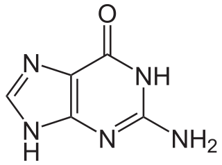
Guanine is one of the four main nucleotide bases found in the nucleic acids DNA and RNA, the others being adenine, cytosine, and thymine. In DNA, guanine is paired with cytosine. The guanine nucleoside is called guanosine.
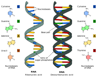
Nucleic acids are large biomolecules that are crucial in all cells and viruses. They are composed of nucleotides, which are the monomer components: a 5-carbon sugar, a phosphate group and a nitrogenous base. The two main classes of nucleic acids are deoxyribonucleic acid (DNA) and ribonucleic acid (RNA). If the sugar is ribose, the polymer is RNA; if the sugar is deoxyribose, a variant of ribose, the polymer is DNA.
Nucleotides are organic molecules composed of a nitrogenous base, a pentose sugar and a phosphate. They serve as monomeric units of the nucleic acid polymers – deoxyribonucleic acid (DNA) and ribonucleic acid (RNA), both of which are essential biomolecules within all life-forms on Earth. Nucleotides are obtained in the diet and are also synthesized from common nutrients by the liver.
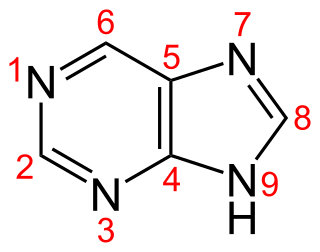
Purine is a heterocyclic aromatic organic compound that consists of two rings fused together. It is water-soluble. Purine also gives its name to the wider class of molecules, purines, which include substituted purines and their tautomers. They are the most widely occurring nitrogen-containing heterocycles in nature.
Pyrimidine is an aromatic, heterocyclic, organic compound similar to pyridine. One of the three diazines, it has nitrogen atoms at positions 1 and 3 in the ring. The other diazines are pyrazine and pyridazine.
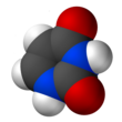
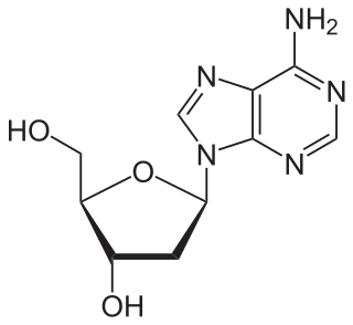
Adenine is a purine nucleotide base. It is one of the four nucleobases in the nucleic acids of DNA, the other three being guanine (G), cytosine (C), and thymine (T). Adenine derivatives have various roles in biochemistry including cellular respiration, in the form of both the energy-rich adenosine triphosphate (ATP) and the cofactors nicotinamide adenine dinucleotide (NAD), flavin adenine dinucleotide (FAD) and Coenzyme A. It also has functions in protein synthesis and as a chemical component of DNA and RNA. The shape of adenine is complementary to either thymine in DNA or uracil in RNA.
Thymine is one of the four nucleotide bases in the nucleic acid of DNA that are represented by the letters G–C–A–T. The others are adenine, guanine, and cytosine. Thymine is also known as 5-methyluracil, a pyrimidine nucleobase. In RNA, thymine is replaced by the nucleobase uracil. Thymine was first isolated in 1893 by Albrecht Kossel and Albert Neumann from calf thymus glands, hence its name.
Nucleotide bases are nitrogen-containing biological compounds that form nucleosides, which, in turn, are components of nucleotides, with all of these monomers constituting the basic building blocks of nucleic acids. The ability of nucleobases to form base pairs and to stack one upon another leads directly to long-chain helical structures such as ribonucleic acid (RNA) and deoxyribonucleic acid (DNA). Five nucleobases—adenine (A), cytosine (C), guanine (G), thymine (T), and uracil (U)—are called primary or canonical. They function as the fundamental units of the genetic code, with the bases A, G, C, and T being found in DNA while A, G, C, and U are found in RNA. Thymine and uracil are distinguished by merely the presence or absence of a methyl group on the fifth carbon (C5) of these heterocyclic six-membered rings. In addition, some viruses have aminoadenine (Z) instead of adenine. It differs in having an extra amine group, creating a more stable bond to thymine.
Nucleosides are glycosylamines that can be thought of as nucleotides without a phosphate group. A nucleoside consists simply of a nucleobase and a five-carbon sugar whereas a nucleotide is composed of a nucleobase, a five-carbon sugar, and one or more phosphate groups. In a nucleoside, the anomeric carbon is linked through a glycosidic bond to the N9 of a purine or the N1 of a pyrimidine. Nucleotides are the molecular building blocks of DNA and RNA.
Uridine (symbol U or Urd) is a glycosylated pyrimidine analog containing uracil attached to a ribose ring (or more specifically, a ribofuranose) via a β-N1-glycosidic bond. The analog is one of the five standard nucleosides which make up nucleic acids, the others being adenosine, thymidine, cytidine and guanosine. The five nucleosides are commonly abbreviated to their symbols, U, A, dT, C, and G, respectively. However, thymidine is more commonly written as 'dT' ('d' represents 'deoxy') as it contains a 2'-deoxyribofuranose moiety rather than the ribofuranose ring found in uridine. This is because thymidine is found in deoxyribonucleic acid (DNA) and usually not in ribonucleic acid (RNA). Conversely, uridine is found in RNA and not DNA. The remaining three nucleosides may be found in both RNA and DNA. In RNA, they would be represented as A, C and G whereas in DNA they would be represented as dA, dC and dG.
In biochemistry, a ribonucleotide is a nucleotide containing ribose as its pentose component. It is considered a molecular precursor of nucleic acids. Nucleotides are the basic building blocks of DNA and RNA. Ribonucleotides themselves are basic monomeric building blocks for RNA. Deoxyribonucleotides, formed by reducing ribonucleotides with the enzyme ribonucleotide reductase (RNR), are essential building blocks for DNA. There are several differences between DNA deoxyribonucleotides and RNA ribonucleotides. Successive nucleotides are linked together via phosphodiester bonds.
A salvage pathway is a pathway in which a biological product is produced from intermediates in the degradative pathway of its own or a similar substance. The term often refers to nucleotide salvage in particular, in which nucleotides are synthesized from intermediates in their degradative pathway.
A nucleoside triphosphate is a nucleoside containing a nitrogenous base bound to a 5-carbon sugar, with three phosphate groups bound to the sugar. They are the molecular precursors of both DNA and RNA, which are chains of nucleotides made through the processes of DNA replication and transcription. Nucleoside triphosphates also serve as a source of energy for cellular reactions and are involved in signalling pathways.
Biosynthesis, i.e., chemical synthesis occurring in biological contexts, is a term most often referring to multi-step, enzyme-catalyzed processes where chemical substances absorbed as nutrients serve as enzyme substrates, with conversion by the living organism either into simpler or more complex products. Examples of biosynthetic pathways include those for the production of amino acids, lipid membrane components, and nucleotides, but also for the production of all classes of biological macromolecules, and of acetyl-coenzyme A, adenosine triphosphate, nicotinamide adenine dinucleotide and other key intermediate and transactional molecules needed for metabolism. Thus, in biosynthesis, any of an array of compounds, from simple to complex, are converted into other compounds, and so it includes both the catabolism and anabolism of complex molecules. Biosynthetic processes are often represented via charts of metabolic pathways. A particular biosynthetic pathway may be located within a single cellular organelle, while others involve enzymes that are located across an array of cellular organelles and structures.
Uridine monophosphate (UMP), also known as 5′-uridylic acid, is a nucleotide that is used as a monomer in RNA. It is an ester of phosphoric acid with the nucleoside uridine. UMP consists of the phosphate group, the pentose sugar ribose, and the nucleobase uracil; hence, it is a ribonucleotide monophosphate. As a substituent or radical its name takes the form of the prefix uridylyl-. The deoxy form is abbreviated dUMP. Covalent attachment of UMP is called uridylylation.
Nucleic acid metabolism is a collective term that refers to the variety of chemical reactions by which nucleic acids are either synthesized or degraded. Nucleic acids are polymers made up of a variety of monomers called nucleotides. Nucleotide synthesis is an anabolic mechanism generally involving the chemical reaction of phosphate, pentose sugar, and a nitrogenous base. Degradation of nucleic acids is a catabolic reaction and the resulting parts of the nucleotides or nucleobases can be salvaged to recreate new nucleotides. Both synthesis and degradation reactions require multiple enzymes to facilitate the event. Defects or deficiencies in these enzymes can lead to a variety of diseases.
Pyrimidine dimers represent molecular lesions originating from thymine or cytosine bases within DNA, resulting from photochemical reactions. These lesions, commonly linked to direct DNA damage, are induced by ultraviolet light (UV), particularly UVC, result in the formation of covalent bonds between adjacent nitrogenous bases along the nucleotide chain near their carbon–carbon double bonds, the photo-coupled dimers are fluorescent. Such dimerization, which can also occur in double-stranded RNA (dsRNA) involving uracil or cytosine, leads to the creation of cyclobutane pyrimidine dimers (CPDs) and 6–4 photoproducts. These pre-mutagenic lesions modify the DNA helix structure, resulting in abnormal non-canonical base pairing and, consequently, adjacent thymines or cytosines in DNA will form a cyclobutane ring when joined together and cause a distortion in the DNA. This distortion prevents DNA replication and transcription mechanisms beyond the dimerization site.
Pyrimidine biosynthesis occurs both in the body and through organic synthesis.
Nucleic acid analogues are compounds which are analogous to naturally occurring RNA and DNA, used in medicine and in molecular biology research. Nucleic acids are chains of nucleotides, which are composed of three parts: a phosphate backbone, a pentose sugar, either ribose or deoxyribose, and one of four nucleobases. An analogue may have any of these altered. Typically the analogue nucleobases confer, among other things, different base pairing and base stacking properties. Examples include universal bases, which can pair with all four canonical bases, and phosphate-sugar backbone analogues such as PNA, which affect the properties of the chain . Nucleic acid analogues are also called xeno nucleic acids and represent one of the main pillars of xenobiology, the design of new-to-nature forms of life based on alternative biochemistries.
Dasselbe stellt sich sonach als Methylderivat der Verbindung: welche ich willkürlich mit dem Namen Uracil belege, dar.[The same compound is therefore represented as the methyl derivative of the compound, which I will arbitrarily endow with the name ‘uracil’.]