Cyclic di-GMP

Last updated
Cyclic di-GMP
Cyclic-di-GMP.svg
Names
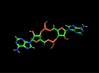
Systematic IUPAC name
(2R,3R,3aS,7aR,9R,10R,10aS,14aR)-2,9-Bis(2-amino-6-oxo-1,6-dihydro-9H-purin-9-yl)-3,5,10,12-tetrahydroxyoctahydro-2H,5H,7H,12H-5λ5,12λ5-difuro[3,2-d:3′,2′-j][1,3,7,9,2,8]tetraoxadiphosphacyclododecine-5,12-dione
Other names
Cyclic diguanylate; 3',5'-Cyclic diguanylic acid; c-di-GMP; 5GP-5GP
Identifiers
3D model (JSmol)
ChemSpider
PubChem CID
  • InChI=1S/C20H24N10O14P2/c21-19-25-13-7(15(33)27-19)23-3-29(13)17-9(31)11-5(41-17)1-39-45(35,36)44-12-6(2-40-46(37,38)43-11)42-18(10(12)32)30-4-24-8-14(30)26-20(22)28-16(8)34/h3-6,9-12,17-18,31-32H,1-2H2,(H,35,36)(H,37,38)(H3,21,25,27,33)(H3,22,26,28,34)/t5-,6-,9-,10-,11-,12-,17-,18-/m1/s1 X mark.svgN
    Key: PKFDLKSEZWEFGL-MHARETSRSA-N X mark.svgN
  • InChI=1/C20H24N10O14P2/c21-19-25-13-7(15(33)27-19)23-3-29(13)17-9(31)11-5(41-17)1-39-45(35,36)44-12-6(2-40-46(37,38)43-11)42-18(10(12)32)30-4-24-8-14(30)26-20(22)28-16(8)34/h3-6,9-12,17-18,31-32H,1-2H2,(H,35,36)(H,37,38)(H3,21,25,27,33)(H3,22,26,28,34)/t5-,6-,9-,10-,11-,12-,17-,18-/m1/s1
    Key: PKFDLKSEZWEFGL-MHARETSRBV
  • O=C7/N=C(/N)Nc1c7ncn1[C@@H]3O[C@@H]4COP(=O)(O)O[C@H]2[C@@H](O)[C@@H](O[C@@H]2COP(=O)(O)O[C@H]4[C@H]3O)n5c6NC(=N/C(=O)c6nc5)\N
Properties
C20H24N10O14P2
Molar mass 690.09 g/mol
Except where otherwise noted, data are given for materials in their standard state (at 25 °C [77 °F], 100 kPa).
X mark.svgN  verify  (what is  Yes check.svgYX mark.svgN ?)

Cyclic di-GMP (also called cyclic diguanylate and c-di-GMP ) is a second messenger used in signal transduction in a wide variety of bacteria. [1] Cyclic di-GMP is not known to be used by archaea, and has only been observed in eukaryotes in Dictyostelium . [2] The biological role of cyclic di-GMP was first uncovered when it was identified as an allosteric activator of a cellulose synthase found in Gluconacetobacter xylinus in order to produce microbial cellulose. [3]

In structure, it is a cycle containing only two guanine bases linked by ribose and phosphate.

Contact with surfaces increases c-di-GMP which increases transcription, translation, and post translation of exopolysaccharides (EPSs) and other extracellular polymeric substance matrix components (see the review by Jenal et al 2017). [4] In bacteria, certain signals are communicated by synthesizing or degrading cyclic di-GMP. Cyclic di-GMP is synthesized by proteins with diguanylate cyclase activity. These proteins typically have a characteristic GGDEF motif, which refers to a conserved sequence of five amino acids. Degradation of cyclic di-GMP is affected by proteins with phosphodiesterase activity. These proteins have either an EAL or an HD-GYP amino acid motif. Processes that are known to be regulated by cyclic di-GMP, at least in some organisms, include biofilm formation (such as EPS matrices found by Steiner et al 2013), [4] motility (especially the motile-to-sessile transition, see the review by Jenal et al 2017) [4] and virulence factor production.

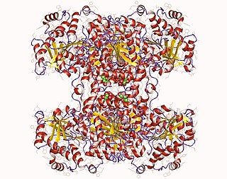
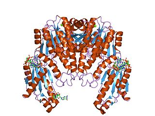
Cyclic di-GMP levels are regulated using a variety of mechanisms. Many proteins with GGDEF, EAL or HD-GYP domains are found with other domains that can receive signals, such as PAS domains. Enzymes that degrade or synthesize cyclic di-GMP are believed to be localized to specific regions of the cell, where they influence receivers in a restricted space. [1] In Gluconacetobacter xylinus, c-di-GMP stimulates the polymerization of glucose into cellulose as a high affinity allosteric activator of the enzyme cellulose synthase. [3] Some diguanylate cyclase enzymes are allosterically inhibited by cyclic di-GMP.

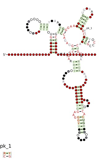
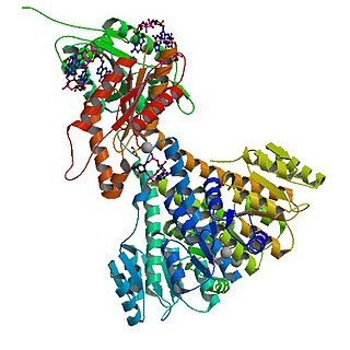
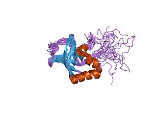
Cyclic di-GMP levels regulate other processes via a number of mechanisms. The Gluconacetobacter xylinus cellulose synthase is allosterically stimulated by cyclic di-GMP, presenting a mechanism by which cyclic di-GMP can regulate cellulose synthase activity. The PilZ domain has been shown to bind cyclic di-GMP and is believed to be involved in cyclic di-GMP-dependent regulation, but the mechanism by which it does this is unknown. Recent structural studies of PilZ domains from two bacterial species have demonstrated that PilZ domains change conformation drastically upon binding to cyclic di-GMP. [5] [6] This leads to the strong inference that conformational changes in PilZ domains allow the activity of targeted effector proteins (such as cellulose synthase) to be regulated by cyclic di-GMP. Riboswitches called the cyclic di-GMP-I riboswitch and cyclic di-GMP-II riboswitch regulate gene expression in response to cyclic di-GMP concentrations in a variety of bacteria, but not all bacteria that are known to use cyclic di-GMP.

For a review of c-di-GMP roles in Caulobacter crescentus , Pseudomonas aeruginosa , Komagataeibacter xylinus /Gluconacetobacter xylinus, Myxococcus xanthus , Bdellovibrio bacteriovorus and Pseudomonas fluorescens see Jenal et al 2017. [4]

See also

Related Research Articles

<span class="mw-page-title-main">Cyclic adenosine monophosphate</span> Cellular second messenger

Cyclic adenosine monophosphate is a second messenger, or cellular signal occurring within cells, that is important in many biological processes. cAMP is a derivative of adenosine triphosphate (ATP) and used for intracellular signal transduction in many different organisms, conveying the cAMP-dependent pathway.

<span class="mw-page-title-main">Allosteric regulation</span> Regulation of enzyme activity

In biochemistry, allosteric regulation is the regulation of an enzyme by binding an effector molecule at a site other than the enzyme's active site.

<span class="mw-page-title-main">Cyclic nucleotide</span> Cyclic nucleic acid

A cyclic nucleotide (cNMP) is a single-phosphate nucleotide with a cyclic bond arrangement between the sugar and phosphate groups. Like other nucleotides, cyclic nucleotides are composed of three functional groups: a sugar, a nitrogenous base, and a single phosphate group. As can be seen in the cyclic adenosine monophosphate (cAMP) and cyclic guanosine monophosphate (cGMP) images, the 'cyclic' portion consists of two bonds between the phosphate group and the 3' and 5' hydroxyl groups of the sugar, very often a ribose.

<span class="mw-page-title-main">Riboswitch</span>

In molecular biology, a riboswitch is a regulatory segment of a messenger RNA molecule that binds a small molecule, resulting in a change in production of the proteins encoded by the mRNA. Thus, an mRNA that contains a riboswitch is directly involved in regulating its own activity, in response to the concentrations of its effector molecule. The discovery that modern organisms use RNA to bind small molecules, and discriminate against closely related analogs, expanded the known natural capabilities of RNA beyond its ability to code for proteins, catalyze reactions, or to bind other RNA or protein macromolecules.

<span class="mw-page-title-main">Guanylate cyclase</span> Lyase enzyme that synthesizes cGMP from GTP

Guanylate cyclase is a lyase enzyme that converts guanosine triphosphate (GTP) to cyclic guanosine monophosphate (cGMP) and pyrophosphate:

<span class="mw-page-title-main">Glycogen synthase</span> Enzyme class, includes all types of glycogen/starch synthases

Glycogen synthase is a key enzyme in glycogenesis, the conversion of glucose into glycogen. It is a glycosyltransferase that catalyses the reaction of UDP-glucose and n to yield UDP and n+1.

<span class="mw-page-title-main">Cobalamin riboswitch</span>

Cobalamin riboswitch is a cis-regulatory element which is widely distributed in 5' untranslated regions of vitamin B12 (Cobalamin) related genes in bacteria.

<span class="mw-page-title-main">Cellulose synthase (UDP-forming)</span> Cellulose synthesizing enzyme in plants and bacteria

The UDP-forming form of cellulose synthase is the main enzyme that produces cellulose. Systematically, it is known as UDP-glucose:(1→4)-β-D-glucan 4-β-D-glucosyltransferase in enzymology. It catalyzes the chemical reaction:

<span class="mw-page-title-main">Cyclic di-GMP-I riboswitch</span>

Cyclic di-GMP-I riboswitches are a class of riboswitch that specifically bind cyclic di-GMP, which is a second messenger that is used in a variety of microbial processes including virulence, motility and biofilm formation. Cyclic di-GMP-I riboswitches were originally identified by bioinformatics as a conserved RNA-like structure called the "GEMM motif". These riboswitches are present in a wide variety of bacteria, and are most common in Clostridia and certain varieties of Pseudomonadota. The riboswitches are present in pathogens such as Clostridium difficile, Vibrio cholerae and Bacillus anthracis. Geobacter uraniumreducens is predicted to have 30 instances of this riboswitch in its genome. A bacteriophage that infects C. difficile is predicted to carry a cyclic di-GMP-I riboswitch, which it might use to detect and exploit the physiological state of bacteria that it infects.

<span class="mw-page-title-main">Cyclic di-GMP-II riboswitch</span>

Cyclic di-GMP-II riboswitches form a class of riboswitches that specifically bind cyclic di-GMP, a second messenger used in multiple bacterial processes such as virulence, motility and biofilm formation. Cyclic di-GMP II riboswitches are structurally unrelated to cyclic di-GMP-I riboswitches, though they have the same function.

<span class="mw-page-title-main">Diguanylate cyclase</span>

In enzymology, diguanylate cyclase, also known as diguanylate kinase, is an enzyme that catalyzes the chemical reaction:

<span class="mw-page-title-main">PilZ domain</span>

The PilZ protein family is named after the type IV pilus control protein first identified in Pseudomonas aeruginosa, expressed as part of the pil operon. It has a cytoplasmic location and is essential for type IV fimbrial, or pilus, biogenesis. PilZ is a c-di-GMP binding domain and PilZ domain-containing proteins represent the best studied class of c-di-GMP effectors. C-di-GMP, cyclic diguanosine monophosphate, the second messenger in cells, is widespread in and unique to the bacterial kingdom. Elevated intracellular levels of c-di-GMP generally cause bacteria to change from a motile single-cell state to a sessile, adhesive surface-attached multicellular state called biofilm.

<span class="mw-page-title-main">GGDEF domain</span>

In molecular biology, the GGDEF domain is a protein domain which appears to be ubiquitous in bacteria and is often linked to a regulatory domain, such as a phosphorylation receiver or oxygen sensing domain. Its function is to act as a diguanylate cyclase and synthesize cyclic di-GMP, which is used as an intracellular signalling molecule in a wide variety of bacteria. Enzymatic activity can be strongly influenced by the adjacent domains. Processes regulated by this domain include exopolysaccharide synthesis, biofilm formation, motility and cell differentiation.

Cyclic-guanylate-specific phosphodiesterase (EC 3.1.4.52, cyclic bis(3′→5')diguanylate phosphodiesterase, c-di-GMP-specific phosphodiesterase, c-di-GMP phosphodiesterase, phosphodiesterase, phosphodiesterase A1, PDEA1, VieA) is an enzyme with systematic name cyclic bis(3′→5′)diguanylate 3-guanylylhydrolase. This enzyme catalyses the following reaction:

<span class="mw-page-title-main">Cyclic guanosine monophosphate–adenosine monophosphate</span> Chemical compound

Cyclic guanosine monophosphate–adenosine monophosphate is the first cyclic di-nucleotide found in metazoa. In mammalian cells, cGAMP is synthesized by cyclic GMP-AMP synthase (cGAS) from ATP and GTP upon cytosolic DNA stimulation. cGAMP produced by cGAS contains mixed phosphodiester linkages, with one between 2'-OH of GMP and 5'-phosphate of AMP and the other between 3'-OH of AMP and 5'-phosphate of GMP.

<span class="mw-page-title-main">Stimulator of interferon genes</span> Protein-coding gene in the species Homo sapiens

Stimulator of interferon genes (STING), also known as transmembrane protein 173 (TMEM173) and MPYS/MITA/ERIS is a protein that in humans is encoded by the STING1 gene.

<span class="mw-page-title-main">Urs Jenal</span>

Urs Jenal is a Swiss Microbiologist and Professor at the Biozentrum University of Basel, Switzerland.

<span class="mw-page-title-main">Cyclic di-AMP</span> Chemical compound

Cyclic di-AMP is a second messenger used in signal transduction in bacteria and archaea. It is present in many Gram-positive bacteria, some Gram-negative species, and archaea of the phylum euryarchaeota.

The cGAS–STING pathway is a component of the innate immune system that functions to detect the presence of cytosolic DNA and, in response, trigger expression of inflammatory genes that can lead to senescence or to the activation of defense mechanisms. DNA is normally found in the nucleus of the cell. Localization of DNA to the cytosol is associated with tumorigenesis, viral infection, and invasion by some intracellular bacteria. The cGAS – STING pathway acts to detect cytosolic DNA and induce an immune response.

Komagataeibacter xylinus is a species of bacteria best known for its ability to produce cellulose, specifically bacterial cellulose.

References

  1. 1 2 Tamayo R, Pratt JT, Camilli A (2007). "Roles of cyclic diguanylate in the regulation of bacterial pathogenesis". Annual Review of Microbiology. 61: 131–148. doi:10.1146/annurev.micro.61.080706.093426. PMC   2776827 . PMID   17480182.
  2. Chen ZH, Schaap P (August 2012). "The prokaryote messenger c-di-GMP triggers stalk cell differentiation in Dictyostelium". Nature. 488 (7413): 680–683. doi:10.1038/nature11313. PMC   3939355 . PMID   22864416.
  3. 1 2 Ross P, Weinhouse H, Aloni Y, Michaeli D, Weinberger-Ohana P, Mayer R, et al. (1987). "Regulation of cellulose synthesis in Acetobacter xylinum by cyclic diguanylic acid". Nature. 325 (6101): 279–281. doi:10.1038/325279a0. PMID   18990795. S2CID   4305512.
  4. 1 2 3 4 Laventie BJ, Jenal U (September 2020). "Surface Sensing and Adaptation in Bacteria" (PDF). Annual Review of Microbiology. Annual Reviews. 74 (1): 735–760. doi:10.1146/annurev-micro-012120-063427. PMID   32905753. S2CID   221622525.
  5. Benach J, Swaminathan SS, Tamayo R, Handelman SK, Folta-Stogniew E, Ramos JE, et al. (December 2007). "The structural basis of cyclic diguanylate signal transduction by PilZ domains". The EMBO Journal. European Molecular Biology Organization. 26 (24): 5153–5166. doi:10.1038/sj.emboj.7601918. PMC   2140105 . PMID   18034161.
  6. Ko J, Ryu KS, Kim H, Shin JS, Lee JO, Cheong C, Choi BS (April 2010). "Structure of PP4397 reveals the molecular basis for different c-di-GMP binding modes by Pilz domain proteins". Journal of Molecular Biology. 398 (1): 97–110. doi:10.1016/j.jmb.2010.03.007. PMID   20226196.