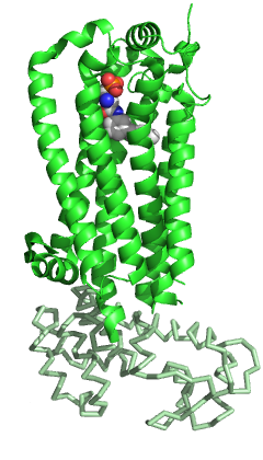
Psychosine receptor is a G protein-coupled receptor (GPCR) protein that in humans is encoded by the GPR65 gene. [5] [6] GPR65 is also referred to as TDAG8.
Psychosine receptor is a G protein-coupled receptor (GPCR) protein that in humans is encoded by the GPR65 gene. [5] [6] GPR65 is also referred to as TDAG8.
GPR65 (TDAG8) is primarily expressed in lymphoid tissues (spleen, lymph nodes, thymus, and leukocytes), [7] and as a GPCR, the protein is localized to the plasma membrane.
In 2001, GPR65 was reported to be a specific receptor for psychosine (d-galactosyl-β-1,1′ sphingosine) as well as several other related glycosphingolipids. [8] However, the specific binding of psychosine to GPR65 has been contested as the reported ligand binding did not satisfy the appropriate pharmacological criteria. [9]
More recently, 3-[(2,4-dichlorophenyl)methylsulfanyl]-1,6-dimethylpyridazino[4,5-e][1,3,4]thiadiazin-5-one (referred to as BTB09089) was found to be a specific agonist for GPR65. [10] Furthermore, [(S)-phenyl(pyridin-4-yl)methyl] 4-methyl-2-pyrimidin-2-yl-1,3-thiazole-5-carboxylate (referred to as ZINC62678696) was found to act as a BTB09089 negative allosteric modulator. [11]
GPR65 senses extracellular pH. [12] Levels of cyclic adenosine monophosphate (cAMP), a secondary messenger associated with activation of GPCRs in the cAMP-dependent pathway, were found to be elevated in neutral to acidic extracellular pH (pH 7.0-6.5) in cells expressing GPR65. In cells with mutated GPR65, this pH-sensing effect was reduced or eliminated. In the presence of psychosine, however, the levels of cAMP increased at a shifted, more acidic pH range. As such, psychosine displayed an inhibitory effect as an antagonist when GPR65 was stimulated with an increasing concentration of protons (increasingly acidic pH). This finding directly contested the previous reporting of psychosine as an activating ligand for GPR65.
The pH-sensing ability of GPR65 was further tested and confirmed, as it was found that cAMP levels increased when GPR65 was stimulated by pH values less than pH 7.2. [13]
GPR65 senses pH by protonation of histidine residues on its extracellular domain, and when activated, GPR65 enables the downstream signaling through the Gq/11, Gs, and G12/13 pathways. [14] The ability of GPR65 to sense pH can modulate several cellular functions in various biological systems including the immune, cardiovascular, respiratory, renal, and nervous systems. [15]
GPR65's ability to sense pH plays a prominent role in tumor development. [16] GPR65 is highly expressed in a variety of human tumors. Tumor development is associated with low extracellular pH due to changes in metabolism of rapidly dividing cells. GPR65 enables tumor growth by sensing the acidic environment. It was found that overexpression of GPR65 prevents tumor cell death in acidic conditions in vitro and facilitates tumor growth in vivo.
GPR65 reduces immune-mediated inflammation by regulating cytokine production of T cells (including IL-6, TNF-α and IL-1β) and macrophages. [17]
After myocardial infarction, anaerobic respiration and severe inflammation occurs—both of which are accompanied by an acidic environment. GPR65 knockout mice showed a decline in survival and cardiac function after myocardial infarction, which indicates that GPR65-mediated pH sensing is physiologically relevant. GPR65 exhibits a cardioprotective effect against myocardial infarction by reducing CCL20 expression and the migration of IL-17A-producing γδT cells that express CCR6, a receptor for CCL20. [18]
Retinal function is sensitive to changes in pH. It was found that GPR65 is overexpressed in the retina of mouse models of retinal degeneration and that the receptor supports the survival of photoreceptors in a degenerating retina by sensing pH and activating microglia after light-injury. [19]
Vagal afferents expressing GPR65 innervate intestinal villi. These GPR65-expressing vagal afferents detect nutrients in the intestinal lumen and also slow gut motility. [20]
GPR65 was identified as a potential target linking inflammation and depression. GPR65 knockout mice exhibited a significant reduction in mobility in a forced swim test as well as higher consumption of sucrose—both of which are behaviors associated with depression. [21]
In 1996, Choi et al. first identified GPR65 (TDAG8) as a G protein-coupled receptor whose expression was induced during activation-induced apoptosis of T cells. [22] The group sought to identify which genes were necessary during T cell receptor-mediated death of immature thymocytes, and using differential mRNA display, they found that TDAG8 expression was induced upon activation of T cells. Because this gene was found to be associated with T-cell death (apoptosis), it was named TDAG8, or T Cell Death Associated Gene 8.

Glutamic acid is an α-amino acid that is used by almost all living beings in the biosynthesis of proteins. It is a non-essential nutrient for humans, meaning that the human body can synthesize enough for its use. It is also the most abundant excitatory neurotransmitter in the vertebrate nervous system. It serves as the precursor for the synthesis of the inhibitory gamma-aminobutyric acid (GABA) in GABAergic neurons.

P2Y receptors are a family of purinergic G protein-coupled receptors, stimulated by nucleotides such as adenosine triphosphate, adenosine diphosphate, uridine triphosphate, uridine diphosphate and UDP-glucose.To date, 8 P2Y receptors have been cloned in humans: P2Y1, P2Y2, P2Y4, P2Y6, P2Y11, P2Y12, P2Y13 and P2Y14.

Jun dimerization protein 2 (JUNDM2) is a protein that in humans is encoded by the JDP2 gene. The Jun dimerization protein is a member of the AP-1 family of transcription factors.

Oncostatin-M specific receptor subunit beta also known as the Oncostatin M receptor (OSMR), is one of the receptor proteins for oncostatin M, that in humans is encoded by the OSMR gene.

Death receptor 4 (DR4), also known as TRAIL receptor 1 (TRAILR1) and tumor necrosis factor receptor superfamily member 10A (TNFRSF10A), is a cell surface receptor of the TNF-receptor superfamily that binds TRAIL and mediates apoptosis.

Sphingosine-1-phosphate receptor 1, also known as endothelial differentiation gene 1 (EDG1) is a protein that in humans is encoded by the S1PR1 gene. S1PR1 is a G-protein-coupled receptor which binds the bioactive signaling molecule sphingosine 1-phosphate (S1P). S1PR1 belongs to a sphingosine-1-phosphate receptor subfamily comprising five members (S1PR1-5). S1PR1 was originally identified as an abundant transcript in endothelial cells and it has an important role in regulating endothelial cell cytoskeletal structure, migration, capillary-like network formation and vascular maturation. In addition, S1PR1 signaling is important in the regulation of lymphocyte maturation, migration and trafficking.

Lysophosphatidic acid receptor 1 also known as LPA1 is a protein that in humans is encoded by the LPAR1 gene. LPA1 is a G protein-coupled receptor that binds the lipid signaling molecule lysophosphatidic acid (LPA).

G-protein coupled receptor 4 is a protein that in humans is encoded by the GPR4 gene.

G-protein coupled receptor 31 also known as 12-(S)-HETE receptor is a protein that in humans is encoded by the GPR31 gene. The human gene is located on chromosome 6q27 and encodes a G-protein coupled receptor protein composed of 319 amino acids.

P2Y purinoceptor 6 is a protein that in humans is encoded by the P2RY6 gene.

Ovarian cancer G-protein coupled receptor 1 is a protein that in humans is encoded by the GPR68 gene.

G protein coupled receptor 132, also termed G2A, is classified as a member of the proton sensing G protein coupled receptor (GPR) subfamily. Like other members of this subfamily, i.e. GPR4, GPR68 (OGR1), and GPR65 (TDAG8), G2A is a G protein coupled receptor that resides in the cell surface membrane, senses changes in extracellular pH, and can alter cellular function as a consequence of these changes. Subsequently, G2A was suggested to be a receptor for lysophosphatidylcholine (LPC). However, the roles of G2A as a pH-sensor or LPC receptor are disputed. Rather, current studies suggest that it is a receptor for certain metabolites of the polyunsaturated fatty acid, linoleic acid.

Free Fatty acid receptor 4 (FFAR4), also termed G-protein coupled receptor 120 (GPR120), is a protein that in humans is encoded by the FFAR4 gene. This gene is located on the long arm of chromosome 10 at position 23.33. G protein-coupled receptors reside on their parent cells' surface membranes, bind any one of the specific set of ligands that they recognize, and thereby are activated to trigger certain responses in their parent cells. FFAR4 is a rhodopsin-like GPR in the broad family of GPRs which in humans are encoded by more than 800 different genes. It is also a member of a small family of structurally and functionally related GPRs that include at least three other free fatty acid receptors (FFARs) viz., FFAR1, FFAR2, and FFAR3. These four FFARs bind and thereby are activated by certain fatty acids.

Coxsackievirus and adenovirus receptor (CAR) is a protein that in humans is encoded by the CXADR gene. The protein encoded by this gene is a type I membrane receptor for group B coxsackie viruses and subgroup C adenoviruses. CAR protein is expressed in several tissues, including heart, brain, and, more generally, epithelial and endothelial cells. In cardiac muscle, CAR is localized to intercalated disc structures, which electrically and mechanically couple adjacent cardiomyocytes. CAR plays an important role in the pathogenesis of myocarditis, dilated cardiomyopathy, and in arrhythmia susceptibility following myocardial infarction or myocardial ischemia. In addition, an isoform of CAR (CAR-SIV) has been recently identified in the cytoplasm of pancreatic beta cells. It's been suggested that CAR-SIV resides in the insulin secreting granules and might be involved in the virus infection of these cells.

CD244 also known as 2B4 or SLAMF4 is a protein that in humans is encoded by the CD244 gene.

Serine/threonine-protein kinase D3 (PKD3) or PKC-nu is an enzyme that in humans is encoded by the PRKD3 gene.

Hepatitis A virus cellular receptor 2 (HAVCR2), also known as T-cell immunoglobulin and mucin-domain containing-3 (TIM-3), is a protein that in humans is encoded by the HAVCR2 (TIM-3)gene. HAVCR2 was first described in 2002 as a cell surface molecule expressed on IFNγ producing CD4+ Th1 and CD8+ Tc1 cells. Later, the expression was detected in Th17 cells, regulatory T-cells, and innate immune cells. HAVCR2 receptor is a regulator of the immune response.

Tumor necrosis factor receptor 2 (TNFR2), also known as tumor necrosis factor receptor superfamily member 1B (TNFRSF1B) and CD120b, is one of two membrane receptors that binds tumor necrosis factor-alpha (TNFα). Like its counterpart, tumor necrosis factor receptor 1 (TNFR1), the extracellular region of TNFR2 consists of four cysteine-rich domains which allow for binding to TNFα. TNFR1 and TNFR2 possess different functions when bound to TNFα due to differences in their intracellular structures, such as TNFR2 lacking a death domain (DD).

Proton-sensing G protein-coupled receptors are transmembrane receptors which sense acidic pH and include GPR132 (G2A), GPR4, GPR68 (OGR1) and GPR65 (TDAG8). These G protein-coupled receptors are activated when extracellular pH falls into the range of 6.4-6.8. The functional role of the low pH sensitivity of the proton-sensing G protein-coupled receptors is being studied in several tissues where cells respond to conditions of low pH including bone and inflamed tissues. The four known proton-sensing G protein-coupled receptors are Class A receptors in subfamily A15.

Acid-sensing ion channels (ASICs) are neuronal voltage-insensitive sodium channels activated by extracellular protons permeable to Na+. ASIC1 also shows low Ca2+ permeability. ASIC proteins are a subfamily of the ENaC/Deg superfamily of ion channels. These genes have splice variants that encode for several isoforms that are marked by a suffix. In mammals, acid-sensing ion channels (ASIC) are encoded by five genes that produce ASIC protein subunits: ASIC1, ASIC2, ASIC3, ASIC4, and ASIC5. Three of these protein subunits assemble to form the ASIC, which can combine into both homotrimeric and heterotrimeric channels typically found in both the central nervous system and peripheral nervous system. However, the most common ASICs are ASIC1a and ASIC1a/2a and ASIC3. ASIC2b is non-functional on its own but modulates channel activity when participating in heteromultimers and ASIC4 has no known function. On a broad scale, ASICs are potential drug targets due to their involvement in pathological states such as retinal damage, seizures, and ischemic brain injury.