Related Research Articles

The medulla oblongata or simply medulla is a long stem-like structure which makes up the lower part of the brainstem. It is anterior and partially inferior to the cerebellum. It is a cone-shaped neuronal mass responsible for autonomic (involuntary) functions, ranging from vomiting to sneezing. The medulla contains the cardiac, respiratory, vomiting and vasomotor centers, and therefore deals with the autonomic functions of breathing, heart rate and blood pressure as well as the sleep wake cycle.
Articles related to anatomy include:
The brainstem is the posterior stalk-like part of the brain that connects the cerebrum with the spinal cord. In the human brain the brainstem is composed of the midbrain, the pons, and the medulla oblongata. The midbrain is continuous with the thalamus of the diencephalon through the tentorial notch, and sometimes the diencephalon is included in the brainstem.

The third ventricle is one of the four connected ventricles of the ventricular system within the mammalian brain. It is a slit-like cavity formed in the diencephalon between the two thalami, in the midline between the right and left lateral ventricles, and is filled with cerebrospinal fluid (CSF).

The midbrain or mesencephalon is the forward-most portion of the brainstem and is associated with vision, hearing, motor control, sleep and wakefulness, arousal (alertness), and temperature regulation. The name comes from the Greek mesos, "middle", and enkephalos, "brain".

The fibers of the oculomotor nerve arise from a nucleus in the midbrain, which lies in the gray substance of the floor of the cerebral aqueduct and extends in front of the aqueduct for a short distance into the floor of the third ventricle. From this nucleus the fibers pass forward through the tegmentum, the red nucleus, and the medial part of the substantia nigra, forming a series of curves with a lateral convexity, and emerge from the oculomotor sulcus on the medial side of the cerebral peduncle.

The midbrain is anatomically delineated into the tectum (roof) and the tegmentum (floor). The midbrain tegmentum extends from the substantia nigra to the cerebral aqueduct in a horizontal section of the midbrain. It forms the floor of the midbrain that surrounds below the cerebral aqueduct as well as the floor of the fourth ventricle while the midbrain tectum forms the roof of the fourth ventricle. The tegmentum contains a collection of tracts and nuclei with movement-related, species-specific, and pain-perception functions. The general structures of midbrain tegmentum include red nucleus and the periaqueductal grey matter.

The fourth ventricle is one of the four connected fluid-filled cavities within the human brain. These cavities, known collectively as the ventricular system, consist of the left and right lateral ventricles, the third ventricle, and the fourth ventricle. The fourth ventricle extends from the cerebral aqueduct to the obex, and is filled with cerebrospinal fluid (CSF).

The lateral ventricles are the two largest ventricles of the brain and contain cerebrospinal fluid (CSF). Each cerebral hemisphere contains a lateral ventricle, known as the left or right ventricle, respectively.
The abducens nucleus is the originating nucleus from which the abducens nerve (VI) emerges—a cranial nerve nucleus. This nucleus is located beneath the fourth ventricle in the caudal portion of the pons, medial to the sulcus limitans.

The subthalamus or prethalamus is a part of the diencephalon. Its most prominent structure is the subthalamic nucleus. The subthalamus connects to the globus pallidus, a basal nucleus of the telencephalon.

The central canal is the cerebrospinal fluid-filled space that runs through the spinal cord. The central canal lies below and is connected to the ventricular system of the brain, from which it receives cerebrospinal fluid, and shares the same ependymal lining. The central canal helps to transport nutrients to the spinal cord as well as protect it by cushioning the impact of a force when the spine is affected.

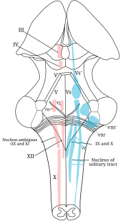
The rhomboid fossa is a rhombus-shaped depression that is the anterior part of the fourth ventricle. Its anterior wall, formed by the back of the pons and the medulla oblongata, constitutes the floor of the fourth ventricle.

Winding around the inferior cerebellar peduncle in the lower part of the fourth ventricle, and crossing the area acustica and the medial eminence are a number of white strands, the medullary striae, which form a portion of the cochlear division of the vestibulocochlear nerve and disappear into the median sulcus.: stria medullaris are axons of arcuate neurons. Courses in the floor of the fourth ventricle. Joins the restiform body to reach the cerebellum.

In the developing nervous system, the basal plate is the region of the neural tube ventral to the sulcus limitans. It extends from the rostral mesencephalon to the end of the spinal cord and contains primarily motor neurons, whereas neurons found in the alar plate are primarily associated with sensory functions. The cell types of the basal plate include lower motor neurons and four types of interneuron.
The sulcus limitans is found in the fourth ventricle of the brain. It separates the cranial nerve motor nuclei (medial) from the sensory nuclei (lateral). It can also be located by searching laterally from the medial eminence. It is parallel to the median sulcus.
The Allothalamus is a division used by some researchers in describing the thalamus.
In the human brain, the rhomboid fossa is divided into symmetrical halves by a median sulcus which reaches from the upper to the lower angles of the fossa and is deeper below than above. On either side of this sulcus is an elevation, the medial eminence, bounded laterally by a sulcus, the sulcus limitans.
Median sulcus can refer to:

The posterior median sulcus of medulla oblongata is a narrow groove; and exists only in the closed part of the medulla oblongata; it becomes gradually shallower from below upward, and finally ends about the middle of the medulla oblongata, where the central canal expands into the cavity of the fourth ventricle.
References
- ↑ Snell, Richard (2010). Clinical Neuroanatomy (7th ed.). Lippincott Williams & Wilkins. p. 206. ISBN 9780781794275.