Accessory cuneate nucleus

Last updated
Accessory cuneate nucleus
Gray694.png
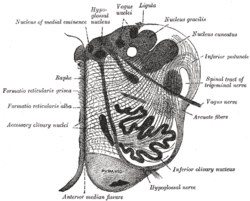
Section of the medulla oblongata at about the middle of the olive. (Accessory cuneate nucleus is not labeled, but cuneate nucleus is labeled at upper right, and the accessory cuneate nucleus would be found lateral to it.)
Details
Part of Medulla oblongata
Identifiers
Latin nucleus cuneatus accessorius
NeuroNames 768
NeuroLex ID birnlex_2634
TA A14.1.04.209
FMA 72603
Anatomical terms of neuroanatomy

The accessory cuneate nucleus is located lateral to the cuneate nucleus in the medulla oblongata at the level of the sensory decussation (the crossing fibers of the posterior column/medial lemniscus tract).

Anatomical terms of location Standard terms for unambiguous description of relative placement of body parts

Standard anatomical terms of location deal unambiguously with the anatomy of animals, including humans.

Medulla oblongata structure of the brain stem

The medulla oblongata is a long stem-like structure located in the brainstem. It is anterior and partially inferior to the cerebellum. It is a cone-shaped neuronal mass responsible for autonomic (involuntary) functions ranging from vomiting to sneezing. The medulla contains the cardiac, respiratory, vomiting and vasomotor centers and therefore deals with the autonomic functions of breathing, heart rate and blood pressure as well as the sleep wake cycle.

Sensory decussation

The sensory decussation or decussation of the lemniscus is a decussation or crossover of axons from the gracile nucleus and cuneate nucleus, which are responsible for fine touch, proprioception and two-point discrimination of the body. The fibres of this decussation are called the internal arcuate fibres and are found at the superior aspect of the closed medulla superior to the motor decussation. It is part of the second neuron in the posterior column–medial lemniscus pathway.

Contents

It receives input from cervical spinal nerves and transmits that information to the cerebellum. [1]

Cervical vertebrae Vertebrae of the neck

In tetrapods, cervical vertebrae are the vertebrae of the neck, immediately below the skull. Truncal vertebrae lie caudal of cervical vertebrae. In sauropsid species, the cervical vertebrae bear cervical ribs. In lizards and saurischian dinosaurs, the cervical ribs are large; in birds, they are small and completely fused to the vertebrae. The vertebral transverse processes of mammals are homologous to the cervical ribs of other amniotes. Most mammals have 7 cervical vertebrae, with the only 3 known exceptions being the manatee with 6, the two-toed sloth with 5–6, and the three-toed sloth with 9.

Spinal nerve nerve that carries signals between the spinal chord and the body

A spinal nerve is a mixed nerve, which carries motor, sensory, and autonomic signals between the spinal cord and the body. In the human body there are 31 pairs of spinal nerves, one on each side of the vertebral column. These are grouped into the corresponding cervical, thoracic, lumbar, sacral and coccygeal regions of the spine. There are eight pairs of cervical nerves, twelve pairs of thoracic nerves, five pairs of lumbar nerves, five pairs of sacral nerves, and one pair of coccygeal nerves. The spinal nerves are part of the peripheral nervous system.

Cerebellum region of the brain that coordinates motor functions and muscle tone

The cerebellum is a major feature of the hindbrain of all vertebrates. Although usually smaller than the cerebrum, in some animals such as the mormyrid fishes it may be as large as or even larger. In humans, the cerebellum plays an important role in motor control. It may also be involved in some cognitive functions such as attention and language as well as in regulating fear and pleasure responses, but its movement-related functions are the most solidly established. The human cerebellum does not initiate movement, but contributes to coordination, precision, and accurate timing: it receives input from sensory systems of the spinal cord and from other parts of the brain, and integrates these inputs to fine-tune motor activity. Cerebellar damage produces disorders in fine movement, equilibrium, posture, and motor learning in humans.

These fibers are called cuneocerebellar (cuneate nucleus → cerebellum) fibers.

In this function, the accessory cuneate nucleus is the upper extremity equivalent of Clarke's column. [1]

Arm forearm and upper arm together

In human anatomy, the arm is the part of the upper limb between the glenohumeral joint and the elbow joint. In common usage, the arm extends to the hand. It can be divided into the upper arm, which extends from the shoulder to the elbow, the forearm which extends from the elbow to the hand, and the hand. Anatomically the shoulder girdle with bones and corresponding muscles is by definition a part of the arm. The Latin term brachium may refer to either the arm as a whole or to the upper arm on its own.

Additional images

Related Research Articles

Pons part of the brainstem

The pons is part of the brainstem, and in humans and other bipeds lies inferior to the midbrain, superior to the medulla oblongata and anterior to the cerebellum.

Brainstem posterior part of the brain, adjoining and structurally continuous with the spinal cord

The brainstem is the posterior part of the brain, continuous with the spinal cord. In the human brain the brainstem includes the midbrain, and the pons and medulla oblongata of the hindbrain. Sometimes the diencephalon, the caudal part of the forebrain, is included.

Dorsal column–medial lemniscus pathway

The dorsal column–medial lemniscus pathway (DCML) is a sensory pathway of the central nervous system that conveys sensations of fine touch, vibration, two-point discrimination, and proprioception (position) from the skin and joints. It transmits information from the body to the primary somatosensory cortex in the postcentral gyrus of the parietal lobe of the brain. The pathway receives information from sensory receptors throughout the body, and carries this in nerve tracts in the white matter of the dorsal columns of the spinal cord, to the medulla where it is continued in the medial lemniscus, on to the thalamus and relayed from there through the internal capsule and transmitted to the somatosensory cortex. The name dorsal-column medial lemniscus comes from the two structures that carry the sensory information: the dorsal columns of the spinal cord, and the medial lemniscus in the brainstem.

Medial lemniscus

The medial lemniscus, also known as Reil's band or Reil's ribbon, is a large ascending bundle of heavily myelinated axons that decussate in the brainstem, specifically in the medulla oblongata. The medial lemniscus is formed by the crossings of the internal arcuate fibers. The internal arcuate fibers are composed of axons of nucleus gracilis and nucleus cuneatus. The axons of the nucleus gracilis and nucleus cuneatus in the medial lemniscus have cell bodies that lie contralaterally.

Inferior olivary nucleus

The inferior olivary nucleus (ION), is a structure found in the medulla oblongata underneath the superior olivary nucleus. In vertebrates, the ION is known to coordinate signals from the spinal cord to the cerebellum to regulate motor coordination and learning. These connections have been shown to be tightly associated, as degeneration of either the cerebellum or the ION results in degeneration of the other.

Spinocerebellar tract set of axonal fibers originating in the spinal cord and terminating in the ipsilateral cerebellum

The spinocerebellar tract is a nerve tract originating in the spinal cord and terminating in the same side (ipsilateral) of the cerebellum.

Inferior cerebellar peduncle

The upper part of the posterior district of the medulla oblongata is occupied by the inferior cerebellar peduncle, a thick rope-like strand situated between the lower part of the fourth ventricle and the roots of the glossopharyngeal and vagus nerves.

The interposed nucleus is part of the deep cerebellar complex and is composed of the globose nucleus and the emboliform nucleus. It is located in the roof of the fourth ventricle, lateral to the fastigial nucleus. It receives its afferent supply from the anterior lobe of the cerebellum and sends output via the superior cerebellar peduncle to the red nucleus.

Flocculus (cerebellar)

The flocculus is a small lobe of the cerebellum at the posterior border of the middle cerebellar peduncle anterior to the biventer lobule. Like other parts of the cerebellum, the flocculus is involved in motor control. It is an essential part of the vestibulo-ocular reflex, and aids in the learning of basic motor skills in the brain.

Gracile fasciculus

The gracile fasciculus is a nerve tract in the dorsal column-medial lemniscus pathway of the spinal cord and carries information from the lower parts of the body. The gracile fasiculus is one of many ascending tracts which carry received sensory information to the brain via the spinal cord. It is also one of the dorsal columns, the other being the cuneate fasciculus.

Cuneate fasciculus

The cuneate fasciculus, fasciculus cuneatus, cuneate tract, is a tract of nerves in the posterior grey column of the spinal cord that primarily transmits information from the upper part of the body. It is part of the dorsal column-medial lemniscus pathway.

Posterior external arcuate fibers

The posterior external arcuate fibers take origin in the accessory cuneate nucleus; they pass to the inferior cerebellar peduncle of the same side.

Dorsal column nuclei

In neuroanatomy, the dorsal column nuclei are a pair of nuclei in the dorsal columns in the brainstem. The name refers collectively to the cuneate nucleus and gracile nucleus, which are present at the junction between the spinal cord and the medulla oblongata. Both nuclei contain second-order neurons of the dorsal column-medial lemniscus pathway, which carries fine touch and proprioceptive information from the body to the brain. Each nucleus has an associated nerve tract, the gracile fasciculus and the cuneate fasciculus.

Superior cerebellar peduncle

In the human brain, the superior cerebellar peduncle is a paired structure of white matter that connects the cerebellum to the midbrain. It consists mainly of efferent fibers, the cerebellothalamic tract that runs from a cerebellar hemisphere to the contralateral thalamus, and the cerebellorubral tract that runs from a cerebellar hemisphere to the red nucleus. It also contains afferent tracts, most prominent of which is the ventral spinocerebellar tract. Other afferent tracts are the trigeminothalamic fibers, tectocerebellar fibers, and noradrenergic fibers from the locus coeruleus. The superior peduncle emerges from the upper and medial parts of the white matter of each hemisphere and is placed under cover of the upper part of the cerebellum.

The cerebellothalamic tract or the tractus cerebellothalamicus, is part of the superior cerebellar peduncle. It originates in the cerebellar nuclei, crosses completely in the decussation of the superior cerebellar peduncle, bypasses the red nucleus, and terminates in posterior division of ventral lateral nucleus of thalamus. The ventrolateral nucleus has different divisions and distinct connections, mostly with frontal and parietal lobes. The primary motor cortex and premotor cortex get information from the ventrolateral nucleus projections originating in the interposed nucleus and dentate nuclei. Other dentate nucleus projections via thalamic pathway transmit information to prefrontal cortex and posterior parietal cortex. The cerebellum sends thalamocortical projections and in addition may also send connections from the thalamus to association areas serving cognitive and affective functions.

Anatomy of the cerebellum

The anatomy of the cerebellum can be viewed at three levels. At the level of gross anatomy, the cerebellum consists of a tightly folded and crumpled layer of cortex, with white matter underneath, several deep nuclei embedded in the white matter, and a fluid-filled ventricle in the middle. At the intermediate level, the cerebellum and its auxiliary structures can be broken down into several hundred or thousand independently functioning modules or "microzones". At the microscopic level, each module consists of the same small set of neuronal elements, laid out with a highly stereotyped geometry.

Granule cell

The name granule cell has been used for a number of different types of neuron whose only common feature is that they all have very small cell bodies. Granule cells are found within the granular layer of the cerebellum, the dentate gyrus of the hippocampus, the superficial layer of the dorsal cochlear nucleus, the olfactory bulb, and the cerebral cortex.

Rhombic lip

The rhombic lip is a posterior section of the developing metencephalon which can be recognized transiently within the vertebrate embryo. It extends posteriorly from the roof of the fourth ventricle to dorsal neuroepithelial cells. The rhombic lip can be divided into eight structural units based on rhombomeres 1-8 (r1-r8), which can be recognized at early stages of hindbrain development. Producing granule cells and five brainstem nuclei, the rhombic lip plays an important role in developing a complex cerebellar neural system.

References

  1. 1 2 "Medical Neurosciences". Archived from the original on 2010-01-25.
Neuroscience Information Framework

The Neuroscience Information Framework is a repository of global neuroscience web resources, including experimental, clinical, and translational neuroscience databases, knowledge bases, atlases, and genetic/genomic resources and provides many authoritative links throughout the neuroscience portal of Wikipedia.