Cranial nerve nucleus

Last updated
Cranial nerve nucleus
Gray696.svg
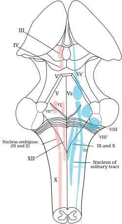
The cranial nerve nuclei schematically represented; dorsal view. Motor nuclei in red; sensory in blue. (The olfactory and optic centers are not represented.)
Details
Identifiers
Latin nucleus nervi cranialis
NeuroLex ID nlx_28532
TA98 A14.1.00.004
TA2 5857
FMA 54501
Anatomical terms of neuroanatomy

A cranial nerve nucleus is a collection of neurons (gray matter) in the brain stem that is associated with one or more of the cranial nerves. Axons carrying information to and from the cranial nerves form a synapse first at these nuclei. Lesions occurring at these nuclei can lead to effects resembling those seen by the severing of nerve(s) they are associated with. All the nuclei except that of the trochlear nerve (CN IV) supply nerves of the same side of the body.

Contents

Structure

Motor and sensory

In general, motor nuclei are closer to the front (ventral), and sensory nuclei and neurons are closer to the back (dorsal). This arrangement mirrors the arrangement of tracts in the spinal cord.

Location

This list documents nuclei by the part of the brain they are found in:

Nuclei present in the Midbrain
Nuclei present in the Pons
Nuclei present in the medulla
Micrograph of the posterior portion of the open part of the medulla oblongata, showing the fourth ventricle (top of image) and the hypoglossal nucleus (medial - left of image) and the dorsal motor nucleus of the vagus nerve (lateral - right of image). H&E-LFB stain. Medulla oblongata - posterior - low mag.jpg
Micrograph of the posterior portion of the open part of the medulla oblongata, showing the fourth ventricle (top of image) and the hypoglossal nucleus (medial - left of image) and the dorsal motor nucleus of the vagus nerve (lateral - right of image). H&E-LFB stain.

Location

Nuclei of the cranial nerves
Olfactory nerve Olfactory bulb
Optic nerve Lateral geniculate nucleus
Oculomotor nerve Oculomotor nucleus
Edinger-Westphal nucleus
Trochlear nerve Trochlear nucleus
Trigeminal nerve Trigeminal nerve nuclei:
Mesencephalic nucleus
Principal sensory nucleus
Spinal trigeminal nucleus
Trigeminal motor nucleus
Abducens nerve Abducens nucleus
Facial nerve Facial motor nucleus
Superior salivatory nucleus
Solitary nucleus
Vestibulocochlear nerve Vestibular nuclei
w. subnuclei
Cochlear nucleus
w. subnuclei
Glossopharyngeal nerve Solitary nucleus
Spinal nucleus of the trigeminal nerve
Lateral nucleus of vagal trigone.
Nucleus ambiguus
Inferior salivatory nucleus
Vagus nerve Dorsal nucleus of vagus nerve
Nucleus ambiguus
Solitary nucleus
Spinal trigeminal nucleus
Accessory nerve Spinal accessory nucleus
Nucleus ambiguus
Hypoglossal nerve Hypoglossal nucleus

Related Research Articles

<span class="mw-page-title-main">Cranial nerves</span> Nerves that emerge directly from the brain and the brainstem

Cranial nerves are the nerves that emerge directly from the brain, of which there are conventionally considered twelve pairs. Cranial nerves relay information between the brain and parts of the body, primarily to and from regions of the head and neck, including the special senses of vision, taste, smell, and hearing.

<span class="mw-page-title-main">Parasympathetic nervous system</span> Division of the autonomic nervous system

The parasympathetic nervous system is one of the three divisions of the autonomic nervous system, the others being the sympathetic nervous system and the enteric nervous system. The enteric nervous system is sometimes considered part of the autonomic nervous system, and sometimes considered an independent system.

<span class="mw-page-title-main">Medulla oblongata</span> Structure of the brain stem

The medulla oblongata or simply medulla is a long stem-like structure which makes up the lower part of the brainstem. It is anterior and partially inferior to the cerebellum. It is a cone-shaped neuronal mass responsible for autonomic (involuntary) functions, ranging from vomiting to sneezing. The medulla contains the cardiac, respiratory, vomiting and vasomotor centers, and therefore deals with the autonomic functions of breathing, heart rate and blood pressure as well as the sleep–wake cycle.

<span class="mw-page-title-main">Facial nerve</span> Cranial nerve VII, for the face and tasting

The facial nerve, also known as the seventh cranial nerve, cranial nerve VII, or simply CN VII, is a cranial nerve that emerges from the pons of the brainstem, controls the muscles of facial expression, and functions in the conveyance of taste sensations from the anterior two-thirds of the tongue. The nerve typically travels from the pons through the facial canal in the temporal bone and exits the skull at the stylomastoid foramen. It arises from the brainstem from an area posterior to the cranial nerve VI and anterior to cranial nerve VIII.

Articles related to anatomy include:

<span class="mw-page-title-main">Pons</span> Part of the brainstem in humans and other bipeds

The pons is part of the brainstem that in humans and other bipeds lies inferior to the midbrain, superior to the medulla oblongata and anterior to the cerebellum.

<span class="mw-page-title-main">Brainstem</span> Posterior part of the brain, adjoining and structurally continuous

The brainstem is the posterior stalk-like part of the brain that connects the cerebrum with the spinal cord. In the human brain the brainstem is composed of the midbrain, the pons, and the medulla oblongata. The midbrain is continuous with the thalamus of the diencephalon through the tentorial notch, and sometimes the diencephalon is included in the brainstem.

<span class="mw-page-title-main">Oculomotor nerve</span> Cranial nerve III, for eye movements

The oculomotor nerve, also known as the third cranial nerve, cranial nerve III, or simply CN III, is a cranial nerve that enters the orbit through the superior orbital fissure and innervates extraocular muscles that enable most movements of the eye and that raise the eyelid. The nerve also contains fibers that innervate the intrinsic eye muscles that enable pupillary constriction and accommodation. The oculomotor nerve is derived from the basal plate of the embryonic midbrain. Cranial nerves IV and VI also participate in control of eye movement.

<span class="mw-page-title-main">Trigeminal nerve</span> Cranial nerve responsible for the faces senses and motor functions

In neuroanatomy, the trigeminal nerve (lit. triplet nerve), also known as the fifth cranial nerve, cranial nerve V, or simply CN V, is a cranial nerve responsible for sensation in the face and motor functions such as biting and chewing; it is the most complex of the cranial nerves. Its name ("trigeminal", from Latin tri- 'three', and -geminus 'twin') derives from each of the two nerves (one on each side of the pons) having three major branches: the ophthalmic nerve (V1), the maxillary nerve (V2), and the mandibular nerve (V3). The ophthalmic and maxillary nerves are purely sensory, whereas the mandibular nerve supplies motor as well as sensory (or "cutaneous") functions. Adding to the complexity of this nerve is that autonomic nerve fibers as well as special sensory fibers (taste) are contained within it.

<span class="mw-page-title-main">Glossopharyngeal nerve</span> Cranial nerve IX, for the tongue and pharynx

The glossopharyngeal nerve, also known as the ninth cranial nerve, cranial nerve IX, or simply CN IX, is a cranial nerve that exits the brainstem from the sides of the upper medulla, just anterior to the vagus nerve. Being a mixed nerve (sensorimotor), it carries afferent sensory and efferent motor information. The motor division of the glossopharyngeal nerve is derived from the basal plate of the embryonic medulla oblongata, whereas the sensory division originates from the cranial neural crest.

<span class="mw-page-title-main">Oculomotor nucleus</span>

The fibers of the oculomotor nerve arise from a nucleus in the midbrain, which lies in the gray substance of the floor of the cerebral aqueduct and extends in front of the aqueduct for a short distance into the floor of the third ventricle. From this nucleus the fibers pass forward through the tegmentum, the red nucleus, and the medial part of the substantia nigra, forming a series of curves with a lateral convexity, and emerge from the oculomotor sulcus on the medial side of the cerebral peduncle.

<span class="mw-page-title-main">Nucleus ambiguus</span>

The nucleus ambiguus is a group of large motor neurons, situated deep in the medullary reticular formation named by Jacob Clarke. The nucleus ambiguus contains the cell bodies of neurons that innervate the muscles of the soft palate, pharynx, and larynx which are associated with speech and swallowing. As well as motor neurons, the nucleus ambiguus contains preganglionic parasympathetic neurons which innervate postganglionic parasympathetic neurons in the heart.

<span class="mw-page-title-main">Corticobulbar tract</span> Motor pathway in the brain connecting the motor cortex to the medullary pyramids

In neuroanatomy, the corticobulbartract is a two-neuron white matter motor pathway connecting the motor cortex in the cerebral cortex to the medullary pyramids, which are part of the brainstem's medulla oblongata region, and are primarily involved in carrying the motor function of the non-oculomotor cranial nerves. The corticobulbar tract is one of the pyramidal tracts, the other being the corticospinal tract.

<span class="mw-page-title-main">Pontine tegmentum</span>

The pontine tegmentum, or dorsal pons, is located within the brainstem, and is one of two parts of the pons, the other being the ventral pons or basilar part of the pons. The pontine tegmentum can be defined in contrast to the basilar pons: basilar pons contains the corticospinal tract running craniocaudally and can be considered the rostral extension of the ventral medulla oblongata; however, basilar pons is distinguished from ventral medulla oblongata in that it contains additional transverse pontine fibres that continue laterally to become the middle cerebellar peduncle. The pontine tegmentum is all the material dorsal from the basilar pons to the fourth ventricle. Along with the dorsal surface of the medulla, it forms part of the rhomboid fossa – the floor of the fourth ventricle.

<span class="mw-page-title-main">Edinger–Westphal nucleus</span> One of two nuclei of the oculomotor nerve

The Edinger–Westphal nucleus is one of two nuclei of the oculomotor nerve. It is located in the midbrain. It contributes the autonomic parasympathetic component to the oculomotor nerve, providing innervation to the iris sphincter muscle and ciliary muscle to mediate the pupillary light reflex and accommodation, respectively. It is composed of parasympathetic pre-ganglionic cell bodies that synapse in the ciliary ganglion.

<span class="mw-page-title-main">Facial motor nucleus</span>

The facial motor nucleus is a collection of neurons in the brainstem that belong to the facial nerve. These lower motor neurons innervate the muscles of facial expression and the stapedius.

<span class="mw-page-title-main">Trochlear nucleus</span>

The nucleus of the trochlear nerve is located in the midbrain, at an intercollicular level between the superior colliculus and inferior colliculus. It is a motor nucleus, and so is located near the midline, embedded within the medial longitudinal fasciculus. The oculomotor nerve and trochlear nerve are the only two cranial nerves with nuclei in the midbrain, other than the trigeminal nerve, which has a midbrain nucleus called the mesencephalic nucleus of trigeminal nerve, which functions in preserving dentition.

<span class="mw-page-title-main">Cochlear nucleus</span> Two cranial nerve nuclei of the human brainstem

The cochlear nuclear (CN) complex comprises two cranial nerve nuclei in the human brainstem, the ventral cochlear nucleus (VCN) and the dorsal cochlear nucleus (DCN). The ventral cochlear nucleus is unlayered whereas the dorsal cochlear nucleus is layered. Auditory nerve fibers, fibers that travel through the auditory nerve carry information from the inner ear, the cochlea, on the same side of the head, to the nerve root in the ventral cochlear nucleus. At the nerve root the fibers branch to innervate the ventral cochlear nucleus and the deep layer of the dorsal cochlear nucleus. All acoustic information thus enters the brain through the cochlear nuclei, where the processing of acoustic information begins. The outputs from the cochlear nuclei are received in higher regions of the auditory brainstem.

<span class="mw-page-title-main">Trigeminal nerve nuclei</span>

The sensory trigeminal nerve nuclei are the largest of the cranial nerve nuclei, and extend through the whole of the midbrain, pons and medulla, and into the high cervical spinal cord.

<span class="mw-page-title-main">Salivatory nuclei</span>

The salivatory nuclei are the superior salivatory nucleus, and the inferior salivatory nucleus that innervate the salivary glands. They are located in the pontine tegmentum in the brainstem. They both are examples of cranial nerve nuclei.

References

Additional images