Periaqueductal gray

Last updated

Periaqueductal gray
Cn3nucleus-en.svg
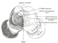
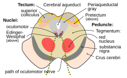
Section through superior colliculus showing path of oculomotor nerve. Periaqueductal gray is the gray area just peripheral to the cerebral aqueduct.
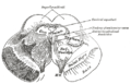
Gray710.png
Details
Identifiers
Latin substantia grisea centralis
MeSH D010487
NeuroNames 1584
NeuroLex ID birnlex_973
TA98 A14.1.06.321
TA2 5909
FMA 83134
Anatomical terms of neuroanatomy

The periaqueductal gray (PAG, also known as the central gray) is a brain region that plays a critical role in autonomic function, motivated behavior and behavioural responses to threatening stimuli. [1] [2] PAG is also the primary control center for descending pain modulation. It has enkephalin-producing cells that suppress pain.

Contents

The periaqueductal gray is the gray matter located around the cerebral aqueduct within the tegmentum of the midbrain. It projects to the nucleus raphe magnus, and also contains descending autonomic tracts. The ascending pain and temperature fibers of the spinothalamic tract send information to the PAG via the spinomesencephalic tract (so-named because the fibers originate in the spine and terminate in the PAG, in the mesencephalon or midbrain).

This region has been used as the target for brain-stimulating implants in patients with chronic pain.

Role in analgesia

Stimulation of the periaqueductal gray matter of the midbrain activates enkephalin-releasing neurons that project to the raphe nuclei in the brainstem. 5-HT (serotonin) released from the raphe nuclei descends to the dorsal horn of the spinal cord where it forms excitatory connections with the inhibitory interneurons located in Laminae II (aka the substantia gelatinosa). When activated, these interneurons release either enkephalin or dynorphin (endogenous opioid peptides), which bind to mu and kappa opioid receptors, respectively, on the axons of incoming C and A-delta fibers carrying pain signals from nociceptors activated in the periphery.

The activation of the mu-opioid receptor inhibits the release of substance P from these incoming first-order neurons and, in turn, inhibits the activation of the second-order neuron that is responsible for transmitting the pain signal up the spinothalamic tract to the ventral posterolateral nucleus (VPL) of the thalamus. The nociceptive signal is thus inhibited before reaching the cortical areas that interpret the signal as pain, such as the anterior cingulate. This is sometimes referred to as the gate control theory of pain and is supported by the fact that electrical stimulation of the PAG results in immediate and profound analgesia. [3] The periaqueductal gray is also activated by viewing distressing images associated with pain. [4]

Notably, the anterior cingulate cortex is thought to be responsible for emotional responses to pain, including perceived social or emotional pain. Reducing nociceptive signaling to this area not only reduces overall pain signaling, but appears to also reduce sensitivity to pain. Furthermore, activation of mu-opioid receptors has been shown to provide an "analgesic" effect for emotional pain. [5] [6]

Role in defensive behavior

Dorsal PAG neurons are activated during various defensive behaviors. [7] Stimulation of the dorsal and lateral aspects of the PAG can provoke defensive responses characterised by freezing immobility, running, jumping, tachycardia, and increases in blood pressure and muscle tonus. In contrast, stimulation of the caudal ventrolateral PAG can result in an immobile, relaxed posture known as quiescence, whereas its inhibition leads to increased locomotor activity.

Lesions of the caudal ventrolateral PAG can greatly reduce conditioned freezing, whereas lesions of the dorsal aspect can reduce innate defensive behavior, virtually "taming" the animal[ citation needed ].

Role in reproductive behavior

Neurons of the PAG are excited by endorphins and by opiate analgesics. It also plays a role in female copulatory behavior (see lordosis behavior) via a pathway from the ventromedial nucleus of the hypothalamus.

Role in maternal behavior

The PAG may be specifically involved in human maternal behavior. The PAG contains a high density of vasopressin and oxytocin receptors, and it has direct connections with the orbitofrontal cortex, which might mediate the role of the PAG in maternal love. The lateral orbitofrontal cortex is activated by pleasant visual, tactile, and olfactory stimuli. Its response depends on pleasantness rather than on intensity of stimulation. Here, its activity is likely to reflect one aspect of the pleasant emotions associated with motherly love. [8]

Additional images

See also

Related Research Articles

In physiology, nociception, also nocioception; from Latin nocere 'to harm/hurt') is the sensory nervous system's process of encoding noxious stimuli. It deals with a series of events and processes required for an organism to receive a painful stimulus, convert it to a molecular signal, and recognize and characterize the signal to trigger an appropriate defensive response.

<span class="mw-page-title-main">Hypothalamus</span> Area of the brain below the thalamus

The hypothalamus is a small part of the brain that contains a number of nuclei with a variety of functions. One of the most important functions is to link the nervous system to the endocrine system via the pituitary gland. The hypothalamus is located below the thalamus and is part of the limbic system. It forms the ventral part of the diencephalon. All vertebrate brains contain a hypothalamus. In humans, it is the size of an almond.

<span class="mw-page-title-main">Brainstem</span> Posterior part of the brain, adjoining and structurally continuous

The brainstem is the stalk-like part of the brain that interconnects the cerebrum and diencephalon with the spinal cord. In the human brain, the brainstem is composed of the midbrain, the pons, and the medulla oblongata. The midbrain is continuous with the thalamus of the diencephalon through the tentorial notch.

<span class="mw-page-title-main">Midbrain</span> Forward-most portion of the brainstem

The midbrain or mesencephalon is the rostral-most portion of the brainstem connecting the diencephalon and cerebrum with the pons. It consists of the cerebral peduncles, tegmentum, and tectum.

<span class="mw-page-title-main">Raphe nuclei</span> Moderate-size cluster of nuclei found in brain stem

The raphe nuclei are a moderate-size cluster of nuclei found in the brain stem. They have 5-HT1 receptors which are coupled with Gi/Go-protein-inhibiting adenyl cyclase. They function as autoreceptors in the brain and decrease the release of serotonin. The anxiolytic drug Buspirone acts as partial agonist against these receptors. Selective serotonin reuptake inhibitor (SSRI) antidepressants are believed to act in these nuclei, as well as at their targets.

<span class="mw-page-title-main">Nociceptor</span> Sensory neuron that detects pain

A nociceptor is a sensory neuron that responds to damaging or potentially damaging stimuli by sending "possible threat" signals to the spinal cord and the brain. The brain creates the sensation of pain to direct attention to the body part, so the threat can be mitigated; this process is called nociception.

<span class="mw-page-title-main">Spinothalamic tract</span> Sensory pathway from the skin to the thalamus

The spinothalamic tract is a part of the anterolateral system or the ventrolateral system, a sensory pathway to the thalamus. From the ventral posterolateral nucleus in the thalamus, sensory information is relayed upward to the somatosensory cortex of the postcentral gyrus.

<span class="mw-page-title-main">Ventral tegmental area</span> Group of neurons on the floor of the midbrain

The ventral tegmental area (VTA), also known as the ventral tegmental area of Tsai, or simply ventral tegmentum, is a group of neurons located close to the midline on the floor of the midbrain. The VTA is the origin of the dopaminergic cell bodies of the mesocorticolimbic dopamine system and other dopamine pathways; it is widely implicated in the drug and natural reward circuitry of the brain. The VTA plays an important role in a number of processes, including reward cognition and orgasm, among others, as well as several psychiatric disorders. Neurons in the VTA project to numerous areas of the brain, ranging from the prefrontal cortex to the caudal brainstem and several regions in between.

β-Endorphin Peptide hormone in humans

β-Endorphin (beta-endorphin) is an endogenous opioid neuropeptide and peptide hormone that is produced in certain neurons within the central nervous system and peripheral nervous system. It is one of three endorphins that are produced in humans, the others of which include α-endorphin and γ-endorphin.

<span class="mw-page-title-main">Pretectal area</span> Structure in the midbrain which mediates responses to ambient light

In neuroanatomy, the pretectal area, or pretectum, is a midbrain structure composed of seven nuclei and comprises part of the subcortical visual system. Through reciprocal bilateral projections from the retina, it is involved primarily in mediating behavioral responses to acute changes in ambient light such as the pupillary light reflex, the optokinetic reflex, and temporary changes to the circadian rhythm. In addition to the pretectum's role in the visual system, the anterior pretectal nucleus has been found to mediate somatosensory and nociceptive information.

<span class="mw-page-title-main">Reticular formation</span> Spinal trigeminal nucleus

The reticular formation is a set of interconnected nuclei that are located throughout the brainstem. It is not anatomically well defined, because it includes neurons located in different parts of the brain. The neurons of the reticular formation make up a complex set of networks in the core of the brainstem that extend from the upper part of the midbrain to the lower part of the medulla oblongata. The reticular formation includes ascending pathways to the cortex in the ascending reticular activating system (ARAS) and descending pathways to the spinal cord via the reticulospinal tracts.

Some philosophers, such as Jeremy Bentham, Baruch Spinoza, and Descartes, have hypothesized that the feelings of pain and pleasure are part of a continuum.

The zona incerta (ZI) is a horizontally elongated region of gray matter in the subthalamus below the thalamus. Its connections project extensively over the brain from the cerebral cortex down into the spinal cord.

<span class="mw-page-title-main">Nucleus raphe magnus</span>

The nucleus raphe magnus is one of the seven raphe nuclei. It is situated in the pons in the brainstem, just rostral to the nucleus raphe obscurus.

<span class="mw-page-title-main">Lateral hypothalamus</span>

The lateral hypothalamus (LH), also called the lateral hypothalamic area (LHA), contains the primary orexinergic nucleus within the hypothalamus that widely projects throughout the nervous system; this system of neurons mediates an array of cognitive and physical processes, such as promoting feeding behavior and arousal, reducing pain perception, and regulating body temperature, digestive functions, and blood pressure, among many others. Clinically significant disorders that involve dysfunctions of the orexinergic projection system include narcolepsy, motility disorders or functional gastrointestinal disorders involving visceral hypersensitivity, and eating disorders.

The ventrobasal complex (VB) is a relay nucleus of the thalamus for nociceptive stimuli received from nociceptive nerves. The VB consists of the ventral posteromedial nucleus (VPM) and the ventral posterolateral nucleus (VPL). In some species, the ventral posterolateral nucleus, pars caudalis is also a part of the VB. The VB gets inputs from the spinothalamic tract, medial lemniscus, and corticothalamic tract. The main output of the VB is the primary somatosensory cortex.

Sleep onset is the transition from wakefulness into sleep. Sleep onset usually transmits into non-rapid eye movement sleep but under certain circumstances it is possible to transit from wakefulness directly into rapid eye movement sleep.

<span class="mw-page-title-main">Rostral ventromedial medulla</span> Group of neurons in medulla of brain

The rostral ventromedial medulla (RVM), or ventromedial nucleus of the spinal cord, is a group of neurons located close to the midline on the floor of the medulla oblongata. The rostral ventromedial medulla sends descending inhibitory and excitatory fibers to the dorsal horn spinal cord neurons. There are 3 categories of neurons in the RVM: on-cells, off-cells, and neutral cells. They are characterized by their response to nociceptive input. Off-cells show a transitory decrease in firing rate right before a nociceptive reflex, and are theorized to be inhibitory. Activation of off-cells, either by morphine or by any other means, results in antinociception. On-cells show a burst of activity immediately preceding nociceptive input, and are theorized to be contributing to the excitatory drive. Neutral cells show no response to nociceptive input.

The rostromedial tegmental nucleus (RMTg), also known as the tail of the ventral tegmental area (tVTA), is a GABAergic nucleus which functions as a "master brake" for the midbrain dopamine system. This region was discovered by the researchers, M. Barrot, J.Kaufling and T. Jhou. It is poorly differentiated from the rest of the ventral tegmental area (VTA) and possesses robust functional and structural links to the dopamine pathways. Notably, both acute and chronic exposure to psychostimulants have been shown to induce FosB and ΔFosB expression in the RMTg; no other drug type has been shown to induce these proteins in the RMTg.

References

  1. Faull, Olivia K.; Subramanian, Hari H.; Ezra, Martyn; Pattinson, Kyle T. S. (2019). "The midbrain periaqueductal gray as an integrative and interoceptive neural structure for breathing". Neuroscience and Biobehavioral Reviews. 98: 135–144. doi: 10.1016/j.neubiorev.2018.12.020 . hdl: 20.500.11850/317617 . ISSN   1873-7528. PMID   30611797.
  2. Silva, Carlos; McNaughton, Neil (2019-02-17). "Are periaqueductal grey and dorsal raphe the foundation of appetitive and aversive control? A comprehensive review". Progress in Neurobiology. 177: 33–72. doi:10.1016/j.pneurobio.2019.02.001. ISSN   1873-5118. PMID   30786258. S2CID   73478335.
  3. Basbaum AI, Fields HL (November 1978). "Endogenous pain control mechanisms: review and hypothesis". Ann. Neurol. 4 (5): 451–62. doi:10.1002/ana.410040511. PMID   216303. S2CID   72620829.
  4. Jenkins, Dacher Keltner, Keith Oatley, Jennifer M. (2013-01-29). Understanding emotions (3rd ed.). Hoboken, N.J.: Wiley. ISBN   9781118147436.{{cite book}}: CS1 maint: multiple names: authors list (link)
  5. Eisenberger NI, Lieberman MD, Williams KD (October 2003). "Does rejection hurt? An FMRI study of social exclusion". Science. 302 (5643): 290–2. Bibcode:2003Sci...302..290E. doi:10.1126/science.1089134. PMID   14551436. S2CID   21253445.
  6. Gorka SM, Fitzgerald DA, de Wit H, Angstadt M, Phan KL (December 2014). "Opioid modulation of resting-state anterior cingulate cortex functional connectivity". J Psychopharmacol. 28 (12): 1115–24. doi:10.1177/0269881114548436. PMC   5613932 . PMID   25237122.
  7. Deng H, Xiao X, Wang Z (2016). "Periaqueductal Gray Neuronal Activities Underlie Different Aspects of Defensive Behaviors". J Neurosci. 36 (29): 7580–8. doi:10.1523/JNEUROSCI.4425-15.2016. PMC   6705556 . PMID   27445137.{{cite journal}}: CS1 maint: multiple names: authors list (link)
  8. Andreas Bartels; Semir Zeki (March 2004). "The neural correlates of maternal and romantic love" (PDF). NeuroImage. 21 (3): 1155–1166. doi:10.1016/j.neuroimage.2003.11.003. PMID   15006682. S2CID   15237043. Archived from the original (PDF) on 2017-08-29. Retrieved 2013-01-27.