| |
| Content | |
|---|---|
| Description | Interactive zoomable high-resolution digital brain atlas |
| Data types captured | Neuroanatomy, Histology |
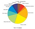
| Organisms | includes: Man, monkey, cat, mouse, opossum, goldfish, platypus, dog, owl, chicken, rat, and more |
| Access | |
| Website | http://brainmaps.org/ |
| Miscellaneous | |
| License | All image dataset is copyrighted to their respective owners, if none indicated, to the UC Regents Davis campus. [1] |

a: choosing from some hundreds of coronal sections.
b: certain coronal section shown.
c: zooming up of insular cortex region.
d: further zooming up of insular cortex. Nissl stained neurons are visible. This slice can be accessed through this link.
BrainMaps is an NIH-funded interactive zoomable high-resolution digital brain atlas and virtual microscope that is based on more than 140 million megapixels (140 terabytes) of scanned images of serial sections of both primate and non-primate brains and that is integrated with a high-speed database for querying and retrieving data about brain structure and function over the internet.
Contents
Currently featured are complete brain atlas datasets for 16 species; a few of which are: Macaca mulatta , Chlorocebus aethiops , Felis silvestris catus , Mus musculus , Rattus norvegicus , and Tyto alba .
The project's principal investigator was UC Davis neuroscientist Ted Jones from 2005 through 2011, after which the role was taken by W. Martin Usrey.