Mouse brain

Last updated
Mouse brain, lateral view Mouse brain.jpg
Mouse brain, lateral view

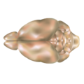
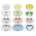
The mouse brain refers to the brain of Mus musculus . Various brain atlases exist.

Contents

For reasons of reproducibility, genetically characterized, stable strains like C57BL/6 were chosen to produce high-resolution images and databases. [1] Well known online resources include:

Despite superficial differences, especially in size and weight, the mouse brain and its function can serve as a powerful animal model for study of human brain diseases or mental disorders (see e.g. Reeler, Chakragati mouse). This is because the genes responsible for building and operating both mouse and human brain are 90% identical. [4] Transgenic mouse lines also allow neuroscientists to specifically target the labeling of certain cell types to probe the neural basis of fundamental processes. [5] [6]

Anatomy

The cerebral cortex of a mouse has around 8–14 million neurons while in those humans there are more than 10–15 billion. [7] [8] The olfactory bulb volume takes about 2% of the mouse brain by volume in contrast to about 0.01% of the human brain. [9] [10]

Development

See also

Related Research Articles

<span class="mw-page-title-main">Reelin</span> Large secreted extracellular matrix glycoprotein involved in neuronal migration

Reelin, encoded by the RELN gene, is a large secreted extracellular matrix glycoprotein that helps regulate processes of neuronal migration and positioning in the developing brain by controlling cell–cell interactions. Besides this important role in early development, reelin continues to work in the adult brain. It modulates synaptic plasticity by enhancing the induction and maintenance of long-term potentiation. It also stimulates dendrite and dendritic spine development and regulates the continuing migration of neuroblasts generated in adult neurogenesis sites like the subventricular and subgranular zones. It is found not only in the brain but also in the liver, thyroid gland, adrenal gland, Fallopian tube, breast and in comparatively lower levels across a range of anatomical regions.

The development of the nervous system, or neural development (neurodevelopment), refers to the processes that generate, shape, and reshape the nervous system of animals, from the earliest stages of embryonic development to adulthood. The field of neural development draws on both neuroscience and developmental biology to describe and provide insight into the cellular and molecular mechanisms by which complex nervous systems develop, from nematodes and fruit flies to mammals.

<span class="mw-page-title-main">Pyramidal cell</span>

Pyramidal cells, or pyramidal neurons, are a type of multipolar neuron found in areas of the brain including the cerebral cortex, the hippocampus, and the amygdala. Pyramidal cells are the primary excitation units of the mammalian prefrontal cortex and the corticospinal tract. Pyramidal neurons are also one of two cell types where the characteristic sign, Negri bodies, are found in post-mortem rabies infection. Pyramidal neurons were first discovered and studied by Santiago Ramón y Cajal. Since then, studies on pyramidal neurons have focused on topics ranging from neuroplasticity to cognition.

<span class="mw-page-title-main">Motor cortex</span> Region of the cerebral cortex

The motor cortex is the region of the cerebral cortex involved in the planning, control, and execution of voluntary movements. The motor cortex is an area of the frontal lobe located in the posterior precentral gyrus immediately anterior to the central sulcus.

Brain mapping is a set of neuroscience techniques predicated on the mapping of (biological) quantities or properties onto spatial representations of the brain resulting in maps.

<span class="mw-page-title-main">BrainMaps</span>

BrainMaps is an NIH-funded interactive zoomable high-resolution digital brain atlas and virtual microscope that is based on more than 140 million megapixels of scanned images of serial sections of both primate and non-primate brains and that is integrated with a high-speed database for querying and retrieving data about brain structure and function over the internet.

<span class="mw-page-title-main">Reeler</span> Mouse mutant

A reeler is a mouse mutant, so named because of its characteristic "reeling" gait. This is caused by the profound underdevelopment of the mouse's cerebellum, a segment of the brain responsible for locomotion. The mutation is autosomal and recessive, and prevents the typical cerebellar folia from forming.

The development of the nervous system in humans, or neural development or neurodevelopment involves the studies of embryology, developmental biology, and neuroscience to describe the cellular and molecular mechanisms by which the complex nervous system forms in humans, develops during prenatal development, and continues to develop postnatally.

<span class="mw-page-title-main">Brainbow</span> Neuroimaging technique to differentiate neurons

Brainbow is a process by which individual neurons in the brain can be distinguished from neighboring neurons using fluorescent proteins. By randomly expressing different ratios of red, green, and blue derivatives of green fluorescent protein in individual neurons, it is possible to flag each neuron with a distinctive color. This process has been a major contribution to the field of neural connectomics.

Optogenetics is a biological technique to control the activity of neurons or other cell types with light. This is achieved by expression of light-sensitive ion channels, pumps or enzymes specifically in the target cells. On the level of individual cells, light-activated enzymes and transcription factors allow precise control of biochemical signaling pathways. In systems neuroscience, the ability to control the activity of a genetically defined set of neurons has been used to understand their contribution to decision making, learning, fear memory, mating, addiction, feeding, and locomotion. In a first medical application of optogenetic technology, vision was partially restored in a blind patient.

<span class="mw-page-title-main">EMX1</span> Protein-coding gene in the species Homo sapiens

Homeobox protein EMX1 is a protein that in humans is encoded by the EMX1 gene. The transcribed EMX1 gene is a member of the EMX family of transcription factors. The EMX1 gene, along with its family members, are expressed in the developing cerebrum. EMX1 plays a role in specification of positional identity, the proliferation of neural stem cells, differentiation of layer-specific neuronal phenotypes and commitment to a neuronal or glial cell fate.

<span class="mw-page-title-main">Connectome</span> Comprehensive map of neural connections in the brain

A connectome is a comprehensive map of neural connections in the brain, and may be thought of as its "wiring diagram". An organism's nervous system is made up of neurons which communicate through synapses. A connectome is constructed by tracing the neuron in a nervous system and mapping where neurons are connected through synapses.

<span class="mw-page-title-main">Genetically modified mouse</span>

A genetically modified mouse or genetically engineered mouse model (GEMM) is a mouse that has had its genome altered through the use of genetic engineering techniques. Genetically modified mice are commonly used for research or as animal models of human diseases, and are also used for research on genes. Together with patient-derived xenografts (PDXs), GEMMs are the most common in vivo models in cancer research. Both approaches are considered complementary and may be used to recapitulate different aspects of disease. GEMMs are also of great interest for drug development, as they facilitate target validation and the study of response, resistance, toxicity and pharmacodynamics.

Connectomics is the production and study of connectomes: comprehensive maps of connections within an organism's nervous system. More generally, it can be thought of as the study of neuronal wiring diagrams with a focus on how structural connectivity, individual synapses, cellular morphology, and cellular ultrastructure contribute to the make up of a network. The nervous system is a network made of billions of connections and these connections are responsible for our thoughts, emotions, actions, memories, function and dysfunction. Therefore, the study of connectomics aims to advance our understanding of mental health and cognition by understanding how cells in the nervous system are connected and communicate. Because these structures are extremely complex, methods within this field use a high-throughput application of functional and structural neural imaging, most commonly magnetic resonance imaging (MRI), electron microscopy, and histological techniques in order to increase the speed, efficiency, and resolution of these nervous system maps. To date, tens of large scale datasets have been collected spanning the nervous system including the various areas of cortex, cerebellum, the retina, the peripheral nervous system and neuromuscular junctions.

Gyrification is the process of forming the characteristic folds of the cerebral cortex.

<span class="mw-page-title-main">Eomesodermin</span> Protein-coding gene in the species Homo sapiens

Eomesodermin also known as T-box brain protein 2 (Tbr2) is a protein that in humans is encoded by the EOMES gene.

Corticogenesis is the process during which the cerebral cortex of the brain is formed as part of the development of the nervous system of mammals including its development in humans. The cortex is the outer layer of the brain and is composed of up to six layers. Neurons formed in the ventricular zone migrate to their final locations in one of the six layers of the cortex. The process occurs from embryonic day 10 to 17 in mice and between gestational weeks seven to 18 in humans.

Cajal–Retzius cells are a heterogeneous population of morphologically and molecularly distinct reelin-producing cell types in the marginal zone/layer I of the developmental cerebral cortex and in the immature hippocampus of different species and at different times during embryogenesis and postnatal life.

Neurogenesis is the process by which nervous system cells, the neurons, are produced by neural stem cells (NSCs). It occurs in all species of animals except the porifera (sponges) and placozoans. Types of NSCs include neuroepithelial cells (NECs), radial glial cells (RGCs), basal progenitors (BPs), intermediate neuronal precursors (INPs), subventricular zone astrocytes, and subgranular zone radial astrocytes, among others.

<span class="mw-page-title-main">Radial unit hypothesis</span> Conceptual theory of cerebral cortex development

The Radial Unit Hypothesis (RUH) is a conceptual theory of cerebral cortex development, first described by Pasko Rakic. The RUH states that the cerebral cortex develops during embryogenesis as an array of interacting cortical columns, or 'radial units', each of which originates from a transient stem cell layer called the ventricular zone, which contains neural stem cells known as radial glial cells.

References

  1. MacKenzie-Graham, Allan; Jones, Eagle S.; Shattuck, David W.; Dinov, Ivo D.; Bota, Mihail; Toga, Arthur W. (2003). "The Informatics of a C57BL/6J Mouse Brain Atlas". Neuroinformatics. Springer Nature. 1 (4): 397–410. doi:10.1385/ni:1:4:397. ISSN   1539-2791. PMID   15043223. S2CID   81057.
  2. "ISH Data :: Allen Brain Atlas: Mouse Brain". ISH Data. Retrieved 2019-02-07.
  3. "Search the library". The Mouse Brain Library. 2003-06-05. Retrieved 2019-02-07.
  4. Park, Alice (19 January 2007). "The Brain: What the Mouse Brain Tells Us". Time.
  5. Gordon, J. W.; Scangos, G. A.; Plotkin, D. J.; Barbosa, J. A.; Ruddle, F. H. (1980-12-01). "Genetic transformation of mouse embryos by microinjection of purified DNA". Proceedings of the National Academy of Sciences. 77 (12): 7380–7384. Bibcode:1980PNAS...77.7380G. doi: 10.1073/pnas.77.12.7380 . ISSN   0027-8424. PMC   350507 . PMID   6261253.
  6. Haruyama, Naoto; Cho, Andrew; Kulkarni, Ashok B. (2009). "Overview: Engineering Transgenic Constructs and Mice". Current Protocols in Cell Biology. 42 (1): 19.10.1–19.10.9. doi:10.1002/0471143030.cb1910s42. ISSN   1934-2616. PMC   2743315 . PMID   19283728.
  7. "Brain size and intelligence- why a human is smarter than a mouse". DNALC Blogs «. 2012-11-13. Retrieved 2019-02-07.
  8. Herculano-Houzel, Suzana; Catania, Kenneth; Manger, Paul R.; Kaas, Jon H. (2015). "Mammalian Brains Are Made of These: A Dataset of the Numbers and Densities of Neuronal and Nonneuronal Cells in the Brain of Glires, Primates, Scandentia, Eulipotyphlans, Afrotherians and Artiodactyls, and Their Relationship with Body Mass". Brain, Behavior and Evolution. S. Karger AG. 86 (3–4): 145–163. doi: 10.1159/000437413 . ISSN   0006-8977. PMID   26418466.
  9. McGann, John P. (2017-05-11). "Poor human olfaction is a 19th-century myth". Science. American Association for the Advancement of Science (AAAS). 356 (6338): eaam7263. doi:10.1126/science.aam7263. ISSN   0036-8075. PMC   5512720 . PMID   28495701.
  10. "Brain Facts and Figures". faculty.washington.edu. Retrieved 2019-02-07.
  11. Dura-Bernal, Salvador; Neymotin, Samuel A.; Suter, Benjamin A.; Dacre, Joshua; Moreira, Joao V.S.; Urdapilleta, Eugenio; Schiemann, Julia; Duguid, Ian; Shepherd, Gordon M.G.; Lytton, William W. (June 2023). "Multiscale model of primary motor cortex circuits predicts in vivo cell-type-specific, behavioral state-dependent dynamics". Cell Reports. 42 (6): 112574. doi:10.1016/j.celrep.2023.112574 . Retrieved 21 June 2023.