The putamen is a subcortical nucleus with a rounded structure, in the basal ganglia nuclear group. It is located at the base of the forebrain and above the midbrain.

The striatum or corpus striatum is a cluster of interconnected nuclei that make up the largest structure of the subcortical basal ganglia. The striatum is a critical component of the motor and reward systems; receives glutamatergic and dopaminergic inputs from different sources; and serves as the primary input to the rest of the basal ganglia.
The basal ganglia (BG) or basal nuclei are a group of subcortical nuclei found in the brains of vertebrates. In humans and other primates, differences exist, primarily in the division of the globus pallidus into external and internal regions, and in the division of the striatum. Positioned at the base of the forebrain and the top of the midbrain, they have strong connections with the cerebral cortex, thalamus, brainstem and other brain areas. The basal ganglia are associated with a variety of functions, including regulating voluntary motor movements, procedural learning, habit formation, conditional learning, eye movements, cognition, and emotion.
Articles related to anatomy include:
In neuroanatomy, a nucleus is a cluster of neurons in the central nervous system, located deep within the cerebral hemispheres and brainstem. The neurons in one nucleus usually have roughly similar connections and functions. Nuclei are connected to other nuclei by tracts, the bundles (fascicles) of axons extending from the cell bodies. A nucleus is one of the two most common forms of nerve cell organization, the other being layered structures such as the cerebral cortex or cerebellar cortex. In anatomical sections, a nucleus shows up as a region of gray matter, often bordered by white matter. The vertebrate brain contains hundreds of distinguishable nuclei, varying widely in shape and size. A nucleus may itself have a complex internal structure, with multiple types of neurons arranged in clumps (subnuclei) or layers.

The caudate nucleus is one of the structures that make up the corpus striatum, which is part of the basal ganglia in the human brain. Although the caudate nucleus has long been associated with motor processes because of its role in Parkinson's disease, it also plays important roles in nonmotor functions, such as procedural learning, associative learning, and inhibitory control of action. The caudate is also one of the brain structures that compose the reward system, and it functions as part of the cortico-basal ganglia-thalamo-cortical loop.

The globus pallidus (GP), also known as paleostriatum or dorsal pallidum, is a major component of the subcortical basal ganglia in the brain. It consists of two adjacent segments, one external, known in rodents simply as the globus pallidus, and one internal. It is part of the telencephalon, but retains close functional ties with the subthalamus in the diencephalon – both of which are part of the extrapyramidal motor system.

The internal capsule is a paired white matter structure, as a two-way tract, carrying ascending and descending fibers, to and from the cerebral cortex. The internal capsule is situated in the inferomedial part of each cerebral hemisphere of the brain. It carries information past the subcortical basal ganglia. As it courses it separates the caudate nucleus and the thalamus from the putamen and the globus pallidus. It also separates the caudate nucleus and the putamen in the dorsal striatum, a brain region involved in motor and reward pathways.

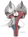
The nigrostriatal pathway is a bilateral dopaminergic pathway in the brain that connects the substantia nigra pars compacta (SNc) in the midbrain with the dorsal striatum in the forebrain. It is one of the four major dopamine pathways in the brain, and is critical in the production of movement as part of a system called the basal ganglia motor loop. Dopaminergic neurons of this pathway release dopamine from axon terminals that synapse onto GABAergic medium spiny neurons (MSNs), also known as spiny projection neurons (SPNs), located in the striatum.

The direct pathway, sometimes known as the direct pathway of movement, is a neural pathway within the central nervous system (CNS) through the basal ganglia which facilitates the initiation and execution of voluntary movement. It works in conjunction with the indirect pathway. Both of these pathways are part of the cortico-basal ganglia-thalamo-cortical loop.

The lateral ventricles are the two largest ventricles of the brain and contain cerebrospinal fluid. Each cerebral hemisphere contains a lateral ventricle, known as the left or right lateral ventricle, respectively.

The external capsule is a series of white matter fiber tracts in the brain. These fibers run between the most lateral segment of the lentiform nucleus and the claustrum.

The subthalamus or ventral thalamus is a part of the diencephalon. Its most prominent structure is the subthalamic nucleus. The subthalamus connects to the globus pallidus, a subcortical nucleus of the basal ganglia.

The substantia innominata, also innominate substance or substantia innominata of Meynert, is a series of layers in the human brain consisting partly of gray and partly of white matter, which lies below the anterior part of the thalamus and lentiform nucleus. It is included as part of the anterior perforated substance. It is part of the basal forebrain structures and includes the nucleus basalis. A portion of the substantia innominata, below the globus pallidus is considered as part of the extended amygdala.

The basal ganglia form a major brain system in all vertebrates, but in primates there are special differentiating features. The basal ganglia include the striatum, globus pallidus, substantia nigra and subthalamic nucleus. In primates the pallidus is divided into an external and internal globus pallidus, the external globus pallidus is present in other mammals but not the internal globus pallidus. Also in primates, the dorsal striatum is divided by a large nerve tract called the internal capsule into two masses named the caudate nucleus and the putamen. These differences contribute to a complex circuitry of connections between the striatum and cortex that is specific to primates, reflecting different functions in primate cortical areas.
The external globus pallidus combines with the internal globus pallidus (GPi) to form the globus pallidus, an anatomical subset of the basal ganglia. Globus pallidus means "pale globe" in Latin, indicating its appearance. The external globus pallidus is the segment of the globus pallidus that is relatively further (lateral) from the midline of the brain.
The internal globus pallidus is one of the two subcortical nuclei that provides inhibitory output in the basal ganglia, the other being the substantia nigra pars reticulata. Together with the external globus pallidus (GPe), it makes up one of the two segments of the globus pallidus, a structure that can decay with certain neurodegenerative disorders and is a target for medical and neurosurgical therapies. The GPi, along with the substantia nigra pars reticulata, comprise the primary output of the basal ganglia, with its outgoing GABAergic neurons having an inhibitory function in the thalamus, the centromedian complex and the pedunculopontine complex.

Blocq's disease was first considered by Paul Blocq (1860–1896), who described this phenomenon as the loss of memory of specialized movements causing the inability to maintain an upright posture, despite normal function of the legs in the bed. The patient is able to stand up, but as soon as the feet are on the ground, the patient cannot hold himself upright nor walk; however when lying down, the subject conserved the integrity of muscular force and the precision of movements of the lower limbs. The motivation of this study came when a fellow student Georges Marinesco (1864) and Paul published a case of parkinsonian tremor (1893) due to a tumor located in the substantia nigra.

Central arteries of the brain are numerous small arteries branching from the Circle of Willis, and adjacent arteries that often enter the substance of the brain through the anterior and posterior perforated substances. They supply structures of the base of the brain and internal structures of the cerebral hemispheres. They are separated into four principal groups: anteromedial central arteries; anterolateral central arteries ; posteromedial central arteries ; and posterolateral central arteries.