
The medulla oblongata or simply medulla is a long stem-like structure which makes up the lower part of the brainstem. It is anterior and partially inferior to the cerebellum. It is a cone-shaped neuronal mass responsible for autonomic (involuntary) functions, ranging from vomiting to sneezing. The medulla contains the cardiovascular center, the respiratory center, vomiting and vasomotor centers, responsible for the autonomic functions of breathing, heart rate and blood pressure as well as the sleep–wake cycle. "Medulla" is from Latin, ‘pith or marrow’. And "oblongata" is from Latin, ‘lengthened or longish or elongated'.
Articles related to anatomy include:

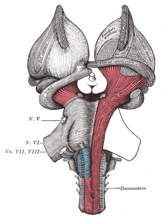
The pons is part of the brainstem that in humans and other mammals, lies inferior to the midbrain, superior to the medulla oblongata and anterior to the cerebellum.
The brainstem is the posterior stalk-like part of the brain that connects the cerebrum with the spinal cord. In the human brain the brainstem is composed of the midbrain, the pons, and the medulla oblongata. The midbrain is continuous with the thalamus of the diencephalon through the tentorial notch, and sometimes the diencephalon is included in the brainstem.

The glossopharyngeal nerve, also known as the ninth cranial nerve, cranial nerve IX, or simply CN IX, is a cranial nerve that exits the brainstem from the sides of the upper medulla, just anterior to the vagus nerve. Being a mixed nerve (sensorimotor), it carries afferent sensory and efferent motor information. The motor division of the glossopharyngeal nerve is derived from the basal plate of the embryonic medulla oblongata, whereas the sensory division originates from the cranial neural crest.
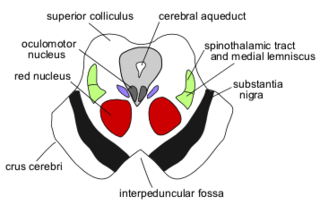
The cerebral peduncles are the two stalks that attach the cerebrum to the brainstem. They are structures at the front of the midbrain which arise from the ventral pons and contain the large ascending (sensory) and descending (motor) tracts that run to and from the cerebrum from the pons. Mainly, the three common areas that give rise to the cerebral peduncles are the cerebral cortex, the spinal cord and the cerebellum. The region includes the tegmentum, crus cerebri and pretectum. By this definition, the cerebral peduncles are also known as the basis pedunculi, while the large ventral bundle of efferent fibers is referred to as the cerebral crus or the pes pedunculi.

The fourth ventricle is one of the four connected fluid-filled cavities within the human brain. These cavities, known collectively as the ventricular system, consist of the left and right lateral ventricles, the third ventricle, and the fourth ventricle. The fourth ventricle extends from the cerebral aqueduct to the obex, and is filled with cerebrospinal fluid (CSF).

The olivary bodies or simply olives are a pair of prominent oval structures on either side of the medullary pyramids in the medulla, the lower portion of the brainstem. They contain the olivary nuclei.

The pontine nuclei are all the neurons of the ventral pons. Corticopontine fibres project from the primary motor cortex to the ipsilateral pontine nucleus; pontocerebellar fibers then relay the information to the contralateral cerebellum via the middle cerebellar peduncle.

The spinocerebellar tracts are nerve tracts originating in the spinal cord and terminating in the same side (ipsilateral) of the cerebellum. The two main tracts are the dorsal spinocerebellar tract, and the ventral spinocerebellar tract. Both of these tracts are located in the peripheral region of the lateral funiculi. Other tracts are the rostral spinocerebellar tract, and the cuneocerebellar tract.

The inferior cerebellar peduncle is formed by fibers of the restiform body that join with fibers from the much smaller juxtarestiform body. The inferior cerebellar peduncle is the smallest of the three cerebellar peduncles.
The interposed nucleus is the combined paired globose and emboliform nuclei, on either side of the cerebellum. It is located in the roof of the fourth ventricle, lateral to the fastigial nucleus. The emboliform nucleus is the anterior interposed nucleus, and the globose nucleus is the posterior interposed nucleus.

The accessory cuneate nucleus is a nucleus situated in the caudal medulla oblongata just lateral to the cuneate nucleus. It relays unconscious proprioceptive sensory information from the upper limb and upper trunk to the cerebellum via the cuneocerebellar fibers.

In the medulla oblongata, the arcuate nucleus is a group of neurons located on the anterior surface of the medullary pyramids. These nuclei are the extension of the pontine nuclei.

The vestibular nuclei (VN) are the cranial nuclei for the vestibular nerve located in the brainstem.

In the human brain, the superior cerebellar peduncle is one of the three paired cerebellar peduncles of bundled fibers that connect the cerebellum to the brainstem. The superior cerebellar peduncle connects to the midbrain. It consists mainly of efferent fibers, the cerebellothalamic tract that runs from a cerebellar hemisphere to the contralateral thalamus, and the cerebellorubral tract that runs from a cerebellar hemisphere to the red nucleus. It also contains afferent tracts, most prominent of which is the ventral spinocerebellar tract. Other afferent tracts are the ventral trigeminal tract, tectocerebellar fibers, and noradrenergic fibers from the locus coeruleus. The superior peduncle emerges from the upper and medial parts of the white matter of each cerebellar hemisphere and is placed under cover of the upper part of the cerebellum.

The middle cerebellar peduncle is one of three paired cerebellar peduncles connecting the brainstem to the cerebellum. The connection is from the pons. It connects the pons to the cerebellum, with fibres originating from the pontine nuclei, and travelling to the opposite cerebellar hemisphere. It is supplied by the anterior inferior cerebellar artery (AICA) and branches from the basilar artery. It conveys information from the cerebrum and the pons to the cerebellum.
The pontocerebellar fibers are the second-order neuron fibers of the corticopontocerebellar tracts that cross to the other side of the pons and run within the middle cerebellar peduncles, from the pons to the contralateral cerebellum. They arise from the pontine nuclei as the second part of the corticopontocerebellar tract, and decussate (cross-over) in the pons before passing through the middle cerebellar peduncles to reach and terminate in the contralateral posterior lobe of the cerebellum (neocerebellum). It is part of a pathway involved in the coordination of voluntary movements.
The central tegmental tract is a tract that carries ascending and descending fibers, situated in the midbrain tegmentum, and the pontine tegmentum. The tract is situated in the central portion of the reticular formation.
