
The pons is part of the brainstem that in humans and other mammals, lies inferior to the midbrain, superior to the medulla oblongata and anterior to the cerebellum.
In neuroanatomy, a nucleus is a cluster of neurons in the central nervous system, located deep within the cerebral hemispheres and brainstem. The neurons in one nucleus usually have roughly similar connections and functions. Nuclei are connected to other nuclei by tracts, the bundles (fascicles) of axons extending from the cell bodies. A nucleus is one of the two most common forms of nerve cell organization, the other being layered structures such as the cerebral cortex or cerebellar cortex. In anatomical sections, a nucleus shows up as a region of gray matter, often bordered by white matter. The vertebrate brain contains hundreds of distinguishable nuclei, varying widely in shape and size. A nucleus may itself have a complex internal structure, with multiple types of neurons arranged in clumps (subnuclei) or layers.

The inferior olivary nucleus (ION) is a structure found in the medulla oblongata underneath the superior olivary nucleus. In vertebrates, the ION is known to coordinate signals from the spinal cord to the cerebellum to regulate motor coordination and learning. These connections have been shown to be tightly associated, as degeneration of either the cerebellum or the ION results in degeneration of the other.
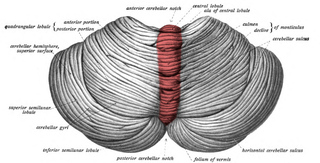
The cerebellar vermis is located in the medial, cortico-nuclear zone of the cerebellum, which is in the posterior fossa of the cranium. The primary fissure in the vermis curves ventrolaterally to the superior surface of the cerebellum, dividing it into anterior and posterior lobes. Functionally, the vermis is associated with bodily posture and locomotion. The vermis is included within the spinocerebellum and receives somatic sensory input from the head and proximal body parts via ascending spinal pathways.

The lentiform nucleus are the putamen (laterally) and the globus pallidus (medially), collectively. Due to their proximity, these two structures were formerly considered one, however, the two are separated by a thin layer of white matter - the external medullary lamina - and are functionally and connectionally distinct.

The spinocerebellar tracts are nerve tracts originating in the spinal cord and terminating in the same side (ipsilateral) of the cerebellum. The two main tracts are the dorsal spinocerebellar tract, and the ventral spinocerebellar tract. Both of these tracts are located in the peripheral region of the lateral funiculi. Other tracts are the rostral spinocerebellar tract, and the cuneocerebellar tract.

The arbor vitae is the cerebellar white matter, so called for its branched, tree-like appearance. In some ways it more resembles a fern and is present in both cerebellar hemispheres. It brings sensory and motor information to and from the cerebellum. The arbor vitae is located deep in the cerebellum. Situated within the arbor vitae are the deep cerebellar nuclei; the dentate, globose, emboliform and the fastigial nuclei. These four different structures lead to the efferent projections of the cerebellum.

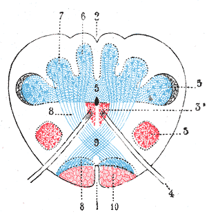
There are four paired deep cerebellar nuclei embedded in the white matter centre of the cerebellum. The nuclei are the fastigial, globose, emboliform, and dentate nuclei.
The dentate nucleus is a cluster of neurons, or nerve cells, in the central nervous system that has a dentate – tooth-like or serrated – edge. It is located within the deep white matter of each cerebellar hemisphere, and it is the largest single structure linking the cerebellum to the rest of the brain. It is the largest and most lateral, or farthest from the midline, of the four pairs of deep cerebellar nuclei, the others being the globose and emboliform nuclei, which together are referred to as the interposed nucleus, and the fastigial nucleus. The dentate nucleus is responsible for the planning, initiation and control of voluntary movements. The dorsal region of the dentate nucleus contains output channels involved in motor function, which is the movement of skeletal muscle, while the ventral region contains output channels involved in nonmotor function, such as conscious thought and visuospatial function.

The globose nucleus is one of the deep cerebellar nuclei. It is located medial to the emboliform nucleus, and lateral to the fastigial nucleus. The globose nucleus and emboliform nucleus are known collectively as the interposed nuclei.
The interposed nucleus is the combined globose and emboliform nuclei on either side. The interposed nucleus is one of the paired cerebellar nuclei. It is located in the roof of the fourth ventricle, lateral to the fastigial nucleus. The emboliform nucleus is the anterior interposed nucleus, and the globose nucleus is the posterior interposed nucleus.
The fastigial nucleus is located in each hemisphere of the cerebellum. It is one of the four deep cerebellar nuclei.

The vestibular nuclei (VN) are the cranial nuclei for the vestibular nerve located in the brainstem.

The cerebellar peduncles are three paired bundles of fibres that connect the cerebellum to the brain stem.

The posterior thoracic nucleus, is a group of interneurons found in the medial part of lamina VII, also known as the intermediate zone, of the spinal cord. It is located from the cervical segment C8 to lumbar segment L3 of the spinal cord and is an important structure for proprioception of the lower limb.
The dorsal column nuclei are a pair of nuclei in the dorsal columns of the dorsal column–medial lemniscus pathway (DCML) in the brainstem. The name refers collectively to the cuneate nucleus and gracile nucleus, which are situated at the lower end of the medulla oblongata. Both nuclei contain second-order neurons of the DCML, which convey fine touch and proprioceptive information from the body to the brain via the thalamus.

In the human brain, the superior cerebellar peduncle is one of the three paired cerebellar peduncles of bundled fibers that connect the cerebellum to the brainstem. The superior cerebellar peduncle connects to the midbrain. It consists mainly of efferent fibers, the cerebellothalamic tract that runs from a cerebellar hemisphere to the contralateral thalamus, and the cerebellorubral tract that runs from a cerebellar hemisphere to the red nucleus. It also contains afferent tracts, most prominent of which is the ventral spinocerebellar tract. Other afferent tracts are the ventral trigeminal tract, tectocerebellar fibers, and noradrenergic fibers from the locus coeruleus. The superior peduncle emerges from the upper and medial parts of the white matter of each cerebellar hemisphere and is placed under cover of the upper part of the cerebellum.

The medial vestibular nucleus is one of the vestibular nuclei. It is located in the medulla oblongata.

The anatomy of the cerebellum can be viewed at three levels. At the level of gross anatomy, the cerebellum consists of a tightly folded and crumpled layer of cortex, with white matter underneath, several deep nuclei embedded in the white matter, and a fluid-filled ventricle in the middle. At the intermediate level, the cerebellum and its auxiliary structures can be broken down into several hundred or thousand independently functioning modules or compartments known as microzones. At the microscopic level, each module consists of the same small set of neuronal elements, laid out with a highly stereotyped geometry.
