Lingula of cerebellum

Last updated
Lingula of cerebellum
Gray704.png
Sagittal section of the cerebellum, near the junction of the vermis with the hemisphere. (Lingula labeled at upper right.)
Details
Identifiers
Latin lingula cerebelli
NeuroNames 657
NeuroLex ID birnlex_932
TA A14.1.07.103
FMA 83884
Anatomical terms of neuroanatomy

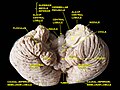
The lingula is a small tongue-shaped process, consisting of four or five folia; it lies in front of the lobulus centralis, and is concealed by it.

Contents

Anteriorly, it rests on the dorsal surface of the anterior medullary velum, and its white substance is continuous with that of the velum.

Additional images

Related Research Articles

Velum may refer to:

Fourth ventricle

The fourth ventricle is one of the four connected fluid-filled cavities within the human brain. These cavities, known collectively as the ventricular system, consist of the left and right lateral ventricles, the third ventricle, and the fourth ventricle. The fourth ventricle extends from the cerebral aqueduct to the obex, and is filled with cerebrospinal fluid (CSF).

Basilar artery artery that supplies the brain with oxygen-rich blood

In human anatomy, the basilar artery is one of the arteries that supplies the brain with oxygen-rich blood.

Posterior inferior cerebellar artery

The posterior inferior cerebellar artery (PICA), the largest branch of the vertebral artery, is one of the three main arterial blood supplies for the cerebellum, part of the brain. Occlusion of the posterior inferior cerebellar artery or one of its branches, or of the vertebral artery leads to lateral medullary syndrome also called Wallenberg syndrome.

Anterior inferior cerebellar artery

The anterior inferior cerebellar artery (AICA) is one of three pairs of arteries that supplies blood to the cerebellum.

Superior cerebellar artery

The superior cerebellar artery (SCA) arises near the termination of the basilar artery.

Inferior petrosal sinus

The inferior petrosal sinuses are two small sinuses situated on the inferior border of the petrous part of the temporal bone, one on each side. Each inferior petrosal sinus drains the cavernous sinus into the internal jugular vein.

Vestibular nuclei cranial nuclei for the vestibular nerve

The vestibular nuclei (VN) are the cranial nuclei for the vestibular nerve.

Cerebellar peduncles connect the cerebellum to the brain stem. There are six cerebellar peduncles in total, three on each side:

Superior cerebellar peduncle

In the human brain, the superior cerebellar peduncle is a paired structure of white matter that connects the cerebellum to the midbrain. It consists mainly of efferent fibers, the cerebellothalamic tract that runs from a cerebellar hemisphere to the contralateral thalamus, and the cerebellorubral tract that runs from a cerebellar hemisphere to the red nucleus. It also contains afferent tracts, most prominent of which is the ventral spinocerebellar tract. Other afferent tracts are the trigeminothalamic fibers, tectocerebellar fibers, and noradrenergic fibers from the locus coeruleus. The superior peduncle emerges from the upper and medial parts of the white matter of each hemisphere and is placed under cover of the upper part of the cerebellum.

Middle cerebellar peduncle

The middle cerebellar peduncles are paired structures that connect the cerebellum to the pons and are composed entirely of centripetal fibers, i.e. incoming fibers. The fibers arise from the pontine nucleus to the opposite hemisphere of the cerebellar cortex. The fibers are arranged in three fasciculi: superior, inferior, and deep.

Superior medullary velum

The superior medullary velum is a thin, transparent lamina of white matter, which stretches between the superior cerebellar peduncles; on the dorsal surface of its lower half the folia and lingula are prolonged.

Cerebellar tonsil

The cerebellar tonsil is analogous to a rounded lobule on the undersurface of each cerebellar hemisphere, continuous medially with the uvula of the cerebellar vermis and superiorly by the flocculonodular lobe. Synonyms include: tonsilla cerebelli, amygdala cerebelli, the latter of which is not to be confused with the cerebral tonsils or amygdala nuclei located deep within the medial temporal lobes of the cerebral cortex. The flocculonodular lobe of the cerebellum which can also be confused for the cerebellar tonsils, is one of three lobes that make up the overall composition of the cerebellum. The cerebellum consists of three anatomical and functional lobes: anterior lobe, posterior lobe, and flocculonodular lobe.

Inferior medullary velum

The inferior medullary velum is a thin layer of white substance, prolonged from the white center of the cerebellum, above and on either side of the nodule; it forms the infero-posterior part of the fourth ventricle.

Nodule of vermis

The nodule, or anterior end of the inferior vermis, abuts against the roof of the fourth ventricle, and can only be distinctly seen after the cerebellum has been separated from the medulla oblongata and pons.

Biventer lobule

The biventer lobule is a region of the cerebellum. It is triangular in shape; its apex points backward, and is joined by the gray band to the pyramid.

Central lobule

The central lobule is a small square lobule, situated in the anterior cerebellar notch. It overlaps the lingula, from which it is separated by the precentral fissure; laterally, it extends along the upper and anterior part of each hemisphere, where it forms a wing-like prolongation (ala), on each side, as the alae of the central lobule or alae lobuli centralis.

Culmen (cerebellum) structure in the brain

The culmen is the portion of the anterior vermis adjacent to the primary fissure of cerebellum.

Uvula of cerebellum

The uvula forms a considerable portion of the inferior vermis; it is separated on either side from the tonsil by the sulcus vallecula, at the bottom of which it is connected to the tonsil by a ridge of gray matter, indented on its surface by shallow furrows, and hence called the furrowed band.

Roof of fourth ventricle

The roof of fourth ventricle is the dorsal surface of the fourth ventricle.

References

This article incorporates text in the public domain from page 789 of the 20th edition of Gray's Anatomy (1918)

The public domain consists of all the creative works to which no exclusive intellectual property rights apply. Those rights may have expired, been forfeited, expressly waived, or may be inapplicable.

<i>Grays Anatomy</i> English-language textbook of human anatomy

Gray's Anatomy is an English language textbook of human anatomy originally written by Henry Gray and illustrated by Henry Vandyke Carter. Earlier editions were called Anatomy: Descriptive and Surgical, Anatomy of the Human Body and Gray's Anatomy: Descriptive and Applied, but the book's name is commonly shortened to, and later editions are titled, Gray's Anatomy. The book is widely regarded as an extremely influential work on the subject, and has continued to be revised and republished from its initial publication in 1858 to the present day. The latest edition of the book, the 41st, was published in September 2015.