The cingulate cortex is a part of the brain situated in the medial aspect of the cerebral cortex. The cingulate cortex includes the entire cingulate gyrus, which lies immediately above the corpus callosum, and the continuation of this in the cingulate sulcus. The cingulate cortex is usually considered part of the limbic lobe.

A Brodmann area is a region of the cerebral cortex, in the human or other primate brain, defined by its cytoarchitecture, or histological structure and organization of cells. The concept was first introduced by the German anatomist Korbinian Brodmann in the early 20th century. Brodmann mapped the human brain based on the varied cellular structure across the cortex and identified 52 distinct regions, which he numbered 1 to 52. These regions, or Brodmann areas, correspond with diverse functions including sensation, motor control, and cognition.

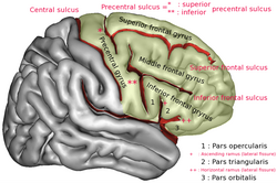
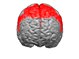
The frontal lobe is the largest of the four major lobes of the brain in mammals, and is located at the front of each cerebral hemisphere. It is parted from the parietal lobe by a groove between tissues called the central sulcus and from the temporal lobe by a deeper groove called the lateral sulcus. The most anterior rounded part of the frontal lobe is known as the frontal pole, one of the three poles of the cerebrum.
Brodmann area 6 (BA6) is part of the frontal cortex in the human brain. Situated just anterior to the primary motor cortex (BA4), it is composed of the premotor cortex and, medially, the supplementary motor area (SMA). This large area of the frontal cortex is believed to play a role in planning complex, coordinated movements.

Brodmann area 44, or BA44, is part of the frontal cortex in the human brain. Situated just anterior to premotor cortex (BA6) and on the lateral surface, inferior to BA9.

Brodmann area 11 is one of Brodmann's cytologically defined regions of the brain. It is in the orbitofrontal cortex which is above the eye sockets (orbitae). It is involved in decision making, processing rewards, and encoding new information into long-term memory.

The inferior frontal gyrus (IFG),, is the lowest positioned gyrus of the frontal gyri, of the frontal lobe, and is part of the prefrontal cortex.

Brodmann area 4 refers to the primary motor cortex of the human brain. It is located in the posterior portion of the frontal lobe.
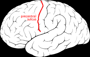
The precentral sulcus is a part of the human brain that lies parallel to, and in front of, the central sulcus. A sulcus is one of the prominent grooves on the surface of the human brain.
The precentral gyrus is a prominent gyrus on the surface of the posterior frontal lobe of the brain. It is the site of the primary motor cortex that in humans is cytoarchitecturally defined as Brodmann area 4.
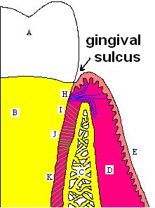
In biological morphology and anatomy, a sulcus is a furrow or fissure. It may be a groove, natural division, deep furrow, elongated cleft, or tear in the surface of a limb or an organ, most notably on the surface of the brain, but also in the lungs, certain muscles, as well as in bones, and elsewhere. Many sulci are the product of a surface fold or junction, such as in the gums, where they fold around the neck of the tooth.

The middle cerebral artery (MCA) is one of the three major paired cerebral arteries that supply blood to the cerebrum. The MCA arises from the internal carotid artery and continues into the lateral sulcus where it then branches and projects to many parts of the lateral cerebral cortex. It also supplies blood to the anterior temporal lobes and the insular cortices.

The lobes of the brain are the major identifiable zones of the human cerebral cortex, and they comprise the surface of each hemisphere of the cerebrum. The two hemispheres are roughly symmetrical in structure, and are connected by the corpus callosum. They traditionally have been divided into four lobes, but are today considered as having six lobes each. The lobes are large areas that are anatomically distinguishable, and are also functionally distinct to some degree. Each lobe of the brain has numerous ridges, or gyri, and furrows, the sulci that constitute further subzones of the cortex. The expression "lobes of the brain" usually refers only to those of the cerebrum, not to the distinct areas of the cerebellum.

In neuroanatomy, a sulcus is a depression or groove in the cerebral cortex. It surrounds a gyrus, creating the characteristic folded appearance of the brain in humans and other mammals. The larger sulci are usually called fissures.

The olfactory tract is a bilateral bundle of afferent nerve fibers from the mitral and tufted cells of the olfactory bulb that connects to several target regions in the brain, including the piriform cortex, amygdala, and entorhinal cortex. It is a narrow white band, triangular on coronal section, the apex being directed upward.
The sensory cortex can refer sometimes to the primary somatosensory cortex, or it can be used as a term for the primary and secondary cortices of the different senses : the visual cortex on the occipital lobes, the auditory cortex on the temporal lobes, the primary olfactory cortex on the uncus of the piriform region of the temporal lobes, the gustatory cortex on the insular lobe, and the primary somatosensory cortex on the anterior parietal lobes. Just posterior to the primary somatosensory cortex lies the somatosensory association cortex or area, which integrates sensory information from the primary somatosensory cortex to construct an understanding of the object being felt. Inferior to the frontal lobes are found the olfactory bulbs, which receive sensory input from the olfactory nerves and route those signals throughout the brain. Not all olfactory information is routed to the olfactory cortex: some neural fibers are routed to the supraorbital region of the frontal lobe, while others are routed directly to limbic structures. The direct limbic connection makes the olfactory sense unique.

The superior longitudinal fasciculus (SLF) is an association tract in the brain that is composed of three separate components. It is present in both hemispheres and can be found lateral to the centrum semiovale and connects the frontal, occipital, parietal, and temporal lobes. This bundle of tracts (fasciculus) passes from the frontal lobe through the operculum to the posterior end of the lateral sulcus where they either radiate to and synapse on neurons in the occipital lobe, or turn downward and forward around the putamen and then radiate to and synapse on neurons in anterior portions of the temporal lobe.
The inferior or orbital surface of the frontal lobe is concave, and rests on the orbital plate of the frontal bone. It is divided into four orbital gyri by a well-marked H-shaped orbital sulcus. These are named, from their position, the medial, anterior, lateral, and posterior, orbital gyri. The medial orbital gyrus presents a well-marked antero-posterior sulcus, the olfactory sulcus, for the olfactory tract; the portion medial to this is named the straight gyrus, and is continuous with the superior frontal gyrus on the medial surface.
The orbital part of inferior frontal gyrus also known as the pars orbitalis is the orbital part of the inferior frontal gyrus.