T-cadherin

Last updated
CDH13
Available structures
PDB Ortholog search: PDBe RCSB
Identifiers
Aliases CDH13 , CDHH, P105, cadherin 13
External IDs OMIM: 601364 MGI: 99551 HomoloGene: 20335 GeneCards: CDH13
Orthologs
SpeciesHumanMouse
Entrez
Ensembl
UniProt
RefSeq (mRNA)

NM_019707

RefSeq (protein)

NP_062681

Location (UCSC) Chr 16: 82.63 – 83.8 Mb Chr 8: 119.01 – 120.05 Mb
PubMed search [3] [4]
Wikidata
View/Edit Human View/Edit Mouse

T-cadherin, also known as cadherin 13, H-cadherin (heart), and CDH13, is a unique member of the cadherin superfamily of proteins because it lacks the transmembrane and cytoplasmic domains common to all other cadherins and is instead anchored to the cell's plasma membrane by the GPI anchor.

Contents

Unlike classical cadherins, which are necessary for cell–cell adhesion, dynamic regulation of morphogenetic processes in embryos, and tissue integrity in adult organisms, and function as membrane receptors mediating signals received from the extracellular space, activate small GTPases and the beta-catenin/Wnt pathway, and play important roles in dynamic cytoskeleton reorganization, the GPI-anchored T-cadherin lacks direct contact with the cytoskeleton and therefore is not involved in cell–cell adhesion. It is instead involved in low-density lipoprotein (LDL) hormone-like effects on Ca2+ mobilization and increased cell migration as well as phenotypic changes. The exact signaling partners and adaptor proteins recognized by T-cadherin remain to be elucidated.

Mediation of intracellular signaling in vascular cells

Though T-cadherin can mediate weak adhesion in aggregation assays in vitro, the lack of intracellular domain suggests that T-cadherin is not involved in stable cell-cell adhesion. In vivo T-cadherin was detected on the apical cell surface of the chick intestinal epithelium. In cultures of transfected MDCS cells, T-cadherin was also expressed apically, whereas N-cadherin located basolaterally corresponded to the zone of cell contacts.

The apical cell surface distribution of T-cadherin was proposed to possibly endow T-cadherin with recognition functions. In confluent cultures of vascular cells, T-cadherin was distributed equally over the entire cell surface, in contrast to VE-cadherin, which was restricted to the cell junctions. In migrating vascular cells, T-cadherin was located at the leading edge as revealed by confocal microscopy. The distribution of T-cadherin on the cell membrane is restricted to lipid rafts where it co-localizes with signal-transducing molecules. These data strongly implicates T-cadherin in intracellular signaling rather than adhesion.

Studying signaling effects of low density lipoproteins (LDL) in vascular smooth muscles (VSMCs), T-cadherin was isolated and identified as new LDL receptor using human aortic media and the ligand-blotting method. The properties of T-cadherin as an LDL receptor were markedly different from the presently known types of LDL receptors. LDL binding to T-cadherin leads to the activation of Erk 1/2 tyrosine kinase and the nuclear translocation of NF-kappaB.

T-cadherin overexpression in ECs facilitates spontaneous cell migration, formation of stress fibers and change of the phenotype from quiescent to promigratory. T-cadherin expression results in LDL-induced migration of T-cadherin expressing cells compared to control. It is likely that T-cadherin regulates cell migration and phenotype via activation of small G-proteins with subsequent actin reorganization. RhoA/ROCK activation is necessary for cell contraction, stress fiber assembly and inhibition of spreading, while Rac is required for the formation of membrane protrusions and actin-rich lamellopodia at the leading edge of migrating cells.

Functions in the vasculature

The function of T-cadherin in situ, in normal conditions, and in pathology is still largely unknown. T-cadherin is highly expressed in the heart, aortic wall, neurons of the brain cortex and spinal cord and also in the small blood vessels in spleen and other organs.

Expression of T-cadherin is upregulated in atherosclerotic lesions and post-angioplasty restenosis —conditions associated with pathological angiogenesis. T-cadherin expression is upregulated in ECs, pericytes and VSMC of atherosclerotic lesions.

T-cadherin expression in arterial wall after balloon angioplasty correlates with late stages of neointima formation and coincidentally with the peak in proliferation and differentiation of vascular cells. It is highly expressed in adventitial vasa vasorum of injured arteries suggesting the involvement of T-cadherin in the processes of angiogenesis after vessel injury. These data implicate T-cadherin to be involved in regulation of vascular functioning and remodeling; however, the exact role of T-cadherin in neointima formation and atherosclerosis development is poorly understood.

LDL is not the only ligand for T-cadherin. High-molecular weight (HMW) complexes of adiponectin were suggested to be a specific ligand for T-cadherin. Adiponectin (adipocyte complement-related protein of 30 kDa) is a cytokine produced by adipose tissue and its deficiency is associated with metabolic syndrome, obesity, type II diabetes and atherosclerosis. Adiponectin binding to T-cadherin on vascular cells is associated with NF-kappa B activation. Two membrane adiponectin receptors with distant homology to seven-transmembrane spanning G-protein-coupled receptors, namely AdipoR1 and AdipoR2 were identified in several tissues, but the University of Tokyo announced it was launching an investigation into anonymously made claims of fabricated and falsified data on the identification of AdipoR1 and AdipoR2 in 2016. [5]

Regulation of cell growth

In vitro T-cadherin is implicated in regulation of cell growth, survival and proliferation. In cultured VSMC and primary astrocytes, the expression of T-cadherin depends on proliferation status with maximum at confluency suggesting its regulation of cell growth by contact inhibition. Known mitogens such as platelet-derived growth factor (PDGF)-BB, epidermal growth factor (EGF) or insulin-like growth factor (IGF) elicit a reversible dose- and time-dependent decrease in T-cadherin expression in cultured VSMCs.

Expression of T-cadherin leads to complete inhibition of subcutaneous tumor growth in nude mice. Seeding T-cadherin expressing cells on plastic coated with recombinant aminoterminal fragments of T-cadherin resulted in suppression of cell growth and was found to be associated with increased expression of p21. In T-cadherin deficient C6 glioma cell lines, its overexpression results in growth suppression involving p21CIP1/WAF1 production and G2 arrest.

T-cadherin loss in tumor cells is associated with tumor malignancy, invasiveness and metastasis. Thus, tumor progression in basal cell carcinoma, cutaneous squamous carcinoma, non-small cell lung carcinoma (NSCLC), ovarian cancer, pancreatic cancer, colorectal cancer correlates with downregulation of T-cadherin expression. In psoriasis vulgaris the hyperproliferation of keratinocytes also correlates with the downregulation of T-cadherin expression. The mechanism for T-cadherin suppression is associated with allelic loss or hypermethylation of the T-cadherin gene promoter region.

Transfection of T-cadherin negative neuroblastoma TGW and NH-12 cells with T-cadherin results in their loss of mitogenic proliferative response to epidermal growth factor (EGF) growth stimulation. Re-expression of T-cadherin in human breast cancer cells (MDAMB435) in culture, which originally do not express T-cadherin, results in the change of the phenotype from invasive to normal epithelial-like morphology. Thus, it was hypothesized that T-cadherin functions as a tumor-suppressor factor; inactivation of T-cadherin is associated with tumor malignancy, invasiveness and metastasis.

However, in other tumors T-cadherin expression could promote tumor growth and metastasis. In primary lung tumors the loss of T-cadherin was not attributed to the presence of metastasis in lymph nodes, and in osteosarcomas T-cadherin expression was correlated with metastasis. Furthermore, T-cadherin overexpression was found to be a common feature of human high grade astrocytomas and associated with malignant transformation of astrocytes. Hetezygosity for NF1 (neurofibromatosis 1) tumor suppressor resulting in reduced attachment and spreading and increased motility also coincides with upregulated T-cadherin expression.

Data show that HUVEC cells overexpressing T-cadherin after adenovirus infection enter S-phase more rapidly and exhibit increased proliferation potential. T-cadherin expression increases in HUVEC under conditions of oxidative stress, and production of reactive oxygen species (ROS) contributes to T-cadherin elevated expression. T-cadherin overexpression in HUVEC leads to higher phosphorylation of Phosphatidylinositol 3-kinase (PIK3) – target of Akt, and mTOR – target p70S6K (survival pathway regulator), resulting in reduced levels of caspase activation and increased survival after exposure to oxidative stress.[ clarification needed ] It was suggested that in vascular cells T-cadherin performs a protective role against stress-induced apoptosis.

Tumor cells can regulate gene expression in growing vessels and the surrounding stroma during tumor neovascularization. T-cadherin expression was found to be altered in tumor vessels: in Lewis carcinoma lung metastasis the expression of T-cadherin was upregulated in blood vessels penetrating the tumor, while T-cadherin was not detected in the surrounding tumor tissue. In tumor neovascularization of hepatocellular carcinoma (HCC) T-cadherin is upregulated in intratumoral capillary endothelial cells, whereas in surrounding tumor tissue as well as in normal liver no T-cadherin could be detected. The increase in T-cadherin expression in endothelial cell in HCC was shown to correlate with tumors progression. Presumably, T-cadherin could play a navigating role in the growing tumor vessels, which in the absence of contact inhibition from the stromal cells, grow into the surrounding tumor tissue.

Guiding molecules in vascular and nervous systems

T-cadherin was originally cloned from chick embryo brain, where it was implicated as a negative guiding cue for motor axon projecting through the somitic sclerotome and presumably for migrating neural crest cells . As a substrate or in soluble form, T-cadherin inhibits neurite outgrowth by motor neurons in vitro supporting the assumption that T-cadherin acts as a negative guiding molecule in the developing nervous system.

Considering that the maximal expression of T-cadherin has been observed in nervous and cardiovascular systems, it is likely that T-cadherin is involved in guiding the growing vessel as well. The mechanism of T-cadherin mediated negative guidance in nervous system involves homophilic interaction and contact inhibition; in vascular system it is supposed that T-cadherin expressing blood vessels would avoid T-cadherin expressing tissues.

Related Research Articles

<span class="mw-page-title-main">Metastasis</span> Spread of a disease inside a body

Metastasis is a pathogenic agent's spread from an initial or primary site to a different or secondary site within the host's body; the term is typically used when referring to metastasis by a cancerous tumor. The newly pathological sites, then, are metastases (mets). It is generally distinguished from cancer invasion, which is the direct extension and penetration by cancer cells into neighboring tissues.

<span class="mw-page-title-main">Adiponectin</span> Mammalian protein found in Homo sapiens

Adiponectin is a protein hormone and adipokine, which is involved in regulating glucose levels as well as fatty acid breakdown. In humans it is encoded by the ADIPOQ gene and it is produced in primarily in adipose tissue, but also in muscle, and even in the brain.

<span class="mw-page-title-main">Cadherin</span>

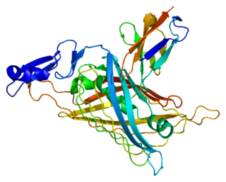
Cadherins (named for "calcium-dependent adhesion") are a type of cell adhesion molecule (CAM) that is important in the formation of adherens junctions to allow cells to adhere to each other. Cadherins are a class of type-1 transmembrane proteins, and they are dependent on calcium (Ca2+) ions to function, hence their name. Cell-cell adhesion is mediated by extracellular cadherin domains, whereas the intracellular cytoplasmic tail associates with numerous adaptors and signaling proteins, collectively referred to as the cadherin adhesome.

<span class="mw-page-title-main">Catenin</span>

Catenins are a family of proteins found in complexes with cadherin cell adhesion molecules of animal cells. The first two catenins that were identified became known as α-catenin and β-catenin. α-Catenin can bind to β-catenin and can also bind filamentous actin (F-actin). β-Catenin binds directly to the cytoplasmic tail of classical cadherins. Additional catenins such as γ-catenin and δ-catenin have been identified. The name "catenin" was originally selected because it was suspected that catenins might link cadherins to the cytoskeleton.

The epithelial–mesenchymal transition (EMT) is a process by which epithelial cells lose their cell polarity and cell–cell adhesion, and gain migratory and invasive properties to become mesenchymal stem cells; these are multipotent stromal cells that can differentiate into a variety of cell types. EMT is essential for numerous developmental processes including mesoderm formation and neural tube formation. EMT has also been shown to occur in wound healing, in organ fibrosis and in the initiation of metastasis in cancer progression.

<span class="mw-page-title-main">Perlecan</span>

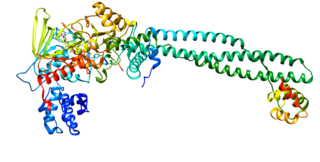
Perlecan (PLC) also known as basement membrane-specific heparan sulfate proteoglycan core protein (HSPG) or heparan sulfate proteoglycan 2 (HSPG2), is a protein that in humans is encoded by the HSPG2 gene.

<span class="mw-page-title-main">E-selectin</span>

E-selectin, also known as CD62 antigen-like family member E (CD62E), endothelial-leukocyte adhesion molecule 1 (ELAM-1), or leukocyte-endothelial cell adhesion molecule 2 (LECAM2), is a selectin cell adhesion molecule expressed only on endothelial cells activated by cytokines. Like other selectins, it plays an important part in inflammation. In humans, E-selectin is encoded by the SELE gene.

<span class="mw-page-title-main">Catenin beta-1</span> Mammalian protein found in Homo sapiens

Catenin beta-1, also known as beta-catenin (β-catenin), is a protein that in humans is encoded by the CTNNB1 gene.

<span class="mw-page-title-main">Syndecan</span>

Syndecans are single transmembrane domain proteins that are thought to act as coreceptors, especially for G protein-coupled receptors. More specifically, these core proteins carry three to five heparan sulfate and chondroitin sulfate chains, i.e. they are proteoglycans, which allow for interaction with a large variety of ligands including fibroblast growth factors, vascular endothelial growth factor, transforming growth factor-beta, fibronectin and antithrombin-1. Interactions between fibronectin and some syndecans can be modulated by the extracellular matrix protein tenascin C.
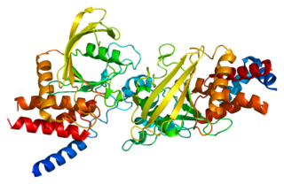
<span class="mw-page-title-main">Basal-like carcinoma</span> Breast cancer subtype

The basal-like carcinoma is a recently proposed subtype of breast cancer defined by its gene expression and protein expression profile.

<span class="mw-page-title-main">CDH3 (gene)</span> Protein-coding gene in the species Homo sapiens

Cadherin-3, also known as P-Cadherin, is a protein that in humans is encoded by the CDH3 gene.

<span class="mw-page-title-main">Epithelial cell adhesion molecule</span> Transmembrane glycoprotein

Epithelial cell adhesion molecule (EpCAM), also known as CD326 among other names, is a transmembrane glycoprotein mediating Ca2+-independent homotypic cell–cell adhesion in epithelia. EpCAM is also involved in cell signaling, migration, proliferation, and differentiation. Additionally, EpCAM has oncogenic potential via its capacity to upregulate c-myc, e-fabp, and cyclins A & E. Since EpCAM is expressed exclusively in epithelia and epithelial-derived neoplasms, EpCAM can be used as diagnostic marker for various cancers. It appears to play a role in tumorigenesis and metastasis of carcinomas, so it can also act as a potential prognostic marker and as a potential target for immunotherapeutic strategies.

<span class="mw-page-title-main">PTPRM</span> Protein-coding gene in the species Homo sapiens

Receptor-type tyrosine-protein phosphatase mu is an enzyme that in humans is encoded by the PTPRM gene.

<span class="mw-page-title-main">CDH6</span> Protein-coding gene in the species Homo sapiens

Cadherin-6 is a protein that in humans is encoded by the CDH6 gene.

<span class="mw-page-title-main">CDH16</span>

Cadherin-16 is a protein that in humans is encoded by the CDH16 gene.

<span class="mw-page-title-main">CDH17</span> Protein-coding gene in the species Homo sapiens

Cadherin-17 is a protein that in humans is encoded by the CDH17 gene.

<span class="mw-page-title-main">PTPRT</span>

Receptor-type tyrosine-protein phosphatase T is an enzyme that in humans is encoded by the PTPRT gene.

<span class="mw-page-title-main">Cadherin-1</span> Human protein-coding gene

Cadherin-1 or Epithelial cadherin (E-cadherin), is a protein that in humans is encoded by the CDH1 gene. Mutations are correlated with gastric, breast, colorectal, thyroid, and ovarian cancers. CDH1 has also been designated as CD324. It is a tumor suppressor gene.
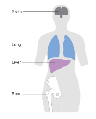
A mesenchymal–epithelial transition (MET) is a reversible biological process that involves the transition from motile, multipolar or spindle-shaped mesenchymal cells to planar arrays of polarized cells called epithelia. MET is the reverse process of epithelial–mesenchymal transition (EMT) and it has been shown to occur in normal development, induced pluripotent stem cell reprogramming, cancer metastasis and wound healing.

Tumor-associated macrophages (TAMs) are a class of immune cells present in high numbers in the microenvironment of solid tumors. They are heavily involved in cancer-related inflammation. Macrophages are known to originate from bone marrow-derived blood monocytes or yolk sac progenitors, but the exact origin of TAMs in human tumors remains to be elucidated. The composition of monocyte-derived macrophages and tissue-resident macrophages in the tumor microenvironment depends on the tumor type, stage, size, and location, thus it has been proposed that TAM identity and heterogeneity is the outcome of interactions between tumor-derived, tissue-specific, and developmental signals.

References

  1. 1 2 3 GRCh38: Ensembl release 89: ENSG00000140945 - Ensembl, May 2017
  2. 1 2 3 GRCm38: Ensembl release 89: ENSMUSG00000031841 - Ensembl, May 2017
  3. "Human PubMed Reference:". National Center for Biotechnology Information, U.S. National Library of Medicine.
  4. "Mouse PubMed Reference:". National Center for Biotechnology Information, U.S. National Library of Medicine.
  5. University of Tokyo to investigate data manipulation charges against six prominent research groups ScienceInsider, Dennis Normile, Sep 20, 2016

Bibliography

Further reading