Plasmacytoma

Last updated
Plasmacytoma
Plasmacytoma ultramini1.jpg
Micrograph of a plasmacytoma. H&E stain.
Specialty Hematology and oncology

Plasmacytoma is a plasma cell dyscrasia in which a plasma cell tumour grows within soft tissue or within the axial skeleton.

Contents

The International Myeloma Working Group lists three types: solitary plasmacytoma of bone (SPB); extramedullary plasmacytoma (EP), and multiple plasmacytomas that are either primary or recurrent. [1] The most common of these is SPB, accounting for 3–5% of all plasma cell malignancies. [2] SPBs occur as lytic lesions within the axial skeleton and extramedullary plasmacytomas most often occur in the upper respiratory tract (85%), but can occur in any soft tissue. Approximately half of all cases produce paraproteinemia. SPBs and extramedullary plasmacytomas are mostly treated with radiotherapy, but surgery is used in some cases of extramedullary plasmacytoma. The skeletal forms frequently progress to multiple myeloma over the course of 2–4 years. [3]

Due to their cellular similarity, plasmacytomas have to be differentiated from multiple myeloma. For SPB and extramedullary plasmacytoma the distinction is the presence of only one lesion (either in bone or soft tissue), normal bone marrow (<5% plasma cells), normal skeletal survey, absent or low paraprotein and no end organ damage. [1]

Signs and symptoms

For SPB the most common presenting symptom is that of pain in the affected bone. Back pain and other consequences of the bone lesion may occur such as spinal cord compression or pathological fracture. Around 85% of extramedullary plasmacytoma presents within the upper respiratory tract mucosa, causing possible symptoms such as epistaxis, rhinorrhoea and nasal obstruction. In some tissues it may be found as a palpable mass. [1] [2] [3]

Diagnosis

Serum protein electrophoresis of an individual with polyclonal antibodies (top) and an individual with a large paraprotein (bottom). Serum protein electrophoresis normal and paraprotein.png
Serum protein electrophoresis of an individual with polyclonal antibodies (top) and an individual with a large paraprotein (bottom).

The diagnosis of plasmacytoma uses a diverse range of interdisciplinary techniques including serum protein electrophoresis, bone marrow biopsy, urine analysis for Bence Jones protein and complete blood count, plain film radiography, MRI and PET-CT. [4] [5]

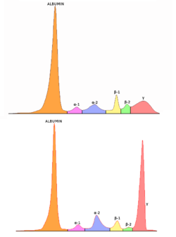
Serum protein electrophoresis separates the proteins in the liquid part of the blood (serum), allowing the analysis of antibodies. Normal blood serum contains a range of antibodies and are said to be polyclonal, whereas serum from a person with plasmacytoma may show a monoclonal spike. This is due to an outgrowth of a single type of plasma cell that forms the plasmacytoma and produces a single type of antibody. The plasma cells are said to be monoclonal and the excessively produced antibody is known as monoclonal protein or paraprotein. [6] Paraproteins are present in 60% of SPB and less than 25% of extramedullary plasmacytoma. [2]

Bone marrow biopsies are performed to ensure the disease is localised; and in SPB or extramedullary plasmacytoma there will not be an increase of monoclonal plasma cells. Tissue biopsies of SPB and extramedullary plasmacytoma are used to assess the phenotype of the plasma cells. Histological analyses can be performed on these biopsies to see what cluster of differentiation (CD) markers are present and to assess monoclonality of the cells. CD markers can aid in the distinction of extramedullary plasmacytoma from lymphomas. [3] [7]

Skeletal surveys are used to ensure there are no other primary tumors within the axial skeleton. MRI can be used to assess tumor status and may be advantageous in detecting primary tumors that are not detected by plain film radiography. PET-CT may also be beneficial in detecting extramedullary tumours in individuals diagnosed with SPB. CT imaging may be better than plain film radiography for assessing bone damage. [4] [5]

An important distinction to be made is that a true plasmacytoma is present and not a systemic plasma cell disorder, such as multiple myeloma. The difference between plasmacytoma and multiple myeloma is that plasmacytoma lacks increased blood calcium, decreased kidney function, too few red blood cells in the bloodstream, and multiple bone lesions (collectively termed CRAB). [4]

Classification

Micrograph of a plasmacytoma. Plasmacytoma1.jpg
Micrograph of a plasmacytoma.

Plasmacytoma is a tumor of plasma cells. The cells are identical to those seen in multiple myeloma, but they form discrete masses of cells in the skeleton (solitary plasmacytoma of bone; SPB) or in soft tissues (extramedullary plasmacytoma; EP). They do not present with systemic disease, which would classify them as another systemic plasma cell disorder. [8]

The International Myeloma Working Group (IMWG) has published criteria for the diagnosis of plasmacytomas. [1] They recognise three distinct entities: SPB, extramedullary plasmacytoma and multiple solitary plasmacytomas (+/- recurrent). The proposed criteria for SPB is the presence of a single bone lesion, normal bone marrow (less than 5% plasma cells), small or no paraprotein, no related organ involvement/damage and a normal skeletal survey (other than the single bone lesion). The criteria for extramedullary plasmacytoma are the same but the tumor is located in soft tissue. No bone lesions should be present. Criteria for multiple solitary plasmacytomas (+/- recurrent) are the same except either multiple solitary bone or soft tissue lesions must be present. They may occur as multiple primary tumors or as a recurrence from a previous plasmacytoma.[ citation needed ]

Association with the Epstein–Barr virus

Rarely, the Epstein–Barr virus (EBV) is associated with multiple myeloma and plasmacytomas, particularly in individuals who have an immunodeficiency due to e.g. HIV/AIDS, organ transplantation, or a chronic inflammatory condition such as rheumatoid arthritis. [9] EBV-positive multiple myeloma and plasmacytoma are classified together by the World Health Organization (2016) as Epstein–Barr virus-associated lymphoproliferative diseases and termed Epstein–Barr virus-associated plasma cell myeloma. EBV-positivity is more common in plasmacytoma than multiple myeloma. The tissues involved in EBV+ plasmacytoma typically show foci of EBV+ cells with the appearance of rapidly proliferating immature or poorly differentiated plasma cells. [10] These cells express products of EBV genes such as EBER1 and EBER2. EBV-positive plasmacytoma(s) is more likely to progress to multiple myeloma than EBV-negative plasmacytoma(s) suggesting that the virus may play a role in the progression of plasmacytoma to multiple myeloma. [11]

Treatment

Radiotherapy is the main choice of treatment for both SPB and extramedullary plasmacytoma, and local control rates of >80% can be achieved. This form of treatment can be used with curative intent because plasmacytoma is a radiosensitive tumor. Surgery is an option for extramedullary plasmacytoma, but for cosmetic reasons it is generally used when the lesion is not present within the head and neck region. [3] [4] [7] Another option is the possible combination of radiotherapy with anti-multiple myeloma treatment. In a study that included 68 patients, a group of 8 patients who were treated with radio- and chemotherapy (with or without surgery) were less likey to have a relapse of plasmacytoma, progress to multiple myeloma, or die compared with patients who were treated with radiotherapy and/or surgery alone [progression free survival (PFS), overall median: not reached vs. 48.0 months; respectively]. [12] Concerning that study, [12] a large prospective trial is needed to evaluate the impact of adding systemic anti-myeloma treatment to local radiotherapy. [12]

Prognosis

Most cases of SPB progress to multiple myeloma within 2–4 years of diagnosis, but the overall median survival for SPB is 7–12 years. 30–50% of extramedullary plasmacytoma cases progress to multiple myeloma with a median time of 1.5–2.5 years. 15–45% of SPB and 50–65% of extramedullary plasmacytoma are disease free after 10 years. [3]

Epidemiology

Plasmacytomas are a rare form of cancer. SPB is the most common form of the disease and accounts for 3-5% of all plasma cell malignancies. The median age at diagnosis for all plasmacytomas is 55. Both SPB and extramedullary plasmacytoma are more prevalent in males; with a 2:1 male to female ratio for SPB and a 3:1 ratio for extramedullary plasmacytoma. [2]

Terminology

There can be some ambiguity when using the word. "Plasmacytoma" is sometimes equated with "plasma cell dyscrasia" or "solitary myeloma". [13] It is often used as part of the phrase "solitary plasmacytoma". [14] [15] [16] or as part of the phrase "extramedullary plasmacytoma". [17] [18] In this context, "extramedullary" means outside of the bone marrow.

See also

Related Research Articles

<span class="mw-page-title-main">Multiple myeloma</span> Cancer of plasma cells

Multiple myeloma (MM), also known as plasma cell myeloma and simply myeloma, is a cancer of plasma cells, a type of white blood cell that normally produces antibodies. Often, no symptoms are noticed initially. As it progresses, bone pain, anemia, kidney dysfunction, and infections may occur. Complications may include hypercalcemia and amyloidosis.

Post-transplant lymphoproliferative disorder (PTLD) is the name given to a B cell proliferation due to therapeutic immunosuppression after organ transplantation. These patients may develop infectious mononucleosis-like lesions or polyclonal polymorphic B-cell hyperplasia. Some of these B cells may undergo mutations which will render them malignant, giving rise to a lymphoma.

<span class="mw-page-title-main">Tumors of the hematopoietic and lymphoid tissues</span> Tumors that affect the blood, bone marrow, lymph, and lymphatic system

Tumors of the hematopoietic and lymphoid tissues or tumours of the haematopoietic and lymphoid tissues are tumors that affect the blood, bone marrow, lymph, and lymphatic system. Because these tissues are all intimately connected through both the circulatory system and the immune system, a disease affecting one will often affect the others as well, making aplasia, myeloproliferation and lymphoproliferation closely related and often overlapping problems. While uncommon in solid tumors, chromosomal translocations are a common cause of these diseases. This commonly leads to a different approach in diagnosis and treatment of hematological malignancies. Hematological malignancies are malignant neoplasms ("cancer"), and they are generally treated by specialists in hematology and/or oncology. In some centers "hematology/oncology" is a single subspecialty of internal medicine while in others they are considered separate divisions. Not all hematological disorders are malignant ("cancerous"); these other blood conditions may also be managed by a hematologist.

<span class="mw-page-title-main">Hybridoma technology</span> Method for producing lots of identical antibodies

Hybridoma technology is a method for producing large numbers of identical antibodies. This process starts by injecting a mouse with an antigen that provokes an immune response. A type of white blood cell, the B cell, produces antibodies that bind to the injected antigen. These antibody producing B-cells are then harvested from the mouse and, in turn, fused with immortal B cell cancer cells, a myeloma, to produce a hybrid cell line called a hybridoma, which has both the antibody-producing ability of the B-cell and the longevity and reproductivity of the myeloma. The hybridomas can be grown in culture, each culture starting with one viable hybridoma cell, producing cultures each of which consists of genetically identical hybridomas which produce one antibody per culture (monoclonal) rather than mixtures of different antibodies (polyclonal). The myeloma cell line that is used in this process is selected for its ability to grow in tissue culture and for an absence of antibody synthesis. In contrast to polyclonal antibodies, which are mixtures of many different antibody molecules, the monoclonal antibodies produced by each hybridoma line are all chemically identical.

<span class="mw-page-title-main">POEMS syndrome</span> Paraneoplastic syndrome

POEMS syndrome is a rare paraneoplastic syndrome caused by a clone of aberrant plasma cells. The name POEMS is an acronym for some of the disease's major signs and symptoms, as is PEP.

<span class="mw-page-title-main">Monoclonal gammopathy of undetermined significance</span> Medical condition

Monoclonal gammopathy of undetermined significance (MGUS) is a plasma cell dyscrasia in which plasma cells or other types of antibody-producing cells secrete a myeloma protein, i.e. an abnormal antibody, into the blood; this abnormal protein is usually found during standard laboratory blood or urine tests. MGUS resembles multiple myeloma and similar diseases, but the levels of antibodies are lower, the number of plasma cells in the bone marrow is lower, and it rarely has symptoms or major problems. However, since MGUS can lead to multiple myeloma, which develops at the rate of about 1.5% a year, or other symptomatic conditions, yearly monitoring is recommended.

<span class="mw-page-title-main">Monoclonal gammopathy</span> Excess myeloma protein or monoclonal gamma globulin in the blood

Monoclonal gammopathy, also known as paraproteinemia, is the presence of excessive amounts of myeloma protein or monoclonal gamma globulin in the blood. It is usually due to an underlying immunoproliferative disorder or hematologic neoplasms, especially multiple myeloma. It is sometimes considered equivalent to plasma cell dyscrasia. The most common form of the disease is monoclonal gammopathy of undetermined significance.

<span class="mw-page-title-main">Primary effusion lymphoma</span> Medical condition

Primary effusion lymphoma (PEL) is classified as a diffuse large B cell lymphoma. It is a rare malignancy of plasmablastic cells that occurs in individuals that are infected with the Kaposi's sarcoma-associated herpesvirus. Plasmablasts are immature plasma cells, i.e. lymphocytes of the B-cell type that have differentiated into plasmablasts but because of their malignant nature do not differentiate into mature plasma cells but rather proliferate excessively and thereby cause life-threatening disease. In PEL, the proliferating plasmablastoid cells commonly accumulate within body cavities to produce effusions, primarily in the pleural, pericardial, or peritoneal cavities, without forming a contiguous tumor mass. In rare cases of these cavitary forms of PEL, the effusions develop in joints, the epidural space surrounding the brain and spinal cord, and underneath the capsule which forms around breast implants. Less frequently, individuals present with extracavitary primary effusion lymphomas, i.e., solid tumor masses not accompanied by effusions. The extracavitary tumors may develop in lymph nodes, bone, bone marrow, the gastrointestinal tract, skin, spleen, liver, lungs, central nervous system, testes, paranasal sinuses, muscle, and, rarely, inside the vasculature and sinuses of lymph nodes. As their disease progresses, however, individuals with the classical effusion-form of PEL may develop extracavitary tumors and individuals with extracavitary PEL may develop cavitary effusions.

<span class="mw-page-title-main">Intravascular lymphomas</span> Medical condition

Intravascular lymphomas (IVL) are rare cancers in which malignant lymphocytes proliferate and accumulate within blood vessels. Almost all other types of lymphoma involve the proliferation and accumulation of malignant lymphocytes in lymph nodes, other parts of the lymphatic system, and various non-lymphatic organs but not in blood vessels.

<span class="mw-page-title-main">Myeloma protein</span> Abnormal immunoglobulin fragment

A myeloma protein is an abnormal antibody (immunoglobulin) or a fragment thereof, such as an immunoglobulin light chain, that is produced in excess by an abnormal monoclonal proliferation of plasma cells, typically in multiple myeloma or Monoclonal gammopathy of undetermined significance. Other terms for such a protein are monoclonal protein, M protein, M component, M spike, spike protein, or paraprotein. This proliferation of the myeloma protein has several deleterious effects on the body, including impaired immune function, abnormally high blood viscosity, and kidney damage.

In hematology, plasma cell dyscrasias are a spectrum of progressively more severe monoclonal gammopathies in which a clone or multiple clones of pre-malignant or malignant plasma cells over-produce and secrete into the blood stream a myeloma protein, i.e. an abnormal monoclonal antibody or portion thereof. The exception to this rule is the disorder termed non-secretory multiple myeloma; this disorder is a form of plasma cell dyscrasia in which no myeloma protein is detected in serum or urine of individuals who have clear evidence of an increase in clonal bone marrow plasma cells and/or evidence of clonal plasma cell-mediated tissue injury. Here, a clone of plasma cells refers to group of plasma cells that are abnormal in that they have an identical genetic identity and therefore are descendants of a single genetically distinct ancestor cell.

<span class="mw-page-title-main">Plasma cell leukemia</span> Medical condition

Plasma cell leukemia (PCL) is a plasma cell dyscrasia, i.e. a disease involving the malignant degeneration of a subtype of white blood cells called plasma cells. It is the terminal stage and most aggressive form of these dyscrasias, constituting 2% to 4% of all cases of plasma cell malignancies. PCL may present as primary plasma cell leukemia, i.e. in patients without prior history of a plasma cell dyscrasia or as secondary plasma cell dyscrasia, i.e. in patients previously diagnosed with a history of its predecessor dyscrasia, multiple myeloma. The two forms of PCL appear to be at least partially distinct from each other. In all cases, however, PCL is an extremely serious, life-threatening, and therapeutically challenging disease.

There are several forms of Epstein–Barr virus (EBV) infection. These include asymptomatic infections, the primary infection, infectious mononucleosis, and the progression of asymptomatic or primary infections to: 1) any one of various Epstein–Barr virus-associated lymphoproliferative diseases such as chronic active EBV infection, EBV+ hemophagocytic lymphohistiocytosis, Burkitt's lymphoma, and Epstein–Barr virus positive diffuse large B-cell lymphoma, not otherwise specified); 2) non-lymphoid cancers such as Epstein–Barr virus associated gastric cancer, soft tissue sarcomas, leiomyosarcoma, and nasopharyngeal cancers; and 3) Epstein–Barr virus-associated non-lymphoproliferative diseases such as some cases of the immune disorders of multiple sclerosis and systemic lupus erythematosis and the childhood disorders of Alice in Wonderland Syndrome and acute cerebellar ataxia.

Immunoglobulin light chains that are circulating in serum in a free (unbound) state are called free light chains (FLCs). Measurement of the serum level of FLCs became practical as a clinical blood test in recent decades. These tests are used as an aid in the diagnosis and monitoring of multiple myeloma and related disorders. There are two types of immunoglobulin light chain produced in humans, designated by the Greek letters kappa (κ) and lambda (λ). Comparing the ratio of κ FLCs to λ FLCs in a person's serum against reference ranges indicates whether that person may have a plasma cell tumour such as multiple myeloma or AL amyloidosis.

<span class="mw-page-title-main">Extranodal NK/T-cell lymphoma, nasal type</span> Medical condition

Extranodal NK/T-cell lymphoma, nasal type (ENKTCL-NT) is a rare type of lymphoma that commonly involves midline areas of the nasal cavity, oral cavity, and/or pharynx At these sites, the disease often takes the form of massive, necrotic, and extremely disfiguring lesions. However, ENKTCL-NT can also involve the eye, larynx, lung, gastrointestinal tract, skin, and various other tissues. ENKTCL-NT mainly affects adults; it is relatively common in Asia and to lesser extents Mexico, Central America, and South America but is rare in Europe and North America. In Korea, ENKTCL-NT often involves the skin and is reported to be the most common form of cutaneous lymphoma after mycosis fungoides.

<span class="mw-page-title-main">Plasmablastic lymphoma</span> Type of large B-cell lymphoma

Plasmablastic lymphoma (PBL) is a type of large B-cell lymphoma recognized by the World Health Organization (WHO) in 2017 as belonging to a subgroup of lymphomas termed lymphoid neoplasms with plasmablastic differentiation. The other lymphoid neoplasms within this subgroup are: plasmablastic plasma cell lymphoma ; primary effusion lymphoma that is Kaposi's sarcoma-associated herpesvirus positive or Kaposi's sarcoma-associated Herpesvirus negative; anaplastic lymphoma kinase-positive large B-cell lymphoma; and human herpesvirus 8-positive diffuse large B-cell lymphoma, not otherwise specified. All of these lymphomas are malignancies of plasmablasts, i.e. B-cells that have differentiated into plasmablasts but because of their malignant nature: fail to differentiate further into mature plasma cells; proliferate excessively; and accumulate in and injure various tissues and organs.

Smouldering myeloma is a disease classified as intermediate in a spectrum of step-wise progressive diseases termed plasma cell dyscrasias. In this spectrum of diseases, a clone of plasma cells secreting monoclonal paraprotein causes the relatively benign disease of monoclonal gammopathy of undetermined significance. This clone proliferates and may slowly evolve into more aggressive sub-clones that cause smouldering multiple myeloma. Further and more rapid evolution causes the overtly malignant stage of multiple myeloma and can subsequently lead to the extremely malignant stage of secondary plasma cell leukemia. Thus, some patients with smouldering myeloma progress to multiple myeloma and plasma cell leukemia. Smouldering myeloma, however, is not a malignant disease. It is characterised as a pre-malignant disease that lacks symptoms but is associated with bone marrow biopsy showing the presence of an abnormal number of clonal myeloma cells, blood and/or urine containing a myeloma protein, and a significant risk of developing into a malignant disease.

Epstein–Barr virus–associated lymphoproliferative diseases are a group of disorders in which one or more types of lymphoid cells, i.e. B cells, T cells, NK cells, and histiocytic-dendritic cells, are infected with the Epstein–Barr virus (EBV). This causes the infected cells to divide excessively, and is associated with the development of various non-cancerous, pre-cancerous, and cancerous lymphoproliferative disorders (LPDs). These LPDs include the well-known disorder occurring during the initial infection with the EBV, infectious mononucleosis, and the large number of subsequent disorders that may occur thereafter. The virus is usually involved in the development and/or progression of these LPDs although in some cases it may be an "innocent" bystander, i.e. present in, but not contributing to, the disease.

Diffuse large B-cell lymphoma associated with chronic inflammation (DLBCL-CI) is a subtype of the Diffuse large B-cell lymphomas and a rare form of the Epstein–Barr virus-associated lymphoproliferative diseases, i.e. conditions in which lymphocytes infected with the Epstein-Barr virus (EBV) proliferate excessively in one or more tissues. EBV infects ~95% of the world's population to cause no symptoms, minor non-specific symptoms, or infectious mononucleosis. The virus then enters a latency phase in which the infected individual becomes a lifetime asymptomatic carrier of the virus. Some weeks, months, years, or decades thereafter, a very small fraction of these carriers, particularly those with an immunodeficiency, develop any one of various EBV-associated benign or malignant diseases.

Fibrin-associated diffuse large B-cell lymphoma (FA-DLBCL) is an extremely rare form of the diffuse large B-cell lymphomas (DLBCL). DLBCL are lymphomas in which a particular type of lymphocyte, the B-cell, proliferates excessively, invades multiple tissues, and often causes life-threatening tissue damage. DLBCL have various forms as exemplified by one of its subtypes, diffuse large B-cell lymphoma associated with chronic inflammation (DLBCL-CI). DLBCL-CI is an aggressive malignancy that develops in sites of chronic inflammation that are walled off from the immune system. In this protected environment, the B-cells proliferate excessively, acquire malignant gene changes, form tumor masses, and often spread outside of the protected environment. In 2016, the World Health Organization provisionally classified FA-DLBCL as a DLBCL-CI. Similar to DLBCL-CI, FA-DLBCL involves the proliferation of EBV-infected large B-cells in restricted anatomical spaces that afford protection from an individual's immune system. However, FA-DLBCL differs from DLBCL-CI in many other ways, including, most importantly, its comparatively benign nature. Some researchers have suggested that this disease should be regarded as a non-malignant or pre-malignant lymphoproliferative disorder rather than a malignant DLBCL-CI.

References

  1. 1 2 3 4 International Myeloma Working Group (June 2003). "Criteria for the classification of monoclonal gammopathies, multiple myeloma and related disorders: a report of the International Myeloma Working Group". Br. J. Haematol. 121 (5): 749–57. doi: 10.1046/j.1365-2141.2003.04355.x . PMID   12780789.
  2. 1 2 3 4 Soutar R, Lucraft H, Jackson G, et al. (March 2004). "Guidelines on the diagnosis and management of solitary plasmacytoma of bone and solitary extramedullary plasmacytoma". Br. J. Haematol. 124 (6): 717–26. doi: 10.1111/j.1365-2141.2004.04834.x . PMID   15009059.
  3. 1 2 3 4 5 Weber DM (2005). "Solitary bone and extramedullary plasmacytoma". Hematology Am Soc Hematol Educ Program. 2005: 373–6. doi: 10.1182/asheducation-2005.1.373 . PMID   16304406.
  4. 1 2 3 4 Kilciksiz S, Karakoyun-Celik O, Agaoglu FY, Haydaroglu A (2012). "A review for solitary plasmacytoma of bone and extramedullary plasmacytoma". ScientificWorldJournal. 2012: 1–6. doi: 10.1100/2012/895765 . PMC   3354668 . PMID   22654647.
  5. 1 2 Dimopoulos M, Terpos E, Comenzo RL, et al. (September 2009). "International myeloma working group consensus statement and guidelines regarding the current role of imaging techniques in the diagnosis and monitoring of multiple Myeloma". Leukemia. 23 (9): 1545–56. doi:10.1038/leu.2009.89. PMID   19421229. S2CID   13264295.
  6. O'Connell TX, Horita TJ, Kasravi B (January 2005). "Understanding and interpreting serum protein electrophoresis". Am Fam Physician. 71 (1): 105–12. PMID   15663032.
  7. 1 2 M. Hughes; R. Soutar; H. Lucraft; R. Owen; J. Bird. "Guidelines on the diagnosis and management of solitary plasmacytoma of bone, extramedullary plasmacytoma and multiple solitary plasmacytomas: 2009 update" (PDF). Retrieved January 2, 2019.
  8. Pathology and Genetics of Haemo (World Health Organization Classification of Tumours S.). Oxford Univ Pr. 2003. ISBN   978-92-832-2411-2.
  9. Sekiguchi Y, Shimada A, Ichikawa K, Wakabayashi M, Sugimoto K, Ikeda K, Sekikawa I, Tomita S, Izumi H, Nakamura N, Sawada T, Ohta Y, Komatsu N, Noguchi M (2015). "Epstein–Barr virus-positive multiple myeloma developing after immunosuppressant therapy for rheumatoid arthritis: a case report and review of literature". International Journal of Clinical and Experimental Pathology. 8 (2): 2090–102. PMC   4396324 . PMID   25973110.
  10. Rezk SA, Zhao X, Weiss LM (June 2018). "Epstein - Barr virus - associated lymphoid proliferations, a 2018 update". Human Pathology. 79: 18–41. doi:10.1016/j.humpath.2018.05.020. PMID   29885408. S2CID   47010934.
  11. Yan J, Wang J, Zhang W, Chen M, Chen J, Liu W (April 2017). "Solitary plasmacytoma associated with Epstein-Barr virus: a clinicopathologic, cytogenetic study and literature review". Annals of Diagnostic Pathology. 27: 1–6. doi:10.1016/j.anndiagpath.2016.09.002. PMID   28325354.
  12. 1 2 3 Ganzel C, Trestman S, Levi S, et al. (September 2022). "Clinical features, therapy patterns, outcomes and prognostic factors of solitary plasmacytomas: a report of the Israeli Myeloma Study Group". Leuk Lymphoma. 63 (14): 3448–3455. doi:10.1080/10428194.2022.2118535. PMID   36075048. S2CID   252161148.
  13. " plasmacytoma " at Dorland's Medical Dictionary
  14. Dimopoulos MA, Moulopoulos LA, Maniatis A, Alexanian R (September 2000). "Solitary plasmacytoma of bone and asymptomatic multiple myeloma". Blood. 96 (6): 2037–44. doi:10.1182/blood.V96.6.2037. PMID   10979944.
  15. Di Micco P, Di Micco B (March 2005). "Up-date on solitary plasmacytoma and its main differences with multiple myeloma". Exp. Oncol. 27 (1): 7–12. PMID   15812350. Archived from the original on 2009-01-07.
  16. Dingli D, Kyle RA, Rajkumar SV, et al. (September 2006). "Immunoglobulin free light chains and solitary plasmacytoma of bone". Blood. 108 (6): 1979–83. doi:10.1182/blood-2006-04-015784. PMC   1895544 . PMID   16741249.
  17. Megat Shiraz MA, Jong YH, Primuharsa Putra SH (November 2008). "Extramedullary plasmacytoma in the maxillary sinus" (PDF). Singapore Med J. 49 (11): e310–1. PMID   19037537.
  18. Bink K, Haralambieva E, Kremer M, et al. (April 2008). "Primary extramedullary plasmacytoma: similarities with and differences from multiple myeloma revealed by interphase cytogenetics". Haematologica. 93 (4): 623–6. doi: 10.3324/haematol.12005 . PMID   18326524.