Interleukin 21

Last updated
IL21
Available structures
PDB Ortholog search: PDBe RCSB
Identifiers
Aliases IL21 , CVID11, IL-21, Za11, interleukin 21
External IDs OMIM: 605384 MGI: 1890474 HomoloGene: 11032 GeneCards: IL21
Orthologs
SpeciesHumanMouse
Entrez
Ensembl
UniProt
RefSeq (mRNA)

NM_021803
NM_001207006

NM_021782
NM_001291041

RefSeq (protein)

NP_001193935
NP_068575

NP_001277970
NP_068554

Location (UCSC) Chr 4: 122.61 – 122.62 Mb Chr 3: 37.28 – 37.29 Mb
PubMed search [3] [4]
Wikidata
View/Edit Human View/Edit Mouse

Interleukin 21 (IL-21) is a protein that in humans is encoded by the IL21 gene. [5] [6] [7]

Contents

Interleukin-21 is a cytokine that has potent regulatory effects on cells of the immune system, including natural killer (NK) cells and cytotoxic T cells that can destroy virally infected or cancerous cells. [5] [8] This cytokine induces cell division/proliferation in its target cells.

Gene

The human IL-21 gene is about 8.43kb, mapped to chromosome 4 and 180kb from IL-2 gene, and the mRNA product is 616 nucleotides long. [5] [7]

Tissue and cell distribution

IL-21 is expressed in activated human CD4 + T cells but not in most other tissues. [5] In addition, IL-21 expression is up-regulated in Th2 and Th17 subsets of T helper cells, as well as T follicular cells. [9] [10] [11] In fact, it was shown that IL-21 can be used to identify peripheral T follicular helper cells. [12] Furthermore, IL-21 is expressed in NK T cells regulating the function of these cells. [13]

Interleukin-21 is also produced by Hodgkin's lymphoma (HL) cancer cells (which is surprising because IL-21 was thought to be produced only in T cells). This observation may explain a great deal of the behavior of classical Hodgkin's lymphoma including clusters of other immune cells gathered around HL cells in cultures. Targeting IL-21 may be a potential treatment or possibly a test for HL. [14]

Receptor

The IL-21 receptor (IL-21R) is expressed on the surface of T, B and NK cells. IL-21r is similar in structure to the receptors for other type I cytokines like IL-2R [15] or IL-15 and requires dimerization with the common gamma chain (γc) in order to bind IL-21. [16] [17] When bound to IL-21, the IL-21 receptor acts through the Jak/STAT pathway, utilizing Jak1 and Jak3 and a STAT3 homodimer to activate its target genes. [17]

Clinical relevance

Role in allergies

It has been shown that IL-21R knock-out mice express higher levels of IgE and lower levels of IgG1 than normal mice after antigen exposure. IgE levels decreased after mice were injected with IL-21. This has implications for the role of IL-21 in controlling allergic responses because of the role of IgE in hypersensitivity type 1 responses. [18] IL-21 has been tried as therapy for alleviating allergic responses. It was shown to be successful in decreasing pro-inflammatory cytokines produced by T cells in addition to decreasing IgE levels in a mouse model for rhinitis (nasal passage inflammation). [19] A study using mice with peanut allergies showed that systemic treatment of IL-21 was an effective means of mitigating the allergic response. [20] This has strong implications for the pharmacological development of IL-21 for controlling both localized and systemic allergies.

Role in cancer immunotherapy

A role for IL-21 in modulating the differentiation programming of human T cells was first reported by Li et al., where it was shown to enrich for a population of central memory-type CTL with a unique CD28+ CD127hi CD45RO+ phenotype with IL-2 producing capacity. Tumor-reactive antigen-specific CTL generated by priming in the presence of IL-21 led to a stable, 'helper-independent' phenotype. [21] IL-21 is also noted to have anti-tumour effects through continued and increased CD8+ cell response to achieve enduring tumor immunity. [22]

IL-21 was approved for Phase 1 clinical trials in metastatic melanoma (MM) and renal cell carcinoma (RCC) patients. It was shown to be safe for administration with flu-like symptoms as side effects. Dose-limiting toxicities included low lymphocyte, neutrophil, and thrombocyte count as well as hepatotoxicity. According to the Response Evaluation Criteria in Solid Tumors (RECIST) response scale, 2 out of 47 MM patients and 4 out of 19 RCC patients showed complete and partial responses, respectively. In addition, there was an increase of perforin, granzyme B, IFN-γ, and CXCR3 mRNA in peripheral NK cells and CD8+ T cells. This suggested that IL-21 enhances the CD8+ effector functions thus leading to anti-tumor response. IL-21 proceeded to Phase 2 clinical trials where it was administered alone or coupled with drugs as sorafinib and rituximab. [23]

Role in viral infections

IL-21 may be a critical factor in the control of persistent viral infections. IL-21 (or IL-21R) knock-out mice infected with chronic LCMV (lymphocytic choriomeningitis virus) were not able to overcome chronic infection compared to normal mice. Besides, these mice with impaired IL-21 signaling had more dramatic exhaustion of LCMV-specific CD8+ T cells, suggesting that IL-21 produced by CD4+ T cells is required for sustained CD8+ T cell effector activity and then, for maintaining immunity to resolve persistent viral infection. [24] Thus, IL-21 may contribute to the mechanism by which CD4+ T helper cells orchestrate the immune system response to viral infections.

In HIV infected subjects, IL-21 has been reported to critically improve the HIV-specific cytotoxic T cell responses [25] [26] and NK cell functions. [27] It has also been shown that HIV-specific CD4 T cells from “HIV controllers” (rare individuals who don’t progress to AIDS by controlling the virus replication without treatment) are able to produce significantly more IL-21 than those of progressors. [26] In addition, IL-21 producing virus specific CD8 T cells were also preferentially found in HIV controllers. [28] These data and the fact that IL-21 stimulated CD8 or NK cells are able to inhibit HIV viral replication in vitro, [26] [27] show that this cytokine could potentially be useful for anti-HIV therapeutics.

Drug development

An antibody to IL-21 is in development for multiple inflammatory conditions (Clinicaltrials.gov entries).

Related Research Articles

<span class="mw-page-title-main">T helper cell</span> Type of immune cell

The T helper cells (Th cells), also known as CD4+ cells or CD4-positive cells, are a type of T cell that play an important role in the adaptive immune system. They aid the activity of other immune cells by releasing cytokines. They are considered essential in B cell antibody class switching, breaking cross-tolerance in dendritic cells, in the activation and growth of cytotoxic T cells, and in maximizing bactericidal activity of phagocytes such as macrophages and neutrophils. CD4+ cells are mature Th cells that express the surface protein CD4. Genetic variation in regulatory elements expressed by CD4+ cells determines susceptibility to a broad class of autoimmune diseases.

<span class="mw-page-title-main">Cell-mediated immunity</span> Immune response that does not involve antibodies

Cellular immunity, also known as cell-mediated immunity, is an immune response that does not rely on the production of antibodies. Rather, cell-mediated immunity is the activation of phagocytes, antigen-specific cytotoxic T-lymphocytes, and the release of various cytokines in response to an antigen.

Interleukins (ILs) are a group of cytokines that are expressed and secreted by white blood cells (leukocytes) as well as some other body cells. The human genome encodes more than 50 interleukins and related proteins.

<span class="mw-page-title-main">Interleukin 10</span> Anti-inflammatory cytokine

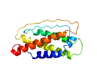
Interleukin 10 (IL-10), also known as human cytokine synthesis inhibitory factor (CSIF), is an anti-inflammatory cytokine. In humans, interleukin 10 is encoded by the IL10 gene. IL-10 signals through a receptor complex consisting of two IL-10 receptor-1 and two IL-10 receptor-2 proteins. Consequently, the functional receptor consists of four IL-10 receptor molecules. IL-10 binding induces STAT3 signalling via the phosphorylation of the cytoplasmic tails of IL-10 receptor 1 + IL-10 receptor 2 by JAK1 and Tyk2 respectively.

The regulatory T cells (Tregs or Treg cells), formerly known as suppressor T cells, are a subpopulation of T cells that modulate the immune system, maintain tolerance to self-antigens, and prevent autoimmune disease. Treg cells are immunosuppressive and generally suppress or downregulate induction and proliferation of effector T cells. Treg cells express the biomarkers CD4, FOXP3, and CD25 and are thought to be derived from the same lineage as naïve CD4+ cells. Because effector T cells also express CD4 and CD25, Treg cells are very difficult to effectively discern from effector CD4+, making them difficult to study. Research has found that the cytokine transforming growth factor beta (TGF-β) is essential for Treg cells to differentiate from naïve CD4+ cells and is important in maintaining Treg cell homeostasis.

<span class="mw-page-title-main">Interleukin 12</span> Interleukin

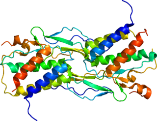
Interleukin 12 (IL-12) is an interleukin that is naturally produced by dendritic cells, macrophages, neutrophils, helper T cells and human B-lymphoblastoid cells (NC-37) in response to antigenic stimulation. IL-12 belongs to the family of interleukin-12. IL-12 family is unique in comprising the only heterodimeric cytokines, which includes IL-12, IL-23, IL-27 and IL-35. Despite sharing many structural features and molecular partners, they mediate surprisingly diverse functional effects.

<span class="mw-page-title-main">Interleukin 4</span> Mammalian protein found in Mus musculus

The interleukin 4 is a cytokine that induces differentiation of naive helper T cells (Th0 cells) to Th2 cells. Upon activation by IL-4, Th2 cells subsequently produce additional IL-4 in a positive feedback loop. IL-4 is produced primarily by mast cells, Th2 cells, eosinophils and basophils. It is closely related and has functions similar to IL-13.

<span class="mw-page-title-main">Fc receptor</span> Surface protein important to the immune system

In immunology, an Fc receptor is a protein found on the surface of certain cells – including, among others, B lymphocytes, follicular dendritic cells, natural killer cells, macrophages, neutrophils, eosinophils, basophils, human platelets, and mast cells – that contribute to the protective functions of the immune system. Its name is derived from its binding specificity for a part of an antibody known as the Fc region. Fc receptors bind to antibodies that are attached to infected cells or invading pathogens. Their activity stimulates phagocytic or cytotoxic cells to destroy microbes, or infected cells by antibody-mediated phagocytosis or antibody-dependent cell-mediated cytotoxicity. Some viruses such as flaviviruses use Fc receptors to help them infect cells, by a mechanism known as antibody-dependent enhancement of infection.

<span class="mw-page-title-main">Interleukin 7</span> Growth factor secreted by stromal cells in the bone marrow and thymus.

Interleukin 7 (IL-7) is a protein that in humans is encoded by the IL7 gene.

Memory T cells are a subset of T lymphocytes that might have some of the same functions as memory B cells. Their lineage is unclear.

<span class="mw-page-title-main">Interleukin 15</span> Cytokine with structural similarity to Interleukin-2

Interleukin-15 (IL-15) is a protein that in humans is encoded by the IL15 gene. IL-15 is an inflammatory cytokine with structural similarity to Interleukin-2 (IL-2). Like IL-2, IL-15 binds to and signals through a complex composed of IL-2/IL-15 receptor beta chain (CD122) and the common gamma chain. IL-15 is secreted by mononuclear phagocytes following infection by virus(es). This cytokine induces the proliferation of natural killer cells, i.e. cells of the innate immune system whose principal role is to kill virally infected cells.

<span class="mw-page-title-main">Interleukin 26</span>

Interleukin-26 (IL-26) is a protein that in humans is encoded by the IL26 gene.

CD16, also known as FcγRIII, is a cluster of differentiation molecule found on the surface of natural killer cells, neutrophils, monocytes, macrophages, and certain T cells. CD16 has been identified as Fc receptors FcγRIIIa (CD16a) and FcγRIIIb (CD16b), which participate in signal transduction. The most well-researched membrane receptor implicated in triggering lysis by NK cells, CD16 is a molecule of the immunoglobulin superfamily (IgSF) involved in antibody-dependent cellular cytotoxicity (ADCC). It can be used to isolate populations of specific immune cells through fluorescent-activated cell sorting (FACS) or magnetic-activated cell sorting, using antibodies directed towards CD16.

CD4 immunoadhesin is a recombinant fusion protein consisting of a combination of CD4 and the fragment crystallizable region, similarly known as immunoglobulin. It belongs to the antibody (Ig) gene family. CD4 is a surface receptor for human immunodeficiency virus (HIV). The CD4 immunoadhesin molecular fusion allow the protein to possess key functions from each independent subunit. The CD4 specific properties include the gp120-binding and HIV-blocking capabilities. Properties specific to immunoglobulin are the long plasma half-life and Fc receptor binding. The properties of the protein means that it has potential to be used in AIDS therapy as of 2017. Specifically, CD4 immunoadhesin plays a role in antibody-dependent cell-mediated cytotoxicity (ADCC) towards HIV-infected cells. While natural anti-gp120 antibodies exhibit a response towards uninfected CD4-expressing cells that have a soluble gp120 bound to the CD4 on the cell surface, CD4 immunoadhesin, however, will not exhibit a response. One of the most relevant of these possibilities is its ability to cross the placenta.

<span class="mw-page-title-main">CD244</span> Protein found in humans

CD244 also known as 2B4 or SLAMF4 is a protein that in humans is encoded by the CD244 gene.

<span class="mw-page-title-main">Interleukin-17A</span> Protein-coding gene in the species Homo sapiens

Interleukin-17A is a protein that in humans is encoded by the IL17A gene. In rodents, IL-17A used to be referred to as CTLA8, after the similarity with a viral gene.

<span class="mw-page-title-main">IL2RA</span> Mammalian protein found in Homo sapiens

The Interleukin-2 receptor alpha chain is a protein involved in the assembly of the high-affinity Interleukin-2 receptor, consisting of alpha (IL2RA), beta (IL2RB) and the common gamma chain (IL2RG). As the name indicates, this receptor interacts with Interleukin-2, a pleiotropic cytokine which plays an important role in immune homeostasis.

<span class="mw-page-title-main">NKG2D</span> Protein-coding gene in the species Homo sapiens

NKG2D is an activating receptor (transmembrane protein) belonging to the NKG2 family of C-type lectin-like receptors. NKG2D is encoded by KLRK1 (killer cell lectin like receptor K1) gene which is located in the NK-gene complex (NKC) situated on chromosome 6 in mice and chromosome 12 in humans. In mice, it is expressed by NK cells, NK1.1+ T cells, γδ T cells, activated CD8+ αβ T cells and activated macrophages. In humans, it is expressed by NK cells, γδ T cells and CD8+ αβ T cells. NKG2D recognizes induced-self proteins from MIC and RAET1/ULBP families which appear on the surface of stressed, malignant transformed, and infected cells.

Regulatory B cells (Bregs or Breg cells) represent a small population of B cells that participates in immunomodulation and in the suppression of immune responses. The population of Bregs can be further separated into different human or murine subsets such as B10 cells, marginal zone B cells, Br1 cells, GrB+B cells, CD9+ B cells, and even some plasmablasts or plasma cells. Bregs regulate the immune system by different mechanisms. One of the main mechanisms is the production of anti-inflammatory cytokines such as interleukin 10 (IL-10), IL-35, or transforming growth factor beta (TGF-β). Another known mechanism is the production of cytotoxic Granzyme B. Bregs also express various inhibitory surface markers such as programmed death-ligand 1 (PD-L1), CD39, CD73, and aryl hydrocarbon receptor. The regulatory effects of Bregs were described in various models of inflammation, autoimmune diseases, transplantation reactions, and in anti-tumor immunity.

<span class="mw-page-title-main">CD28 family receptor</span> Group of regulatory cell surface receptors

CD28 family receptors are a group of regulatory cell surface receptors expressed on immune cells. The CD28 family in turn is a subgroup of the immunoglobulin superfamily.

References

  1. 1 2 3 GRCh38: Ensembl release 89: ENSG00000138684 - Ensembl, May 2017
  2. 1 2 3 GRCm38: Ensembl release 89: ENSMUSG00000027718 - Ensembl, May 2017
  3. "Human PubMed Reference:". National Center for Biotechnology Information, U.S. National Library of Medicine.
  4. "Mouse PubMed Reference:". National Center for Biotechnology Information, U.S. National Library of Medicine.
  5. 1 2 3 4 Parrish-Novak J, Dillon SR, Nelson A, Hammond A, Sprecher C, Gross JA, Johnston J, Madden K, Xu W, West J, Schrader S, Burkhead S, Heipel M, Brandt C, Kuijper JL, Kramer J, Conklin D, Presnell SR, Berry J, Shiota F, Bort S, Hambly K, Mudri S, Clegg C, Moore M, Grant FJ, Lofton-Day C, Gilbert T, Rayond F, Ching A, Yao L, Smith D, Webster P, Whitmore T, Maurer M, Kaushansky K, Holly RD, Foster D (November 2000). "Interleukin 21 and its receptor are involved in NK cell expansion and regulation of lymphocyte function". Nature. 408 (6808): 57–63. Bibcode:2000Natur.408...57P. doi:10.1038/35040504. PMID   11081504. S2CID   4428126.
  6. Kuchen S, Robbins R, Sims GP, Sheng C, Phillips TM, Lipsky PE, Ettinger R (November 2007). "Essential role of IL-21 in B cell activation, expansion, and plasma cell generation during CD4+ T cell-B cell collaboration". Journal of Immunology. 179 (9): 5886–96. doi: 10.4049/jimmunol.179.9.5886 . PMID   17947662.
  7. 1 2 "Entrez Gene: IL21 interleukin 21".
  8. Parrish-Novak J, Foster DC, Holly RD, Clegg CH (November 2002). "Interleukin-21 and the IL-21 receptor: novel effectors of NK and T cell responses". Journal of Leukocyte Biology. 72 (5): 856–63. doi: 10.1189/jlb.72.5.856 . PMID   12429707. S2CID   27480657.
  9. Chtanova T, Tangye SG, Newton R, Frank N, Hodge MR, Rolph MS, Mackay CR (July 2004). "T follicular helper cells express a distinctive transcriptional profile, reflecting their role as non-Th1/Th2 effector cells that provide help for B cells". Journal of Immunology. 173 (1): 68–78. doi: 10.4049/jimmunol.173.1.68 . PMID   15210760.
  10. Wei L, Laurence A, Elias KM, O'Shea JJ (November 2007). "IL-21 is produced by Th17 cells and drives IL-17 production in a STAT3-dependent manner". The Journal of Biological Chemistry. 282 (48): 34605–10. doi: 10.1074/jbc.M705100200 . PMC   2323680 . PMID   17884812.
  11. Wurster AL, Rodgers VL, Satoskar AR, Whitters MJ, Young DA, Collins M, Grusby MJ (October 2002). "Interleukin 21 is a T helper (Th) cell 2 cytokine that specifically inhibits the differentiation of naive Th cells into interferon gamma-producing Th1 cells". The Journal of Experimental Medicine. 196 (7): 969–77. doi:10.1084/jem.20020620. PMC   2194031 . PMID   12370258.
  12. Schultz BT, Teigler JE, Pissani F, Oster AF, Kranias G, Alter G, Marovich M, Eller MA, Dittmer U, Robb ML, Kim JH, Michael NL, Bolton D, Streeck H (January 2016). "Circulating HIV-Specific Interleukin-21(+)CD4(+) T Cells Represent Peripheral Tfh Cells with Antigen-Dependent Helper Functions". Immunity. 44 (1): 167–178. doi: 10.1016/j.immuni.2015.12.011 . PMID   26795249.
  13. Coquet JM, Kyparissoudis K, Pellicci DG, Besra G, Berzins SP, Smyth MJ, Godfrey DI (March 2007). "IL-21 is produced by NKT cells and modulates NKT cell activation and cytokine production". Journal of Immunology. 178 (5): 2827–34. doi: 10.4049/jimmunol.178.5.2827 . PMID   17312126.
  14. Lamprecht B, Kreher S, Anagnostopoulos I, Jöhrens K, Monteleone G, Jundt F, Stein H, Janz M, Dörken B, Mathas S (October 2008). "Aberrant expression of the Th2 cytokine IL-21 in Hodgkin lymphoma cells regulates STAT3 signaling and attracts Treg cells via regulation of MIP-3alpha". Blood. 112 (8): 3339–47. doi: 10.1182/blood-2008-01-134783 . PMID   18684866.
  15. Ozaki K, Kikly K, Michalovich D, Young PR, Leonard WJ (October 2000). "Cloning of a type I cytokine receptor most related to the IL-2 receptor beta chain". Proceedings of the National Academy of Sciences of the United States of America. 97 (21): 11439–44. Bibcode:2000PNAS...9711439O. doi: 10.1073/pnas.200360997 . PMC   17218 . PMID   11016959.
  16. Asao H, Okuyama C, Kumaki S, Ishii N, Tsuchiya S, Foster D, Sugamura K (July 2001). "Cutting edge: the common gamma-chain is an indispensable subunit of the IL-21 receptor complex". Journal of Immunology. 167 (1): 1–5. doi: 10.4049/jimmunol.167.1.1 . PMID   11418623.
  17. 1 2 Habib T, Senadheera S, Weinberg K, Kaushansky K (July 2002). "The common gamma chain (gamma c) is a required signaling component of the IL-21 receptor and supports IL-21-induced cell proliferation via JAK3". Biochemistry. 41 (27): 8725–31. doi:10.1021/bi0202023. PMID   12093291.
  18. Ozaki K, Spolski R, Feng CG, Qi CF, Cheng J, Sher A, Morse HC, Liu C, Schwartzberg PL, Leonard WJ (November 2002). "A critical role for IL-21 in regulating immunoglobulin production". Science. 298 (5598): 1630–4. Bibcode:2002Sci...298.1630O. doi:10.1126/science.1077002. PMID   12446913. S2CID   6210513.
  19. Hiromura Y, Kishida T, Nakano H, Hama T, Imanishi J, Hisa Y, Mazda O (November 2007). "IL-21 administration into the nostril alleviates murine allergic rhinitis". Journal of Immunology. 179 (10): 7157–65. doi: 10.4049/jimmunol.179.10.7157 . PMID   17982108.
  20. Kishida T, Hiromura Y, Shin-Ya M, Asada H, Kuriyama H, Sugai M, Shimizu A, Yokota Y, Hama T, Imanishi J, Hisa Y, Mazda O (December 2007). "IL-21 induces inhibitor of differentiation 2 and leads to complete abrogation of anaphylaxis in mice". Journal of Immunology. 179 (12): 8554–61. doi: 10.4049/jimmunol.179.12.8554 . PMID   18056403.
  21. Li Y, Bleakley M, Yee C (August 2005). "IL-21 influences the frequency, phenotype, and affinity of the antigen-specific CD8 T cell response". Journal of Immunology. 175 (4): 2261–9. doi: 10.4049/jimmunol.175.4.2261 . PMID   16081794.
  22. Moroz A, Eppolito C, Li Q, Tao J, Clegg CH, Shrikant PA (July 2004). "IL-21 enhances and sustains CD8+ T cell responses to achieve durable tumor immunity: comparative evaluation of IL-2, IL-15, and IL-21". Journal of Immunology. 173 (2): 900–9. doi: 10.4049/jimmunol.173.2.900 . PMID   15240677.
  23. Søndergaard H, Skak K (December 2009). "IL-21: roles in immunopathology and cancer therapy". Tissue Antigens. 74 (6): 467–79. doi:10.1111/j.1399-0039.2009.01382.x. PMID   19845910.
  24. Johnson LD, Jameson SC (June 2009). "Immunology. A chronic need for IL-21". Science. 324 (5934): 1525–6. doi:10.1126/science.1176487. PMID   19541985. S2CID   5345355.
  25. White L, Krishnan S, Strbo N, Liu H, Kolber MA, Lichtenheld MG, Pahwa RN, Pahwa S (May 2007). "Differential effects of IL-21 and IL-15 on perforin expression, lysosomal degranulation, and proliferation in CD8 T cells of patients with human immunodeficiency virus-1 (HIV)". Blood. 109 (9): 3873–80. doi:10.1182/blood-2006-09-045278. PMC   1874576 . PMID   17192392.
  26. 1 2 3 Chevalier MF, Jülg B, Pyo A, Flanders M, Ranasinghe S, Soghoian DZ, Kwon DS, Rychert J, Lian J, Muller MI, Cutler S, McAndrew E, Jessen H, Pereyra F, Rosenberg ES, Altfeld M, Walker BD, Streeck H (January 2011). "HIV-1-specific interleukin-21+ CD4+ T cell responses contribute to durable viral control through the modulation of HIV-specific CD8+ T cell function". Journal of Virology. 85 (2): 733–41. doi:10.1128/JVI.02030-10. PMC   3020027 . PMID   21047960.
  27. 1 2 Iannello A, Boulassel MR, Samarani S, Tremblay C, Toma E, Routy JP, Ahmad A (May 2010). "IL-21 enhances NK cell functions and survival in healthy and HIV-infected patients with minimal stimulation of viral replication". Journal of Leukocyte Biology. 87 (5): 857–67. doi:10.1189/jlb.1009701. PMID   20103765. S2CID   21961522.
  28. Williams LD, Bansal A, Sabbaj S, Heath SL, Song W, Tang J, Zajac AJ, Goepfert PA (March 2011). "Interleukin-21-producing HIV-1-specific CD8 T cells are preferentially seen in elite controllers". Journal of Virology. 85 (5): 2316–24. doi:10.1128/JVI.01476-10. PMC   3067790 . PMID   21159862.

Further reading