Phospholipid

Last updated
Phospholipid arrangement in cell membranes. Phospholipid TvanBrussel.edit.jpg
Phospholipid arrangement in cell membranes.
Phosphatidylcholine is the major component of lecithin. It is also a source for choline in the synthesis of acetylcholine in cholinergic neurons. Phosphatidyl-Choline.svg
Phosphatidylcholine is the major component of lecithin. It is also a source for choline in the synthesis of acetylcholine in cholinergic neurons.

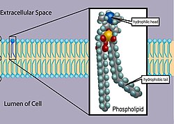
Phospholipids [1] are a class of lipids whose molecule has a hydrophilic "head" containing a phosphate group and two hydrophobic "tails" derived from fatty acids, joined by an alcohol residue (usually a glycerol molecule). Marine phospholipids typically have omega-3 fatty acids EPA and DHA integrated as part of the phospholipid molecule. [2] The phosphate group can be modified with simple organic molecules such as choline, ethanolamine or serine.[ citation needed ]

Contents

Phospholipids are a key component of all cell membranes. They can form lipid bilayers because of their amphiphilic characteristic. In eukaryotes, cell membranes also contain another class of lipid, sterol, interspersed among the phospholipids. The combination provides fluidity in two dimensions combined with mechanical strength against rupture. Purified phospholipids are produced commercially and have found applications in nanotechnology and materials science. [3]

The first phospholipid identified in 1847 as such in biological tissues was lecithin, or phosphatidylcholine, in the egg yolk of chickens by the French chemist and pharmacist Theodore Nicolas Gobley.

Phospholipids in biological membranes

Arrangement

The phospholipids are amphiphilic. The hydrophilic end usually contains a negatively charged phosphate group, and the hydrophobic end usually consists of two "tails" that are long fatty acid residues. [4]

In aqueous solutions, phospholipids are driven by hydrophobic interactions, which result in the fatty acid tails aggregating to minimize interactions with the water molecules. The result is often a phospholipid bilayer: a membrane that consists of two layers of oppositely oriented phospholipid molecules, with their heads exposed to the liquid on both sides, and with the tails directed into the membrane. That is the dominant structural motif of the membranes of all cells and of some other biological structures, such as vesicles or virus coatings.[ citation needed ]

Phospholipid bilayers are the main structural component of the cell membranes. Cell membrane detailed diagram 4.svg
Phospholipid bilayers are the main structural component of the cell membranes.

In biological membranes, the phospholipids often occur with other molecules (e.g., proteins, glycolipids, sterols) in a bilayer such as a cell membrane. [5] Lipid bilayers occur when hydrophobic tails line up against one another, forming a membrane of hydrophilic heads on both sides facing the water.[ citation needed ]

Dynamics

These specific properties allow phospholipids to play an important role in the cell membrane. Their movement can be described by the fluid mosaic model, which describes the membrane as a mosaic of lipid molecules that act as a solvent for all the substances and proteins within it, so proteins and lipid molecules are then free to diffuse laterally through the lipid matrix and migrate over the membrane. Sterols contribute to membrane fluidity by hindering the packing together of phospholipids. However, this model has now been superseded, as through the study of lipid polymorphism it is now known that the behaviour of lipids under physiological (and other) conditions is not simple.[ citation needed ]

Main phospholipids

Diacylglyceride structures

See: Glycerophospholipid

Phosphosphingolipids

See Sphingolipid

Applications

Phospholipids have been widely used to prepare liposomal, ethosomal and other nanoformulations of topical, oral and parenteral drugs for differing reasons like improved bio-availability, reduced toxicity and increased permeability across membranes. Liposomes are often composed of phosphatidylcholine-enriched phospholipids and may also contain mixed phospholipid chains with surfactant properties. The ethosomal formulation of ketoconazole using phospholipids is a promising option for transdermal delivery in fungal infections. [6] Advances in phospholipid research lead to exploring these biomolecules and their conformations using lipidomics.[ citation needed ]

Simulations

Computational simulations of phospholipids are often performed using molecular dynamics with force fields such as GROMOS, CHARMM, or AMBER.[ citation needed ]

Characterization

Phospholipids are optically highly birefringent, i.e. their refractive index is different along their axis as opposed to perpendicular to it. Measurement of birefringence can be achieved using cross polarisers in a microscope to obtain an image of e.g. vesicle walls or using techniques such as dual polarisation interferometry to quantify lipid order or disruption in supported bilayers.[ citation needed ]

Analysis

There are no simple methods available for analysis of phospholipids, since the close range of polarity between different phospholipid species makes detection difficult. Oil chemists often use spectroscopy to determine total phosphorus abundance and then calculate approximate mass of phospholipids based on molecular weight of expected fatty acid species. Modern lipid profiling employs more absolute methods of analysis, with NMR spectroscopy, particularly 31P-NMR, [7] [8] while HPLC-ELSD [9] provides relative values.

Phospholipid synthesis

Phospholipid synthesis occurs in the cytosolic side of ER membrane [10] that is studded with proteins that act in synthesis (GPAT and LPAAT acyl transferases, phosphatase and choline phosphotransferase) and allocation (flippase and floppase). Eventually a vesicle will bud off from the ER containing phospholipids destined for the cytoplasmic cellular membrane on its exterior leaflet and phospholipids destined for the exoplasmic cellular membrane on its inner leaflet. [11] [12]

Sources

Common sources of industrially produced phospholipids are soya, rapeseed, sunflower, chicken eggs, bovine milk, fish eggs etc. Phospholipids for gene delivery such as distearoylphosphatidylcholine, dioleoyl-3-trimethylammonium propane etc. are produced synthetically. Each source has a unique profile of individual phospholipid species, as well as fatty acids, and consequently differing applications in food, nutrition, pharmaceuticals, cosmetics and drug delivery.[ citation needed ]

In signal transduction

Some types of phospholipid can be split to produce products that function as second messengers in signal transduction. Examples include phosphatidylinositol (4,5)-bisphosphate (PIP2), that can be split by the enzyme phospholipase C into inositol triphosphate (IP3) and diacylglycerol (DAG), which both carry out the functions of the Gq type of G protein in response to various stimuli and intervene in various processes from long term depression in neurons [13] to leukocyte signal pathways started by chemokine receptors. [14]

Phospholipids also intervene in prostaglandin signal pathways as the raw material used by lipase enzymes to produce the prostaglandin precursors. In plants they serve as the raw material to produce jasmonic acid, a plant hormone similar in structure to prostaglandins that mediates defensive responses against pathogens.[ citation needed ]

Food technology

Phospholipids can act as emulsifiers, enabling oils to form a colloid with water. Phospholipids are one of the components of lecithin, which is found in egg yolks, as well as being extracted from soybeans, and is used as a food additive in many products and can be purchased as a dietary supplement. Lysolecithins are typically used for water–oil emulsions like margarine, due to their higher HLB ratio.[ citation needed ]

Phospholipid derivatives

See table below for an extensive list.

Abbreviations used and chemical information of glycerophospholipids

AbbreviationCASNameType
DDPC3436-44-01,2-Didecanoyl-sn-glycero-3-phosphocholine Phosphatidylcholine
DEPA-NA 80724-31-81,2-Dierucoyl-sn-glycero-3-phosphate (sodium salt) Phosphatidic acid
DEPC 56649-39-91,2-Dierucoyl-sn-glycero-3-phosphocholine Phosphatidylcholine
DEPE 988-07-21,2-Dierucoyl-sn-glycero-3-phosphoethanolamine Phosphatidylethanolamine
DEPG-NA 1,2-Dierucoyl-sn-glycero-3[phospho-rac-(1-glycerol...) (sodium salt) Phosphatidylglycerol
DLOPC 998-06-11,2-Dilinoleoyl-sn-glycero-3-phosphocholine Phosphatidylcholine
DLPA-NA 1,2-Dilauroyl-sn-glycero-3-phosphate (sodium salt) Phosphatidic acid
DLPC18194-25-71,2-Dilauroyl-sn-glycero-3-phosphocholine Phosphatidylcholine
DLPE 1,2-Dilauroyl-sn-glycero-3-phosphoethanolamine Phosphatidylethanolamine
DLPG-NA 1,2-Dilauroyl-sn-glycero-3[phospho-rac-(1-glycerol...) (sodium salt) Phosphatidylglycerol
DLPG-NH4 1,2-Dilauroyl-sn-glycero-3[phospho-rac-(1-glycerol...) (ammonium salt) Phosphatidylglycerol
DLPS-NA 1,2-Dilauroyl-sn-glycero-3-phosphoserine (sodium salt) Phosphatidylserine
DMPA-NA 80724-31,2-Dimyristoyl-sn-glycero-3-phosphate (sodium salt) Phosphatidic acid
DMPC 18194-24-61,2-Dimyristoyl-sn-glycero-3-phosphocholine Phosphatidylcholine
DMPE 988-07-21,2-Dimyristoyl-sn-glycero-3-phosphoethanolamine Phosphatidylethanolamine
DMPG-NA 67232-80-81,2-Dimyristoyl-sn-glycero-3[phospho-rac-(1-glycerol...) (sodium salt) Phosphatidylglycerol
DMPG-NH4 1,2-Dimyristoyl-sn-glycero-3[phospho-rac-(1-glycerol...) (ammonium salt) Phosphatidylglycerol
DMPG-NH4/NA 1,2-Dimyristoyl-sn-glycero-3[phospho-rac-(1-glycerol...) (sodium/ammonium salt) Phosphatidylglycerol
DMPS-NA 1,2-Dimyristoyl-sn-glycero-3-phosphoserine (sodium salt) Phosphatidylserine
DOPA-NA 1,2-Dioleoyl-sn-glycero-3-phosphate (sodium salt) Phosphatidic acid
DOPC 4235-95-41,2-Dioleoyl-sn-glycero-3-phosphocholine Phosphatidylcholine
DOPE 4004-5-1-1,2-Dioleoyl-sn-glycero-3-phosphoethanolamine Phosphatidylethanolamine
DOPG-NA 62700-69-01,2-Dioleoyl-sn-glycero-3[phospho-rac-(1-glycerol...) (sodium salt) Phosphatidylglycerol
DOPS-NA 70614-14-11,2-Dioleoyl-sn-glycero-3-phosphoserine (sodium salt) Phosphatidylserine
DPPA-NA 71065-87-71,2-Dipalmitoyl-sn-glycero-3-phosphate (sodium salt) Phosphatidic acid
DPPC 63-89-81,2-Dipalmitoyl-sn-glycero-3-phosphocholine Phosphatidylcholine
DPPE 923-61-51,2-Dipalmitoyl-sn-glycero-3-phosphoethanolamine Phosphatidylethanolamine
DPPG-NA 67232-81-91,2-Dipalmitoyl-sn-glycero-3[phospho-rac-(1-glycerol...) (sodium salt) Phosphatidylglycerol
DPPG-NH4 73548-70-61,2-Dipalmitoyl-sn-glycero-3[phospho-rac-(1-glycerol...) (ammonium salt) Phosphatidylglycerol
DPPS-NA 1,2-Dipalmitoyl-sn-glycero-3-phosphoserine (sodium salt) Phosphatidylserine
DSPA-NA 108321-18-21,2-Distearoyl-sn-glycero-3-phosphate (sodium salt) Phosphatidic acid
DSPC 816-94-41,2-Distearoyl-sn-glycero-3-phosphocholine Phosphatidylcholine
DSPE 1069-79-01,2-Distearoyl-sn-glycero-3-phosphoethanolamine Phosphatidylethanolamine
DSPG-NA 67232-82-01,2-Distearoyl-sn-glycero-3[phospho-rac-(1-glycerol...) (sodium salt) Phosphatidylglycerol
DSPG-NH4 108347-80-41,2-Distearoyl-sn-glycero-3[phospho-rac-(1-glycerol...) (ammonium salt) Phosphatidylglycerol
DSPS-NA 1,2-Distearoyl-sn-glycero-3-phosphoserine (sodium salt) Phosphatidylserine
EPC Egg-PC Phosphatidylcholine
HEPCHydrogenated egg PC Phosphatidylcholine
HSPC Hydrogenated soy PC Phosphatidylcholine
LYSOPC MYRISTIC 18194-24-61-Myristoyl-sn-glycero-3-phosphocholine Lysophosphatidylcholine
LYSOPC PALMITIC 17364-16-81-Palmitoyl-sn-glycero-3-phosphocholine Lysophosphatidylcholine
LYSOPC STEARIC 19420-57-61-Stearoyl-sn-glycero-3-phosphocholine Lysophosphatidylcholine
Milk Sphingomyelin MPPC 1-Myristoyl-2-palmitoyl-sn-glycero 3-phosphocholine Phosphatidylcholine
MSPC 1-Myristoyl-2-stearoyl-sn-glycero-3–phosphocholine Phosphatidylcholine
PMPC 1-Palmitoyl-2-myristoyl-sn-glycero-3–phosphocholine Phosphatidylcholine
POPC 26853-31-61-Palmitoyl-2-oleoyl-sn-glycero-3-phosphocholine Phosphatidylcholine
POPE 1-Palmitoyl-2-oleoyl-sn-glycero-3-phosphoethanolamine Phosphatidylethanolamine
POPG-NA 81490-05-31-Palmitoyl-2-oleoyl-sn-glycero-3[phospho-rac-(1-glycerol)...] (sodium salt) Phosphatidylglycerol
PSPC 1-Palmitoyl-2-stearoyl-sn-glycero-3–phosphocholine Phosphatidylcholine
SMPC 1-Stearoyl-2-myristoyl-sn-glycero-3–phosphocholine Phosphatidylcholine
SOPC 1-Stearoyl-2-oleoyl-sn-glycero-3-phosphocholine Phosphatidylcholine
SPPC 1-Stearoyl-2-palmitoyl-sn-glycero-3-phosphocholine Phosphatidylcholine

See also

Related Research Articles

<span class="mw-page-title-main">Biological membrane</span> Enclosing or separating membrane in organisms acting as selective semi-permeable barrier

A biological membrane, biomembrane or cell membrane is a selectively permeable membrane that separates the interior of a cell from the external environment or creates intracellular compartments by serving as a boundary between one part of the cell and another. Biological membranes, in the form of eukaryotic cell membranes, consist of a phospholipid bilayer with embedded, integral and peripheral proteins used in communication and transportation of chemicals and ions. The bulk of lipids in a cell membrane provides a fluid matrix for proteins to rotate and laterally diffuse for physiological functioning. Proteins are adapted to high membrane fluidity environment of the lipid bilayer with the presence of an annular lipid shell, consisting of lipid molecules bound tightly to the surface of integral membrane proteins. The cell membranes are different from the isolating tissues formed by layers of cells, such as mucous membranes, basement membranes, and serous membranes.

<span class="mw-page-title-main">Lipid</span> Substance of biological origin that is soluble in nonpolar solvents

Lipids are a broad group of organic compounds which include fats, waxes, sterols, fat-soluble vitamins, monoglycerides, diglycerides, phospholipids, and others. The functions of lipids include storing energy, signaling, and acting as structural components of cell membranes. Lipids have applications in the cosmetic and food industries, and in nanotechnology.

<span class="mw-page-title-main">Lipid bilayer</span> Membrane of two layers of lipid molecules

The lipid bilayer is a thin polar membrane made of two layers of lipid molecules. These membranes are flat sheets that form a continuous barrier around all cells. The cell membranes of almost all organisms and many viruses are made of a lipid bilayer, as are the nuclear membrane surrounding the cell nucleus, and membranes of the membrane-bound organelles in the cell. The lipid bilayer is the barrier that keeps ions, proteins and other molecules where they are needed and prevents them from diffusing into areas where they should not be. Lipid bilayers are ideally suited to this role, even though they are only a few nanometers in width, because they are impermeable to most water-soluble (hydrophilic) molecules. Bilayers are particularly impermeable to ions, which allows cells to regulate salt concentrations and pH by transporting ions across their membranes using proteins called ion pumps.

<span class="mw-page-title-main">Peripheral membrane protein</span> Membrane proteins that adhere temporarily to membranes with which they are associated

Peripheral membrane proteins, or extrinsic membrane proteins, are membrane proteins that adhere only temporarily to the biological membrane with which they are associated. These proteins attach to integral membrane proteins, or penetrate the peripheral regions of the lipid bilayer. The regulatory protein subunits of many ion channels and transmembrane receptors, for example, may be defined as peripheral membrane proteins. In contrast to integral membrane proteins, peripheral membrane proteins tend to collect in the water-soluble component, or fraction, of all the proteins extracted during a protein purification procedure. Proteins with GPI anchors are an exception to this rule and can have purification properties similar to those of integral membrane proteins.

<span class="mw-page-title-main">Lipid-anchored protein</span> Membrane protein

Lipid-anchored proteins are proteins located on the surface of the cell membrane that are covalently attached to lipids embedded within the cell membrane. These proteins insert and assume a place in the bilayer structure of the membrane alongside the similar fatty acid tails. The lipid-anchored protein can be located on either side of the cell membrane. Thus, the lipid serves to anchor the protein to the cell membrane. They are a type of proteolipids.

<span class="mw-page-title-main">Phosphatidylcholine</span> Class of phospholipids

Phosphatidylcholines (PC) are a class of phospholipids that incorporate choline as a headgroup. They are a major component of biological membranes and can be easily obtained from a variety of readily available sources, such as egg yolk or soybeans, from which they are mechanically or chemically extracted using hexane. They are also a member of the lecithin group of yellow-brownish fatty substances occurring in animal and plant tissues. Dipalmitoylphosphatidylcholine (lecithin) is a major component of the pulmonary surfactant, and is often used in the lecithin–sphingomyelin ratio to calculate fetal lung maturity. While phosphatidylcholines are found in all plant and animal cells, they are absent in the membranes of most bacteria, including Escherichia coli. Purified phosphatidylcholine is produced commercially.

<span class="mw-page-title-main">Sphingolipid</span> Family of chemical compounds

Sphingolipids are a class of lipids containing a backbone of sphingoid bases, which are a set of aliphatic amino alcohols that includes sphingosine. They were discovered in brain extracts in the 1870s and were named after the mythological sphinx because of their enigmatic nature. These compounds play important roles in signal transduction and cell recognition. Sphingolipidoses, or disorders of sphingolipid metabolism, have particular impact on neural tissue. A sphingolipid with a terminal hydroxyl group is a ceramide. Other common groups bonded to the terminal oxygen atom include phosphocholine, yielding a sphingomyelin, and various sugar monomers or dimers, yielding cerebrosides and globosides, respectively. Cerebrosides and globosides are collectively known as glycosphingolipids.

<span class="mw-page-title-main">Sphingomyelin</span> Class of chemical compounds

Sphingomyelin is a type of sphingolipid found in animal cell membranes, especially in the membranous myelin sheath that surrounds some nerve cell axons. It usually consists of phosphocholine and ceramide, or a phosphoethanolamine head group; therefore, sphingomyelins can also be classified as sphingophospholipids. In humans, SPH represents ~85% of all sphingolipids, and typically make up 10–20 mol % of plasma membrane lipids.

<span class="mw-page-title-main">Phosphatidylinositol</span> Signaling molecule

Phosphatidylinositol or inositol phospholipid is a biomolecule. It was initially called "inosite" when it was discovered by Léon Maquenne and Johann Joseph von Scherer in the late 19th century. It was discovered in bacteria but later also found in eukaryotes, and was found to be a signaling molecule.

<span class="mw-page-title-main">Glycerophospholipid</span> Class of lipids

Glycerophospholipids or phosphoglycerides are glycerol-based phospholipids. They are the main component of biological membranes in eukaryotic cells. They are a type of lipid, of which its composition affects membrane structure and properties. Two major classes are known: those for bacteria and eukaryotes and a separate family for archaea.

<span class="mw-page-title-main">Ceramide</span> Family of waxy lipid molecules

Ceramides are a family of waxy lipid molecules. A ceramide is composed of sphingosine and a fatty acid joined by an amide bond. Ceramides are found in high concentrations within the cell membrane of eukaryotic cells, since they are component lipids that make up sphingomyelin, one of the major lipids in the lipid bilayer. Contrary to previous assumptions that ceramides and other sphingolipids found in cell membrane were purely supporting structural elements, ceramide can participate in a variety of cellular signaling: examples include regulating differentiation, proliferation, and programmed cell death (PCD) of cells.

<span class="mw-page-title-main">Dipalmitoylphosphatidylcholine</span> Chemical compound

Dipalmitoylphosphatidylcholine (DPPC) is a phospholipid (and a lecithin) consisting of two C16 palmitic acid groups attached to a phosphatidylcholine head-group.

<span class="mw-page-title-main">Lipid signaling</span> Biological signaling using lipid molecules

Lipid signaling, broadly defined, refers to any biological cell signaling event involving a lipid messenger that binds a protein target, such as a receptor, kinase or phosphatase, which in turn mediate the effects of these lipids on specific cellular responses. Lipid signaling is thought to be qualitatively different from other classical signaling paradigms because lipids can freely diffuse through membranes. One consequence of this is that lipid messengers cannot be stored in vesicles prior to release and so are often biosynthesized "on demand" at their intended site of action. As such, many lipid signaling molecules cannot circulate freely in solution but, rather, exist bound to special carrier proteins in serum.

<span class="mw-page-title-main">Flippase</span>

Flippases are transmembrane lipid transporter proteins located in the membrane which belong to ABC transporter or P4-type ATPase families. They are responsible for aiding the movement of phospholipid molecules between the two leaflets that compose a cell's membrane. This is necessary to continue their normal function of growth and mobility. The possibility of active maintenance of an asymmetric distribution of molecules in the phospholipid bilayer was predicted in the early 1970s by Mark Bretscher. Although phospholipids diffuse rapidly in the plane of the membrane, their polar head groups cannot pass easily through the hydrophobic center of the bilayer, limiting their diffusion in this dimension. Some flippases - often instead called scramblases - are energy-independent and bidirectional, causing reversible equilibration of phospholipid between the two sides of the membrane, whereas others are energy-dependent and unidirectional, using energy from ATP hydrolysis to pump the phospholipid in a preferred direction. Flippases are described as transporters that move lipids from the exoplasmic to the cytosolic face, while floppases transport in the reverse direction.

<span class="mw-page-title-main">Phosphatidylethanolamine</span> Group of chemical compounds

Phosphatidylethanolamine (PE) is a class of phospholipids found in biological membranes. They are synthesized by the addition of cytidine diphosphate-ethanolamine to diglycerides, releasing cytidine monophosphate. S-Adenosyl methionine can subsequently methylate the amine of phosphatidylethanolamines to yield phosphatidylcholines.

<span class="mw-page-title-main">Membrane lipid</span> Lipid molecules on cell membrane

Membrane lipids are a group of compounds which form the lipid bilayer of the cell membrane. The three major classes of membrane lipids are phospholipids, glycolipids, and cholesterol. Lipids are amphiphilic: they have one end that is soluble in water ('polar') and an ending that is soluble in fat ('nonpolar'). By forming a double layer with the polar ends pointing outwards and the nonpolar ends pointing inwards membrane lipids can form a 'lipid bilayer' which keeps the watery interior of the cell separate from the watery exterior. The arrangements of lipids and various proteins, acting as receptors and channel pores in the membrane, control the entry and exit of other molecules and ions as part of the cell's metabolism. In order to perform physiological functions, membrane proteins are facilitated to rotate and diffuse laterally in two dimensional expanse of lipid bilayer by the presence of a shell of lipids closely attached to protein surface, called annular lipid shell.

α-Parinaric acid Chemical compound

α-Parinaric acid is a conjugated polyunsaturated fatty acid. Discovered by Tsujimoto and Koyanagi in 1933, it contains 18 carbon atoms and 4 conjugated double bonds. The repeating single bond-double bond structure of α-parinaric acid distinguishes it structurally and chemically from the usual "methylene-interrupted" arrangement of polyunsaturated fatty acids that have double-bonds and single bonds separated by a methylene unit (−CH2−). Because of the fluorescent properties conferred by the alternating double bonds, α-parinaric acid is commonly used as a molecular probe in the study of biomembranes.

<span class="mw-page-title-main">Phospholipase C</span> Class of enzymes

Phospholipase C (PLC) is a class of membrane-associated enzymes that cleave phospholipids just before the phosphate group (see figure). It is most commonly taken to be synonymous with the human forms of this enzyme, which play an important role in eukaryotic cell physiology, in particular signal transduction pathways. Phospholipase C's role in signal transduction is its cleavage of phosphatidylinositol 4,5-bisphosphate (PIP2) into diacyl glycerol (DAG) and inositol 1,4,5-trisphosphate (IP3), which serve as second messengers. Activators of each PLC vary, but typically include heterotrimeric G protein subunits, protein tyrosine kinases, small G proteins, Ca2+, and phospholipids.

Membrane contact sites (MCS) are close appositions between two organelles. Ultrastructural studies typically reveal an intermembrane distance in the order of the size of a single protein, as small as 10 nm or wider, with no clear upper limit. These zones of apposition are highly conserved in evolution. These sites are thought to be important to facilitate signalling, and they promote the passage of small molecules, including ions, lipids and reactive oxygen species. MCS are important in the function of the endoplasmic reticulum (ER), since this is the major site of lipid synthesis within cells. The ER makes close contact with many organelles, including mitochondria, Golgi, endosomes, lysosomes, peroxisomes, chloroplasts and the plasma membrane. Both mitochondria and sorting endosomes undergo major rearrangements leading to fission where they contact the ER. Sites of close apposition can also form between most of these organelles most pairwise combinations. First mentions of these contact sites can be found in papers published in the late 1950s mainly visualized using electron microscopy (EM) techniques. Copeland and Dalton described them as “highly specialized tubular form of endoplasmic reticulum in association with the mitochondria and apparently in turn, with the vascular border of the cell”.

<span class="mw-page-title-main">Cell membrane</span> Biological membrane that separates the interior of a cell from its outside environment

The cell membrane is a biological membrane that separates and protects the interior of a cell from the outside environment. The cell membrane consists of a lipid bilayer, made up of two layers of phospholipids with cholesterols interspersed between them, maintaining appropriate membrane fluidity at various temperatures. The membrane also contains membrane proteins, including integral proteins that span the membrane and serve as membrane transporters, and peripheral proteins that loosely attach to the outer (peripheral) side of the cell membrane, acting as enzymes to facilitate interaction with the cell's environment. Glycolipids embedded in the outer lipid layer serve a similar purpose. The cell membrane controls the movement of substances in and out of a cell, being selectively permeable to ions and organic molecules. In addition, cell membranes are involved in a variety of cellular processes such as cell adhesion, ion conductivity, and cell signalling and serve as the attachment surface for several extracellular structures, including the cell wall and the carbohydrate layer called the glycocalyx, as well as the intracellular network of protein fibers called the cytoskeleton. In the field of synthetic biology, cell membranes can be artificially reassembled.

References

  1. "Phospholipid". Encyclopedia Britannica. Retrieved 2020-12-22.
  2. Burri, L.; Hoem, N.; Banni, S.; Berge, K. (2012). "Marine Omega-3 Phospholipids: Metabolism and Biological Activities". International Journal of Molecular Sciences. 13 (11): 15401–15419. doi: 10.3390/ijms131115401 . PMC   3509649 . PMID   23203133.
  3. Mashaghi S.; Jadidi T.; Koenderink G.; Mashaghi A. (2013). "Lipid Nanotechnology". Int. J. Mol. Sci. 14 (2): 4242–4282. doi: 10.3390/ijms14024242 . PMC   3588097 . PMID   23429269.
  4. Alberts, Bruce; Johnson, Alexander; Lewis, Julian; Raff, Martin; Roberts, Keith; Walter, Peter (2002), "The Lipid Bilayer", Molecular Biology of the Cell. 4th edition, Garland Science, retrieved 2023-05-25
  5. Campbell, Neil A.; Brad Williamson; Robin J. Heyden (2006). Biology: Exploring Life. Boston, Massachusetts: Pearson Prentice Hall. ISBN   978-0-13-250882-7. Archived from the original on 2014-11-02. Retrieved 2008-12-14.[ page needed ]
  6. Ketoconazole Encapsulated Liposome and Ethosome: GUNJAN TIWARI.
  7. N. Culeddu; M. Bosco; R. Toffanin; P. Pollesello (1998). "High resolution 31P NMR of extracted phospholipids". Magnetic Resonance in Chemistry. 36 (12): 907–912. doi:10.1002/(sici)1097-458x(199812)36:12<907::aid-omr394>3.0.co;2-5. S2CID   85602251.
  8. Furse, Samuel; Liddell, Susan; Ortori, Catharine A.; Williams, Huw; Neylon, D. Cameron; Scott, David J.; Barrett, David A.; Gray, David A. (2013). "The lipidome and proteome of oil bodies from Helianthus annuus (common sunflower)". Journal of Chemical Biology. 6 (2): 63–76. doi:10.1007/s12154-012-0090-1. PMC   3606697 . PMID   23532185.
  9. T. L. Mounts; A. M. Nash (1990). "HPLC analysis of phospholipids in crude oil for evaluation of soybean deterioration". Journal of the American Oil Chemists' Society. 67 (11): 757–760. doi:10.1007/BF02540486. S2CID   84380025.
  10. Prinz, William A.; Choudhary, Vineet; Liu, Li-Ka; Lahiri, Sujoy; Kannan, Muthukumar (2017-03-01). "Phosphatidylserine synthesis at membrane contact sites promotes its transport out of the ER". Journal of Lipid Research. 58 (3): 553–562. doi: 10.1194/jlr.M072959 . ISSN   0022-2275. PMC   5335585 . PMID   28119445.
  11. Lodish H.; Berk A.; et al. (2007). Molecular Cell Biology (6th ed.). W. H. Freeman. ISBN   978-0-7167-7601-7.
  12. Zheng L.; Lin Y.; Lu S.; Zhang J.; Bogdanov M. (November 2017). "Biogenesis, transport and remodeling of lysophospholipids in Gram-negative bacteria". Biochimica et Biophysica Acta (BBA) - Molecular and Cell Biology of Lipids. 1862 (11): 1404–1413. doi:10.1016/j.bbalip.2016.11.015. PMC   6162059 . PMID   27956138.
  13. Choi, S.-Y.; Chang, J.; Jiang, B.; Seol, G. H.; Min, S. S.; Han, J. S.; Shin, H. S.; Gallagher, M.; Kirkwood, A. (2005). "Multiple Receptors Coupled to Phospholipase C Gate Long-Term Depression in Visual Cortex". Journal of Neuroscience. 25 (49): 11433–11443. doi:10.1523/JNEUROSCI.4084-05.2005. PMC   6725895 . PMID   16339037.
  14. Cronshaw, D. G.; Kouroumalis, A.; Parry, R.; Webb, A.; Brown, Z.; Ward, S. G. (2006). "Evidence that phospholipase C-dependent, calcium-independent mechanisms are required for directional migration of T lymphocytes in response to the CCR4 ligands CCL17 and CCL22". Journal of Leukocyte Biology. 79 (6): 1369–1380. doi: 10.1189/jlb.0106035 . PMID   16614259.