Nuclear envelope

Last updated
Nuclear envelope
Diagram human cell nucleus.svg
Human cell nucleus
Identifiers
TH H1.00.01.2.01001
FMA 63888
Anatomical terminology

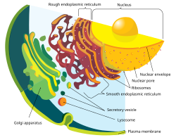
The nuclear envelope, also known as the nuclear membrane, [1] [lower-alpha 1] is made up of two lipid bilayer membranes that in eukaryotic cells surround the nucleus, which encloses the genetic material.

Contents

The nuclear envelope consists of two lipid bilayer membranes: an inner nuclear membrane and an outer nuclear membrane. [4] The space between the membranes is called the perinuclear space. It is usually about 10–50  nm wide. [5] [6] The outer nuclear membrane is continuous with the endoplasmic reticulum membrane. [4] The nuclear envelope has many nuclear pores that allow materials to move between the cytosol and the nucleus. [4] Intermediate filament proteins called lamins form a structure called the nuclear lamina on the inner aspect of the inner nuclear membrane and give structural support to the nucleus. [4]

Structure

The nuclear envelope is made up of two lipid bilayer membranes, an inner nuclear membrane and an outer nuclear membrane. These membranes are connected to each other by nuclear pores. Two sets of intermediate filaments provide support for the nuclear envelope. An internal network forms the nuclear lamina on the inner nuclear membrane. [7] A looser network forms outside to give external support. [4] The actual shape of the nuclear envelope is irregular. It has invaginations and protrusions and can be observed with an electron microscope.

A volumetric surface render (red) of the nuclear envelope of one HeLa cell. The cell was observed in 300 slices of electron microscopy, the nuclear envelope was automatically segmented and rendered. One vertical and one horizontal slice are added for reference. Nuclear envelope of one cancerous HeLa cell.png
A volumetric surface render (red) of the nuclear envelope of one HeLa cell. The cell was observed in 300 slices of electron microscopy, the nuclear envelope was automatically segmented and rendered. One vertical and one horizontal slice are added for reference.

Outer membrane

The outer nuclear membrane also shares a common border with the endoplasmic reticulum. [8] While it is physically linked, the outer nuclear membrane contains proteins found in far higher concentrations than the endoplasmic reticulum. [9] All four nesprin proteins (nuclear envelope spectrin repeat proteins) present in mammals are expressed in the outer nuclear membrane. [10] Nesprin proteins connect cytoskeletal filaments to the nucleoskeleton. [11] Nesprin-mediated connections to the cytoskeleton contribute to nuclear positioning and to the cell’s mechanosensory function. [12] KASH domain proteins of Nesprin-1 and -2 are part of a LINC complex (linker of nucleoskeleton and cytoskeleton) and can bind directly to cystoskeletal components, such as actin filaments, or can bind to proteins in the perinuclear space. [13] [14] Nesprin-3 and -4 may play a role in unloading enormous cargo; Nesprin-3 proteins bind plectin and link the nuclear envelope to cytoplasmic intermediate filaments. [15] Nesprin-4 proteins bind the plus end directed motor kinesin-1. [16] The outer nuclear membrane is also involved in development, as it fuses with the inner nuclear membrane to form nuclear pores. [17]

Inner membrane

The inner nuclear membrane encloses the nucleoplasm, and is covered by the nuclear lamina, a mesh of intermediate filaments which stabilizes the nuclear membrane as well as being involved in chromatin function. [9] It is connected to the outer membrane by nuclear pores which penetrate the membranes. While the two membranes and the endoplasmic reticulum are linked, proteins embedded in the membranes tend to stay put rather than dispersing across the continuum. [18] It is lined with a fiber network called the nuclear lamina which is 10-40 nm thick and provides strength.[ citation needed ]

Mutations in the genes that encode for the inner nuclear membrane proteins can cause several laminopathies.[ citation needed ]

Nuclear pores

Nuclear pores crossing the nuclear envelope Endomembrane system diagram en.svg
Nuclear pores crossing the nuclear envelope

The nuclear envelope is punctured by around a thousand nuclear pore complexes, about 100 nm across, with an inner channel about 40 nm wide. [9] The complexes contain a number of nucleoporins, proteins that link the inner and outer nuclear membranes.[ citation needed ]

Cell division

During the G2 phase of interphase, the nuclear membrane increases its surface area and doubles its number of nuclear pore complexes. [9] In eukaryotes such as yeast which undergo closed mitosis, the nuclear membrane stays intact during cell division. The spindle fibers either form within the membrane, or penetrate it without tearing it apart. [9] In other eukaryotes (animals as well as plants), the nuclear membrane must break down during the prometaphase stage of mitosis to allow the mitotic spindle fibers to access the chromosomes inside. The breakdown and reformation processes are not well understood.

Breakdown

Breakdown and reassembly in mitosis Nuclear envelope breakdown and reassembly in mitosis.jpg
Breakdown and reassembly in mitosis

In mammals, the nuclear membrane can break down within minutes, following a set of steps during the early stages of mitosis. First, M-Cdk's phosphorylate nucleoporin polypeptides and they are selectively removed from the nuclear pore complexes. After that, the rest of the nuclear pore complexes break apart simultaneously. Biochemical evidence suggests that the nuclear pore complexes disassemble into stable pieces rather than disintegrating into small polypeptide fragments. [9] M-Cdk's also phosphorylate elements of the nuclear lamina (the framework that supports the envelope) leading to the disassembly of the lamina and hence the envelope membranes into small vesicles. [19] Electron and fluorescence microscopy has given strong evidence that the nuclear membrane is absorbed by the endoplasmic reticulum—nuclear proteins not normally found in the endoplasmic reticulum show up during mitosis. [9]

In addition to the breakdown of the nuclear membrane during the prometaphase stage of mitosis, the nuclear membrane also ruptures in migrating mammalian cells during the interphase stage of the cell cycle. [20] This transient rupture is likely caused by nuclear deformation. The rupture is rapidly repaired by a process dependent on "endosomal sorting complexes required for transport" (ESCRT) made up of cytosolic protein complexes. [20] During nuclear membrane rupture events, DNA double-strand breaks occur. Thus the survival of cells migrating through confined environments appears to depend on efficient nuclear envelope and DNA repair machineries.

Aberrant nuclear envelope breakdown has also been observed in laminopathies and in cancer cells leading to mislocalization of cellular proteins, the formation of micronuclei and genomic instability. [21] [22] [23]

Reformation

Exactly how the nuclear membrane reforms during telophase of mitosis is debated. Two theories exist [9]

Origin of the nuclear membrane

A study of the comparative genomics, evolution and origins of the nuclear membrane led to the proposal that the nucleus emerged in the primitive eukaryotic ancestor (the “prekaryote”), and was triggered by the archaeo-bacterial symbiosis. [24] Several ideas have been proposed for the evolutionary origin of the nuclear membrane. [25] These ideas include the invagination of the plasma membrane in a prokaryote ancestor, or the formation of a genuine new membrane system following the establishment of proto-mitochondria in the archaeal host. The adaptive function of the nuclear membrane may have been to serve as a barrier to protect the genome from reactive oxygen species (ROS) produced by the cells' pre-mitochondria. [26] [27]

Notes

  1. Less used names include nucleolemma [2] and karyotheca. [3]

Related Research Articles

<span class="mw-page-title-main">Cell nucleus</span> Eukaryotic membrane-bounded organelle containing DNA

The cell nucleus is a membrane-bound organelle found in eukaryotic cells. Eukaryotic cells usually have a single nucleus, but a few cell types, such as mammalian red blood cells, have no nuclei, and a few others including osteoclasts have many. The main structures making up the nucleus are the nuclear envelope, a double membrane that encloses the entire organelle and isolates its contents from the cellular cytoplasm; and the nuclear matrix, a network within the nucleus that adds mechanical support.

Cell biology is a branch of biology that studies the structure, function, and behavior of cells. All living organisms are made of cells. A cell is the basic unit of life that is responsible for the living and functioning of organisms. Cell biology is the study of the structural and functional units of cells. Cell biology encompasses both prokaryotic and eukaryotic cells and has many subtopics which may include the study of cell metabolism, cell communication, cell cycle, biochemistry, and cell composition. The study of cells is performed using several microscopy techniques, cell culture, and cell fractionation. These have allowed for and are currently being used for discoveries and research pertaining to how cells function, ultimately giving insight into understanding larger organisms. Knowing the components of cells and how cells work is fundamental to all biological sciences while also being essential for research in biomedical fields such as cancer, and other diseases. Research in cell biology is interconnected to other fields such as genetics, molecular genetics, molecular biology, medical microbiology, immunology, and cytochemistry.

<span class="mw-page-title-main">Endoplasmic reticulum</span> Cell organelle that synthesizes, folds and processes proteins

The endoplasmic reticulum (ER) is a part of a transportation system of the eukaryotic cell, and has many other important functions such as protein folding. It is a type of organelle made up of two subunits – rough endoplasmic reticulum (RER), and smooth endoplasmic reticulum (SER). The endoplasmic reticulum is found in most eukaryotic cells and forms an interconnected network of flattened, membrane-enclosed sacs known as cisternae, and tubular structures in the SER. The membranes of the ER are continuous with the outer nuclear membrane. The endoplasmic reticulum is not found in red blood cells, or spermatozoa.

<span class="mw-page-title-main">Endomembrane system</span> Membranes in the cytoplasm of a eukaryotic cell

The endomembrane system is composed of the different membranes (endomembranes) that are suspended in the cytoplasm within a eukaryotic cell. These membranes divide the cell into functional and structural compartments, or organelles. In eukaryotes the organelles of the endomembrane system include: the nuclear membrane, the endoplasmic reticulum, the Golgi apparatus, lysosomes, vesicles, endosomes, and plasma (cell) membrane among others. The system is defined more accurately as the set of membranes that forms a single functional and developmental unit, either being connected directly, or exchanging material through vesicle transport. Importantly, the endomembrane system does not include the membranes of plastids or mitochondria, but might have evolved partially from the actions of the latter.

<span class="mw-page-title-main">Nuclear pore</span> Openings in nuclear envelope of eukaryotic cells

A nuclear pore is a channel as part of the nuclear pore complex (NPC), a large protein complex found in the nuclear envelope of eukaryotic cells. The nuclear envelope (NE) surrounds the cell nucleus containing DNA and facilitates the selective membrane transport of various molecules.

Protein targeting or protein sorting is the biological mechanism by which proteins are transported to their appropriate destinations within or outside the cell. Proteins can be targeted to the inner space of an organelle, different intracellular membranes, the plasma membrane, or to the exterior of the cell via secretion. Information contained in the protein itself directs this delivery process. Correct sorting is crucial for the cell; errors or dysfunction in sorting have been linked to multiple diseases.

<span class="mw-page-title-main">Nucleoplasm</span> Protoplasm that permeates a cells nucleus

The nucleoplasm, also known as karyoplasm, is the type of protoplasm that makes up the cell nucleus, the most prominent organelle of the eukaryotic cell. It is enclosed by the nuclear envelope, also known as the nuclear membrane. The nucleoplasm resembles the cytoplasm of a eukaryotic cell in that it is a gel-like substance found within a membrane, although the nucleoplasm only fills out the space in the nucleus and has its own unique functions. The nucleoplasm suspends structures within the nucleus that are not membrane-bound and is responsible for maintaining the shape of the nucleus. The structures suspended in the nucleoplasm include chromosomes, various proteins, nuclear bodies, the nucleolus, nucleoporins, nucleotides, and nuclear speckles.

<span class="mw-page-title-main">Telophase</span> Final stage of a cell division for eukaryotic cells both in mitosis and meiosis

Telophase is the final stage in both meiosis and mitosis in a eukaryotic cell. During telophase, the effects of prophase and prometaphase are reversed. As chromosomes reach the cell poles, a nuclear envelope is re-assembled around each set of chromatids, the nucleoli reappear, and chromosomes begin to decondense back into the expanded chromatin that is present during interphase. The mitotic spindle is disassembled and remaining spindle microtubules are depolymerized. Telophase accounts for approximately 2% of the cell cycle's duration.

<span class="mw-page-title-main">Lamin</span>

Lamins, also known as nuclear lamins are fibrous proteins in type V intermediate filaments, providing structural function and transcriptional regulation in the cell nucleus. Nuclear lamins interact with inner nuclear membrane proteins to form the nuclear lamina on the interior of the nuclear envelope. Lamins have elastic and mechanosensitive properties, and can alter gene regulation in a feedback response to mechanical cues. Lamins are present in all animals but are not found in microorganisms, plants or fungi. Lamin proteins are involved in the disassembling and reforming of the nuclear envelope during mitosis, the positioning of nuclear pores, and programmed cell death. Mutations in lamin genes can result in several genetic laminopathies, which may be life-threatening.

The microtubule-organizing center (MTOC) is a structure found in eukaryotic cells from which microtubules emerge. MTOCs have two main functions: the organization of eukaryotic flagella and cilia and the organization of the mitotic and meiotic spindle apparatus, which separate the chromosomes during cell division. The MTOC is a major site of microtubule nucleation and can be visualized in cells by immunohistochemical detection of γ-tubulin. The morphological characteristics of MTOCs vary between the different phyla and kingdoms. In animals, the two most important types of MTOCs are 1) the basal bodies associated with cilia and flagella and 2) the centrosome associated with spindle formation.

<span class="mw-page-title-main">Nuclear lamina</span>

The nuclear lamina is a dense fibrillar network inside the nucleus of eukaryote cells. It is composed of intermediate filaments and membrane associated proteins. Besides providing mechanical support, the nuclear lamina regulates important cellular events such as DNA replication and cell division. Additionally, it participates in chromatin organization and it anchors the nuclear pore complexes embedded in the nuclear envelope.

<span class="mw-page-title-main">Intermembrane space</span>

The intermembrane space (IMS) is the space occurring between or involving two or more membranes. In cell biology, it is most commonly described as the region between the inner membrane and the outer membrane of a mitochondrion or a chloroplast. It also refers to the space between the inner and outer nuclear membranes of the nuclear envelope, but is often called the perinuclear space. The IMS of mitochondria plays a crucial role in coordinating a variety of cellular activities, such as regulation of respiration and metabolic functions. Unlike the IMS of the mitochondria, the IMS of the chloroplast does not seem to have any obvious function.

<span class="mw-page-title-main">Cellular compartment</span> Closed part in cytosol

Cellular compartments in cell biology comprise all of the closed parts within the cytosol of a eukaryotic cell, usually surrounded by a single or double lipid layer membrane. These compartments are often, but not always, defined as membrane-bound organelles. The formation of cellular compartments is called compartmentalization.

<span class="mw-page-title-main">Nucleoporin 210kDa</span> Protein-coding gene in the species Homo sapiens

Nuclear pore glycoprotein-210 (gp210) is an essential trafficking regulator in the eukaryotic nuclear pore complex. Gp-210 anchors the pore complex to the nuclear membrane. and protein tagging reveals its primarily located on the luminal side of double layer membrane at the pore. A single polypeptide motif of gp210 is responsible for sorting to nuclear membrane, and indicate the carboxyl tail of the protein is oriented toward the cytoplasmic side of the membrane.

<span class="mw-page-title-main">Nucleoporin</span> Family of proteins that form the nuclear pore complex

Nucleoporins are a family of proteins which are the constituent building blocks of the nuclear pore complex (NPC). The nuclear pore complex is a massive structure embedded in the nuclear envelope at sites where the inner and outer nuclear membranes fuse, forming a gateway that regulates the flow of macromolecules between the cell nucleus and the cytoplasm. Nuclear pores enable the passive and facilitated transport of molecules across the nuclear envelope. Nucleoporins, a family of around 30 proteins, are the main components of the nuclear pore complex in eukaryotic cells. Nucleoporin 62 is the most abundant member of this family. Nucleoporins are able to transport molecules across the nuclear envelope at a very high rate. A single NPC is able to transport 60,000 protein molecules across the nuclear envelope every minute.

<span class="mw-page-title-main">Outline of cell biology</span> Overview of and topical guide to cell biology

The following outline is provided as an overview of and topical guide to cell biology:

<span class="mw-page-title-main">Nuclear pore complex protein Nup133</span> Protein-coding gene in the species Homo sapiens

Nuclear pore complex protein Nup133, or Nucleoporin Nup133, is a protein that in humans is encoded by the NUP133 gene.

Nesprins are a family of proteins that are found primarily in the outer nuclear membrane, as well as other subcellular compartments. They contain a C-terminal KASH transmembrane domain and are part of the LINC complex which is a protein network that associates the nuclear envelope to the cytoskeleton, outside the nucleus, and the nuclear lamina, inside the nucleus. Nesprin-1 and -2 bind to the actin filaments. Nesprin-3 binds to plectin, which is bound to the intermediate filaments, while nesprin-4 interacts with kinesin-1.

<span class="mw-page-title-main">Inner nuclear membrane protein</span> Protein embedded in inner membrane of nuclear envelope

Inner nuclear membrane proteins are membrane proteins that are embedded in or associated with the inner membrane of the nuclear envelope. There are about 60 INM proteins, most of which are poorly characterized with respect to structure and function. Among the few well-characterized INM proteins are lamin B receptor (LBR), lamina-associated polypeptide 1 (LAP1), lamina-associated polypeptide-2 (LAP2), emerin and MAN1.

The LINC complex is a protein complex associated with both inner and outer membranes of the nucleus. It is composed of SUN-domain proteins and KASH-domain proteins. The SUN-domain proteins are associated with both nuclear lamins and chromatin and cross the inner nuclear membrane. They interact with the KASH domain proteins in the perinuclear (lumen) space between the two membranes. The KASH domain proteins cross the outer nuclear membrane and interact with actin filaments, microtubule filaments, intermediate filaments, centrosomes and cytoplasmic organelles. The number of SUN-domain and KASH-domain proteins increased in evolution.

References

  1. Georgia State University. "Cell Nucleus and Nuclear Envelope". gsu.edu. Archived from the original on 2018-06-18. Retrieved 2014-01-21.
  2. "Nuclear membrane". Biology Dictionary. Biology Online. Retrieved 7 December 2012.
  3. "nuclear membrane". Merriam Webster. Retrieved 7 December 2012.
  4. 1 2 3 4 5 Alberts, Bruce (2002). Molecular biology of the cell (4th ed.). New York [u.a.]: Garland. p. 197. ISBN   978-0815340720.
  5. "Perinuclear space". Dictionary. Biology Online. Retrieved 7 December 2012.
  6. Berrios, Miguel, ed. (1998). Nuclear structure and function . San Diego: Academic Press. p.  4. ISBN   9780125641555.
  7. Coutinho, Henrique Douglas M; Falcão-Silva, Vivyanne S; Gonçalves, Gregório Fernandes; da Nóbrega, Raphael Batista (20 April 2009). "Molecular ageing in progeroid syndromes: Hutchinson-Gilford progeria syndrome as a model". Immunity & Ageing. 6: 4. doi: 10.1186/1742-4933-6-4 . PMC   2674425 . PMID   19379495.
  8. "Chloride channels in the Nuclear membrane" (PDF). Harvard.edu. Archived from the original (PDF) on 2 August 2010. Retrieved 7 December 2012.
  9. 1 2 3 4 5 6 7 8 Hetzer, Mertin (February 3, 2010). "The Nuclear Envelope". Cold Spring Harbor Perspectives in Biology. 2 (3): a000539. doi:10.1101/cshperspect.a000539. PMC   2829960 . PMID   20300205.
  10. Wilson, Katherine L.; Berk, Jason M. (2010-06-15). "The nuclear envelope at a glance". J Cell Sci. 123 (12): 1973–1978. doi:10.1242/jcs.019042. ISSN   0021-9533. PMC   2880010 . PMID   20519579.
  11. Burke, Brian; Roux, Kyle J. (2009-11-01). "Nuclei take a position: managing nuclear location". Developmental Cell. 17 (5): 587–597. doi: 10.1016/j.devcel.2009.10.018 . ISSN   1878-1551. PMID   19922864.
  12. Uzer, Gunes; Thompson, William R.; Sen, Buer; Xie, Zhihui; Yen, Sherwin S.; Miller, Sean; Bas, Guniz; Styner, Maya; Rubin, Clinton T. (2015-06-01). "Cell Mechanosensitivity to Extremely Low-Magnitude Signals Is Enabled by a LINCed Nucleus". Stem Cells. 33 (6): 2063–2076. doi:10.1002/stem.2004. ISSN   1066-5099. PMC   4458857 . PMID   25787126.
  13. Crisp, Melissa; Liu, Qian; Roux, Kyle; Rattner, J. B.; Shanahan, Catherine; Burke, Brian; Stahl, Phillip D.; Hodzic, Didier (2006-01-02). "Coupling of the nucleus and cytoplasm: role of the LINC complex". The Journal of Cell Biology. 172 (1): 41–53. doi:10.1083/jcb.200509124. ISSN   0021-9525. PMC   2063530 . PMID   16380439.
  14. Zeng, X; et al. (2018). "Nuclear Envelope-Associated Chromosome Dynamics during Meiotic Prophase I." Frontiers in Cell and Developmental Biology. 5: 121. doi: 10.3389/fcell.2017.00121 . PMC   5767173 . PMID   29376050.
  15. Wilhelmsen, Kevin; Litjens, Sandy H. M.; Kuikman, Ingrid; Tshimbalanga, Ntambua; Janssen, Hans; van den Bout, Iman; Raymond, Karine; Sonnenberg, Arnoud (2005-12-05). "Nesprin-3, a novel outer nuclear membrane protein, associates with the cytoskeletal linker protein plectin". The Journal of Cell Biology. 171 (5): 799–810. doi:10.1083/jcb.200506083. ISSN   0021-9525. PMC   2171291 . PMID   16330710.
  16. Roux, Kyle J.; Crisp, Melissa L.; Liu, Qian; Kim, Daein; Kozlov, Serguei; Stewart, Colin L.; Burke, Brian (2009-02-17). "Nesprin 4 is an outer nuclear membrane protein that can induce kinesin-mediated cell polarization". Proceedings of the National Academy of Sciences of the United States of America. 106 (7): 2194–2199. Bibcode:2009PNAS..106.2194R. doi: 10.1073/pnas.0808602106 . ISSN   1091-6490. PMC   2650131 . PMID   19164528.
  17. Fichtman, Boris; Ramos, Corinne; Rasala, Beth; Harel, Amnon; Forbes, Douglass J. (2010-12-01). "Inner/Outer Nuclear Membrane Fusion in Nuclear Pore Assembly". Molecular Biology of the Cell. 21 (23): 4197–4211. doi:10.1091/mbc.E10-04-0309. ISSN   1059-1524. PMC   2993748 . PMID   20926687.
  18. Georgatos, S. D. (April 19, 2001). "The inner nuclear membrane: simple, or very complex?". The EMBO Journal. 20 (12): 2989–2994. doi:10.1093/emboj/20.12.2989. PMC   150211 . PMID   11406575.
  19. Alberts; et al. (2008). "Chapter 17: The Cell Cycle". Molecular Biology of The Cell (5th ed.). New York: Garland Science. pp. 1079–1080. ISBN   978-0-8153-4106-2.
  20. 1 2 Raab M, Gentili M, de Belly H, Thiam HR, Vargas P, Jimenez AJ, Lautenschlaeger F, Voituriez R, Lennon-Duménil AM, Manel N, Piel M (2016). "ESCRT III repairs nuclear envelope ruptures during cell migration to limit DNA damage and cell death". Science. 352 (6283): 359–62. Bibcode:2016Sci...352..359R. doi:10.1126/science.aad7611. PMID   27013426. S2CID   28544308.
  21. Vargas; et al. (2012). "Transient nuclear envelope rupturing during interphase in human cancer cells". Nucleus (Austin, Tex.). 3 (1). Nucleus: 88–100. doi:10.4161/nucl.18954. PMC   3342953 . PMID   22567193.
  22. Lim; et al. (2016). "Nuclear envelope rupture drives genome instability in cancer". Molecular Biology of the Cell. 27 (21). MBoC: 3210–3213. doi:10.1091/mbc.E16-02-0098. PMC   5170854 . PMID   27799497.
  23. Hatch; et al. (2016). "Nuclear envelope rupture is induced by actin-based nucleus confinement". Journal of Cell Biology. 215 (1). JCB: 27–36. doi:10.1083/jcb.201603053. PMC   5057282 . PMID   27697922 . Retrieved 24 March 2019.
  24. Mans BJ, Anantharaman V, Aravind L, Koonin EV (2004). "Comparative genomics, evolution and origins of the nuclear envelope and nuclear pore complex". Cell Cycle. 3 (12): 1612–37. doi: 10.4161/cc.3.12.1316 . PMID   15611647.
  25. Martin W (2005). "Archaebacteria (Archaea) and the origin of the eukaryotic nucleus". Curr. Opin. Microbiol. 8 (6): 630–7. doi:10.1016/j.mib.2005.10.004. PMID   16242992.
  26. Speijer D (2015). "Birth of the eukaryotes by a set of reactive innovations: New insights force us to relinquish gradual models". BioEssays. 37 (12): 1268–76. doi:10.1002/bies.201500107. PMID   26577075. S2CID   20068849.
  27. Bernstein H, Bernstein C. Sexual communication in archaea, the precursor to meiosis. pp. 103-117 in Biocommunication of Archaea (Guenther Witzany, ed.) 2017. Springer International Publishing ISBN   978-3-319-65535-2 DOI 10.1007/978-3-319-65536-9