Peripheral membrane protein

Last updated

Peripheral membrane proteins, or extrinsic membrane proteins, [1] are membrane proteins that adhere only temporarily to the biological membrane with which they are associated. These proteins attach to integral membrane proteins, or penetrate the peripheral regions of the lipid bilayer. The regulatory protein subunits of many ion channels and transmembrane receptors, for example, may be defined as peripheral membrane proteins. In contrast to integral membrane proteins, peripheral membrane proteins tend to collect in the water-soluble component, or fraction, of all the proteins extracted during a protein purification procedure. Proteins with GPI anchors are an exception to this rule and can have purification properties similar to those of integral membrane proteins.

Contents

The reversible attachment of proteins to biological membranes has shown to regulate cell signaling and many other important cellular events, through a variety of mechanisms. [2] For example, the close association between many enzymes and biological membranes may bring them into close proximity with their lipid substrate(s). [3] Membrane binding may also promote rearrangement, dissociation, or conformational changes within many protein structural domains, resulting in an activation of their biological activity. [4] [5] Additionally, the positioning of many proteins are localized to either the inner or outer surfaces or leaflets of their resident membrane. [6] This facilitates the assembly of multi-protein complexes by increasing the probability of any appropriate protein–protein interactions.

Schematic representation of the different types of interaction between monotopic membrane proteins and the cell membrane: 1. interaction by an amphipathic a-helix parallel to the membrane plane (in-plane membrane helix) 2. interaction by a hydrophobic loop 3. interaction by a covalently bound membrane lipid (lipidation) 4. electrostatic or ionic interactions with membrane lipids (e.g. through a calcium ion) Monotopic membrane protein.svg
Schematic representation of the different types of interaction between monotopic membrane proteins and the cell membrane: 1. interaction by an amphipathic α-helix parallel to the membrane plane (in-plane membrane helix) 2. interaction by a hydrophobic loop 3. interaction by a covalently bound membrane lipid (lipidation) 4. electrostatic or ionic interactions with membrane lipids (e.g. through a calcium ion)

Binding to the lipid bilayer

PH domain of phospholipase C delta 1. Middle plane of the lipid bilayer - black dots. Boundary of the hydrocarbon core region - blue dots (intracellular side). Layer of lipid phosphates - yellow dots. 1mai.png
PH domain of phospholipase C delta 1. Middle plane of the lipid bilayer – black dots. Boundary of the hydrocarbon core region – blue dots (intracellular side). Layer of lipid phosphates – yellow dots.

Peripheral membrane proteins may interact with other proteins or directly with the lipid bilayer. In the latter case, they are then known as amphitropic proteins. [4] Some proteins, such as G-proteins and certain protein kinases, interact with transmembrane proteins and the lipid bilayer simultaneously. Some polypeptide hormones, antimicrobial peptides, and neurotoxins accumulate at the membrane surface prior to locating and interacting with their cell surface receptor targets, which may themselves be peripheral membrane proteins.

The phospholipid bilayer that forms the cell surface membrane consists of a hydrophobic inner core region sandwiched between two regions of hydrophilicity, one at the inner surface and one at the outer surface of the cell membrane (see lipid bilayer article for a more detailed structural description of the cell membrane). The inner and outer surfaces, or interfacial regions, of model phospholipid bilayers have been shown to have a thickness of around 8 to 10 Å, although this may be wider in biological membranes that include large amounts of gangliosides or lipopolysaccharides. [7] The hydrophobic inner core region of typical biological membranes may have a thickness of around 27 to 32 Å, as estimated by Small angle X-ray scattering (SAXS). [8] The boundary region between the hydrophobic inner core and the hydrophilic interfacial regions is very narrow, at around 3 Å, (see lipid bilayer article for a description of its component chemical groups). Moving outwards away from the hydrophobic core region and into the interfacial hydrophilic region, the effective concentration of water rapidly changes across this boundary layer, from nearly zero to a concentration of around 2 M. [9] [10] The phosphate groups within phospholipid bilayers are fully hydrated or saturated with water and are situated around 5 Å outside the boundary of the hydrophobic core region. [11]

Some water-soluble proteins associate with lipid bilayers irreversibly and can form transmembrane alpha-helical or beta-barrel channels. Such transformations occur in pore forming toxins such as colicin A, alpha-hemolysin, and others. They may also occur in BcL-2 like protein, in some amphiphilic antimicrobial peptides, and in certain annexins. These proteins are usually described as peripheral as one of their conformational states is water-soluble or only loosely associated with a membrane. [12]

Membrane binding mechanisms

Bee venom phospholipase A2 (1poc). Middle plane of the lipid bilayer - black dots. Boundary of the hydrocarbon core region - red dots (extracellular side). Layer of lipid phosphates - yellow dots. 1poc.png
Bee venom phospholipase A2 (1poc). Middle plane of the lipid bilayer – black dots. Boundary of the hydrocarbon core region – red dots (extracellular side). Layer of lipid phosphates – yellow dots.

The association of a protein with a lipid bilayer may involve significant changes within tertiary structure of a protein. These may include the folding of regions of protein structure that were previously unfolded or a re-arrangement in the folding or a refolding of the membrane-associated part of the proteins. It also may involve the formation or dissociation of protein quaternary structures or oligomeric complexes, and specific binding of ions, ligands, or regulatory lipids.

Typical amphitropic proteins must interact strongly with the lipid bilayer in order to perform their biological functions. These include the enzymatic processing of lipids and other hydrophobic substances, membrane anchoring, and the binding and transfer of small nonpolar compounds between different cellular membranes. These proteins may be anchored to the bilayer as a result of hydrophobic interactions between the bilayer and exposed nonpolar residues at the surface of a protein, [13] by specific non-covalent binding interactions with regulatory lipids , or through their attachment to covalently bound lipid anchors.

It has been shown that the membrane binding affinities of many peripheral proteins depend on the specific lipid composition of the membrane with which they are associated. [14]

amphitropic proteins bind to hydrophobic anchor structures Proteina amfitropica.png
amphitropic proteins bind to hydrophobic anchor structures

Non-specific hydrophobic association

Amphitropic proteins associate with lipid bilayers via various hydrophobic anchor structures. Such as amphiphilic α-helixes, exposed nonpolar loops, post-translationally acylated or lipidated amino acid residues, or acyl chains of specifically bound regulatory lipids such as phosphatidylinositol phosphates. Hydrophobic interactions have been shown to be important even for highly cationic peptides and proteins, such as the polybasic domain of the MARCKS protein or histactophilin, when their natural hydrophobic anchors are present. [15]

Covalently bound lipid anchors

Lipid anchored proteins are covalently attached to different fatty acid acyl chains on the cytoplasmic side of the cell membrane via palmitoylation, myristoylation, or prenylation. On the exoplasmic face of the cell membrane, lipid anchored proteins are covalently attached to the lipids glycosylphosphatidylinositol (GPI) and cholesterol. [16] [17] Protein association with membranes through the use of acylated residues is a reversible process, as the acyl chain can be buried in a protein's hydrophobic binding pocket after dissociation from the membrane. This process occurs within the beta-subunits of G-proteins. Perhaps because of this additional need for structural flexibility, lipid anchors are usually bound to the highly flexible segments of proteins tertiary structure that are not well resolved by protein crystallographic studies.

Specific protein–lipid binding

P40phox PX domain of NADPH oxidase Middle plane of the lipid bilayer - black dots. Boundary of the hydrocarbon core region - blue dots (intracellular side). Layer of lipid phosphates - yellow dots. 1h6h.png
P40phox PX domain of NADPH oxidase Middle plane of the lipid bilayer – black dots. Boundary of the hydrocarbon core region – blue dots (intracellular side). Layer of lipid phosphates – yellow dots.

Some cytosolic proteins are recruited to different cellular membranes by recognizing certain types of lipid found within a given membrane. [18] Binding of a protein to a specific lipid occurs via specific membrane-targeting structural domains that occur within the protein and have specific binding pockets for the lipid head groups of the lipids to which they bind. This is a typical biochemical protein–ligand interaction, and is stabilized by the formation of intermolecular hydrogen bonds, van der Waals interactions, and hydrophobic interactions between the protein and lipid ligand. Such complexes are also stabilized by the formation of ionic bridges between the aspartate or glutamate residues of the protein and lipid phosphates via intervening calcium ions (Ca2+). Such ionic bridges can occur and are stable when ions (such as Ca2+) are already bound to a protein in solution, prior to lipid binding. The formation of ionic bridges is seen in the protein–lipid interaction between both protein C2 type domains and annexins..

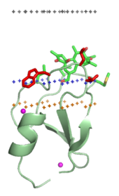
Protein–lipid electrostatic interactions

Any positively charged protein will be attracted to a negatively charged membrane by nonspecific electrostatic interactions. However, not all peripheral peptides and proteins are cationic, and only certain sides of membrane are negatively charged. These include the cytoplasmic side of plasma membranes, the outer leaflet of bacterial outer membranes and mitochondrial membranes. Therefore, electrostatic interactions play an important role in membrane targeting of electron carriers such as cytochrome c, cationic toxins such as charybdotoxin, and specific membrane-targeting domains such as some PH domains, C1 domains, and C2 domains.

Electrostatic interactions are strongly dependent on the ionic strength of the solution. These interactions are relatively weak at the physiological ionic strength (0.14M NaCl): ~3 to 4 kcal/mol for small cationic proteins, such as cytochrome c, charybdotoxin or hisactophilin. [15] [19] [20]

Spatial position in membrane

Orientations and penetration depths of many amphitropic proteins and peptides in membranes are studied using site-directed spin labeling, [21] chemical labeling, measurement of membrane binding affinities of protein mutants, [22] fluorescence spectroscopy, [23] solution or solid-state NMR spectroscopy, [24] ATR FTIR spectroscopy, [25] X-ray or neutron diffraction, [26] and computational methods. [27] [28] [29] [30]

Two distinct membrane-association modes of proteins have been identified. Typical water-soluble proteins have no exposed nonpolar residues or any other hydrophobic anchors. Therefore, they remain completely in aqueous solution and do not penetrate into the lipid bilayer, which would be energetically costly. Such proteins interact with bilayers only electrostatically, for example, ribonuclease and poly-lysine interact with membranes in this mode. However, typical amphitropic proteins have various hydrophobic anchors that penetrate the interfacial region and reach the hydrocarbon interior of the membrane. Such proteins "deform" the lipid bilayer, decreasing the temperature of lipid fluid-gel transition. [31] The binding is usually a strongly exothermic reaction. [32] Association of amphiphilic α-helices with membranes occurs similarly. [26] [33] Intrinsically unstructured or unfolded peptides with nonpolar residues or lipid anchors can also penetrate the interfacial region of the membrane and reach the hydrocarbon core, especially when such peptides are cationic and interact with negatively charged membranes. [34] [35] [36]

Categories

Enzymes

Peripheral enzymes participate in metabolism of different membrane components, such as lipids (phospholipases and cholesterol oxidases), cell wall oligosaccharides (glycosyltransferase and transglycosidases), or proteins (signal peptidase and palmitoyl protein thioesterases). Lipases can also digest lipids that form micelles or nonpolar droplets in water.

ClassFunctionPhysiologyStructure
Alpha/beta hydrolase fold Catalyzes the hydrolysis of chemical bonds. [37] Includes bacterial, fungal, gastric and pancreatic lipases, palmitoyl protein thioesterases, cutinase, and cholinesterases central beta sheet inserted in between two layers of alpha helices [38]
Phospholipase A2 (secretory and cytosolic)Hydrolysis of sn-2 fatty acid bond of phospholipids. [39] Lipid digestion, membrane disruption, and lipid signaling.contains catalytic amino acid triad: aspartic acid, serine, and histidine [40]
Phospholipase C Hydrolyzes PIP2, a phosphatidylinositol, into two second messagers, inositol triphosphate and diacylglycerol. [41] Lipid signaling core structure composed of a split triosephosphate isomerase (TIM) barrel which has an active site, catalytic residues, and a Ca2+ binding site [42]
Cholesterol oxidases Oxidizes and isomerizes cholesterol to cholest-4-en-3-one. [43] Depletes cellular membranes of cholesterol, used in bacterial pathogenesis.two loops of residue which act as a lid on the active site [44]
Carotenoid oxygenase Cleaves carotenoids. [45] Carotenoids function in both plants and animals as hormones (includes vitamin A in humans), pigments, flavors, floral scents and defense compounds.composed of multiple enzymes attached together forming branch-like structures [46]
Lipoxygenases Iron-containing enzymes that catalyze the dioxygenation of polyunsaturated fatty acids. [47] In animals lipoxygenases are involved in the synthesis of inflammatory mediators known as leukotrienes.hundreds of amino acids that makes up a protein are organized into two domains: beta-sheet N terminal and helical C terminal [48]
Alpha toxins Cleave phospholipids in the cell membrane, similar to Phospholipase C. [49] Bacterial pathogenesis, particularly by Clostridium perfringens .soluble monomer with oligomeric pre-pore complexes [50]
Sphingomyelinase CA phosphodiesterase, cleaves phosphodiester bonds. [51] Processing of lipids such as sphingomyelin. saposin domain and connector regions with a metallophosphate catalytic domain [52]
Glycosyltransferases: MurG and TransglycosidasesCatalyzes the transfer of sugar moieties from activated donor molecules to specific acceptor molecules, forming glycosidic bonds. [53] Biosynthesis of disaccharides, oligosaccharides and polysaccharides (glycoconjugates), MurG is involved in bacterial peptidoglycan biosynthesis.three glycine rich loops: one in the C terminal and two in the N terminal [54]
Ferrochelatase Converts protoporphyrin IX into heme. [55] Involved in porphyrin metabolism, protoporphyrins are used to strengthen egg shells. polypeptide folded into two domains that each have a four-stranded parallel beta sheet flanked by alpha helices [56]
Myotubularin-related protein familyLipid phosphatase that dephosphorylates PtdIns3P and PtdIns(3,5)P2. [57] Required for muscle cell differentiation.contains a GRAM domain, SET interacting domain, and a PDZ binding domain [58]
Dihydroorotate dehydrogenases Oxidation of dihydroorotate (DHO) to orotate. [59] Biosynthesis of pyrimidine nucleotides in prokaryotic and eukaryotic cells.composed of two domains: alpha/beta barrel domain that contains the active site and an alpha-helical domain that forms the opening tunnel to the active site [60]
Glycolate oxidase Catalyses the oxidation of α-hydroxycarboxylic acids to the corresponding α-ketoacids. [61] In green plants, the enzyme participates in photorespiration. In animals, the enzyme participates in production of oxalate.β8/α8 fold containing alpha helices, beta strands, and loops and turns [62]

Membrane-targeting domains (“lipid clamps")

C1 domain of PKC-delta (1ptr) Middle plane of the lipid bilayer - black dots. Boundary of the hydrocarbon core region - blue dots (cytoplasmic side). Layer of lipid phosphates - yellow dots. 1ptr.png
C1 domain of PKC-delta (1ptr) Middle plane of the lipid bilayer – black dots. Boundary of the hydrocarbon core region – blue dots (cytoplasmic side). Layer of lipid phosphates – yellow dots.

Membrane-targeting domains associate specifically with head groups of their lipid ligands embedded into the membrane. These lipid ligands are present in different concentrations in distinct types of biological membranes (for example, PtdIns3P can be found mostly in membranes of early endosomes, PtdIns(3,5)P2 in late endosomes, and PtdIns4P in the Golgi). [18] Hence, each domain is targeted to a specific membrane.

Structural domains

Structural domains mediate attachment of other proteins to membranes. Their binding to membranes can be mediated by calcium ions (Ca2+) that form bridges between the acidic protein residues and phosphate groups of lipids, as in annexins or GLA domains.

ClassFunctionPhysiologyStructure
Annexins Calcium-dependent intracellular membrane/ phospholipid binding. [63] Functions include vesicle trafficking, membrane fusion and ion channel formation.
Synapsin I Coats synaptic vesicles and binds to several cytoskeletal elements. [64] Functions in the regulation of neurotransmitter release.
Synuclein Unknown cellular function. [65] Thought to play a role in regulating the stability and/or turnover of the plasma membrane. Associated with both Parkinson's disease and Alzheimer's disease.
GLA-domains of the coagulation system Gamma-carboxyglutamate (GLA) domains are responsible for the high-affinity binding of calcium ions. [66] Involved in function of clotting factors in the blood coagulation cascade.
Spectrin and α-actinin-2Found in several cytoskeletal and microfilament proteins. [67] Maintenance of plasma membrane integrity and cytoskeletal structure.

Transporters of small hydrophobic molecules

These peripheral proteins function as carriers of non-polar compounds between different types of cell membranes or between membranes and cytosolic protein complexes. The transported substances are phosphatidylinositol, tocopherol, gangliosides, glycolipids, sterol derivatives, retinol, fatty acids, water, macromolecules, red blood cells, phospholipids, and nucleotides.

Electron carriers

These proteins are involved in electron transport chains. They include cytochrome c, cupredoxins, high potential iron protein, adrenodoxin reductase, some flavoproteins, and others.

Polypeptide hormones, toxins, and antimicrobial peptides

Many hormones, toxins, inhibitors, or antimicrobial peptides interact specifically with transmembrane protein complexes. They can also accumulate at the lipid bilayer surface, prior to binding their protein targets. Such polypeptide ligands are often positively charged and interact electrostatically with anionic membranes.

Some water-soluble proteins and peptides can also form transmembrane channels. They usually undergo oligomerization, significant conformational changes, and associate with membranes irreversibly. 3D structure of one such transmembrane channel, α-hemolysin, has been determined. In other cases, the experimental structure represents a water-soluble conformation that interacts with the lipid bilayer peripherally, although some of the channel-forming peptides are rather hydrophobic and therefore were studied by NMR spectroscopy in organic solvents or in the presence of micelles.

ClassProteinsPhysiology
Venom toxins Well known types of biotoxins include neurotoxins, cytotoxins, hemotoxins and necrotoxins. Biotoxins have two primary functions: predation (snake, scorpion and cone snail toxins) and defense (honeybee and ant toxins). [68]
Sea anemone toxinsInhibition of sodium and potassium channels and membrane pore formation are the primary actions of over 40 known Sea anemone peptide toxins. Sea anemone are carnivorous animals and use toxins in predation and defense; anemone toxin is of similar toxicity as the most toxic organophosphate chemical warfare agents. [69]
Bacterial toxins Microbial toxins are the primary virulence factors for a variety of pathogenic bacteria. Some toxins, are Pore forming toxins that lyse cellular membranes. Other toxins inhibit protein synthesis or activate second messenger pathways causing dramatic alterations to signal transduction pathways critical in maintaining a variety of cellular functions. Several bacterial toxins can act directly on the immune system, by acting as superantigens and causing massive T cell proliferation, which overextends the immune system. Botulinum toxin is a neurotoxin that prevents neuro-secretory vesicles from docking/fusing with the nerve synapse plasma membrane, inhibiting neurotransmitter release. [70]
Fungal toxinsThese peptides are characterized by the presence of an unusual amino acid, α-aminoisobutyric acid, and exhibit antibiotic and antifungal properties due to their membrane channel-forming activities. [71]
Antimicrobial peptides The modes of action by which antimicrobial peptides kill bacteria is varied and includes disrupting membranes, interfering with metabolism, and targeting cytoplasmic components. In contrast to many conventional antibiotics these peptides appear to be bacteriocidal instead of bacteriostatic.
Defensins Defensins are a type of antimicrobial peptide; and are an important component of virtually all innate host defenses against microbial invasion. Defensins penetrate microbial cell membranes by way of electrical attraction, and form a pore in the membrane allowing efflux, which ultimately leads to the lysis of microorganisms. [72]
Neuronal peptidesThese proteins excite neurons, evoke behavioral responses, are potent vasodilatators, and are responsible for contraction in many types of smooth muscle. [73]
Apoptosis regulatorsMembers of the Bcl-2 family govern mitochondrial outer membrane permeability. Bcl-2 itself suppresses apoptosis in a variety of cell types including lymphocytes and neuronal cells.

See also

Related Research Articles

<span class="mw-page-title-main">Biological membrane</span> Enclosing or separating membrane in organisms acting as selective semi-permeable barrier

A biological membrane, biomembrane or cell membrane is a selectively permeable membrane that separates the interior of a cell from the external environment or creates intracellular compartments by serving as a boundary between one part of the cell and another. Biological membranes, in the form of eukaryotic cell membranes, consist of a phospholipid bilayer with embedded, integral and peripheral proteins used in communication and transportation of chemicals and ions. The bulk of lipids in a cell membrane provides a fluid matrix for proteins to rotate and laterally diffuse for physiological functioning. Proteins are adapted to high membrane fluidity environment of the lipid bilayer with the presence of an annular lipid shell, consisting of lipid molecules bound tightly to the surface of integral membrane proteins. The cell membranes are different from the isolating tissues formed by layers of cells, such as mucous membranes, basement membranes, and serous membranes.

A transmembrane domain (TMD) is a membrane-spanning protein domain. TMDs may consist of one or several alpha-helices or a transmembrane beta barrel. Because the interior of the lipid bilayer is hydrophobic, the amino acid residues in TMDs are often hydrophobic, although proteins such as membrane pumps and ion channels can contain polar residues. TMDs vary greatly in size and hydrophobicity; they may adopt organelle-specific properties.

<span class="mw-page-title-main">Lipid bilayer</span> Membrane of two layers of lipid molecules

The lipid bilayer is a thin polar membrane made of two layers of lipid molecules. These membranes are flat sheets that form a continuous barrier around all cells. The cell membranes of almost all organisms and many viruses are made of a lipid bilayer, as are the nuclear membrane surrounding the cell nucleus, and membranes of the membrane-bound organelles in the cell. The lipid bilayer is the barrier that keeps ions, proteins and other molecules where they are needed and prevents them from diffusing into areas where they should not be. Lipid bilayers are ideally suited to this role, even though they are only a few nanometers in width, because they are impermeable to most water-soluble (hydrophilic) molecules. Bilayers are particularly impermeable to ions, which allows cells to regulate salt concentrations and pH by transporting ions across their membranes using proteins called ion pumps.

<span class="mw-page-title-main">Membrane protein</span> Proteins that are part of, or interact with, biological membranes

Membrane proteins are common proteins that are part of, or interact with, biological membranes. Membrane proteins fall into several broad categories depending on their location. Integral membrane proteins are a permanent part of a cell membrane and can either penetrate the membrane (transmembrane) or associate with one or the other side of a membrane. Peripheral membrane proteins are transiently associated with the cell membrane.

<span class="mw-page-title-main">Transmembrane protein</span> Protein spanning across a biological membrane

A transmembrane protein is a type of integral membrane protein that spans the entirety of the cell membrane. Many transmembrane proteins function as gateways to permit the transport of specific substances across the membrane. They frequently undergo significant conformational changes to move a substance through the membrane. They are usually highly hydrophobic and aggregate and precipitate in water. They require detergents or nonpolar solvents for extraction, although some of them (beta-barrels) can be also extracted using denaturing agents.

<span class="mw-page-title-main">Fluid mosaic model</span> Describe the fluid mosaic model of plasma membrane

The fluid mosaic model explains various characteristics regarding the structure of functional cell membranes. According to this biological model, there is a lipid bilayer in which protein molecules are embedded. The phospholipid bilayer gives fluidity and elasticity to the membrane. Small amounts of carbohydrates are also found in the cell membrane. The biological model, which was devised by Seymour Jonathan Singer and Garth L. Nicolson in 1972, describes the cell membrane as a two-dimensional liquid that restricts the lateral diffusion of membrane components. Such domains are defined by the existence of regions within the membrane with special lipid and protein cocoon that promote the formation of lipid rafts or protein and glycoprotein complexes. Another way to define membrane domains is the association of the lipid membrane with the cytoskeleton filaments and the extracellular matrix through membrane proteins. The current model describes important features relevant to many cellular processes, including: cell-cell signaling, apoptosis, cell division, membrane budding, and cell fusion. The fluid mosaic model is the most acceptable model of the plasma membrane. In this definition of the cell membrane, its main function is to act as a barrier between the contents inside the cell and the extracellular environment.

<span class="mw-page-title-main">Lipid raft</span> Combination in the membranes of cells

The plasma membranes of cells contain combinations of glycosphingolipids, cholesterol and protein receptors organised in glycolipoprotein lipid microdomains termed lipid rafts. Their existence in cellular membranes remains controversial. Indeed, Kervin and Overduin imply that lipid rafts are misconstrued protein islands, which they propose form through a proteolipid code. Nonetheless, it has been proposed that they are specialized membrane microdomains which compartmentalize cellular processes by serving as organising centers for the assembly of signaling molecules, allowing a closer interaction of protein receptors and their effectors to promote kinetically favorable interactions necessary for the signal transduction. Lipid rafts influence membrane fluidity and membrane protein trafficking, thereby regulating neurotransmission and receptor trafficking. Lipid rafts are more ordered and tightly packed than the surrounding bilayer, but float freely within the membrane bilayer. Although more common in the cell membrane, lipid rafts have also been reported in other parts of the cell, such as the Golgi apparatus and lysosomes.

<span class="mw-page-title-main">Antimicrobial peptides</span> Class of peptides that have antimicrobial activity

Antimicrobial peptides (AMPs), also called host defence peptides (HDPs) are part of the innate immune response found among all classes of life. Fundamental differences exist between prokaryotic and eukaryotic cells that may represent targets for antimicrobial peptides. These peptides are potent, broad spectrum antimicrobials which demonstrate potential as novel therapeutic agents. Antimicrobial peptides have been demonstrated to kill Gram negative and Gram positive bacteria, enveloped viruses, fungi and even transformed or cancerous cells. Unlike the majority of conventional antibiotics it appears that antimicrobial peptides frequently destabilize biological membranes, can form transmembrane channels, and may also have the ability to enhance immunity by functioning as immunomodulators.

<span class="mw-page-title-main">ENTH domain</span> InterPro Domain

The epsin N-terminal homology (ENTH) domain is a structural domain that is found in proteins involved in endocytosis and cytoskeletal machinery.

Protein–lipid interaction is the influence of membrane proteins on the lipid physical state or vice versa.

Orientations of Proteins in Membranes (OPM) database provides spatial positions of membrane protein structures with respect to the lipid bilayer. Positions of the proteins are calculated using an implicit solvation model of the lipid bilayer. The results of calculations were verified against experimental studies of spatial arrangement of transmembrane and peripheral proteins in membranes.

<span class="mw-page-title-main">Lipid bilayer fusion</span>

In membrane biology, fusion is the process by which two initially distinct lipid bilayers merge their hydrophobic cores, resulting in one interconnected structure. If this fusion proceeds completely through both leaflets of both bilayers, an aqueous bridge is formed and the internal contents of the two structures can mix. Alternatively, if only one leaflet from each bilayer is involved in the fusion process, the bilayers are said to be hemifused. In hemifusion, the lipid constituents of the outer leaflet of the two bilayers can mix, but the inner leaflets remain distinct. The aqueous contents enclosed by each bilayer also remain separated.

A model lipid bilayer is any bilayer assembled in vitro, as opposed to the bilayer of natural cell membranes or covering various sub-cellular structures like the nucleus. They are used to study the fundamental properties of biological membranes in a simplified and well-controlled environment, and increasingly in bottom-up synthetic biology for the construction of artificial cells. A model bilayer can be made with either synthetic or natural lipids. The simplest model systems contain only a single pure synthetic lipid. More physiologically relevant model bilayers can be made with mixtures of several synthetic or natural lipids.

<span class="mw-page-title-main">WALP peptide</span> Class of peptides used for studying lipid membranes

WALP peptides are a class of synthesized, membrane-spanning α-helices composed of tryptophan (W), alanine (A), and leucine (L) amino acids. They are designed to study properties of proteins in lipid membranes such as orientation, extent of insertion, and hydrophobic mismatch.

<span class="mw-page-title-main">Sec14</span>

Sec14 is a cytosolic protein found in yeast which plays a role in the regulation of several cellular functions, specifically those related to intracellular transport. Encoded by the Sec14 gene, Sec14p may transport phosphatidylinositol and phosphatidylcholine produced in the endoplasmic reticulum and the Golgi body to other cellular membranes. Additionally, Sec14p potentially plays a role in the localization of lipid raft proteins. Sec14p is an essential gene in yeast, and is homologous in function to phosphatidylinositol transfer protein in mammals. A conditional mutant with non-functional Sec14p presents with Berkeley bodies and deficiencies in protein secretion.

The thiol-activated Cholesterol-dependent Cytolysin(CDC) family is a member of the MACPF superfamily. Cholesterol dependent cytolysins are a family of β-barrel pore-forming exotoxins that are secreted by gram-positive bacteria. CDCs are secreted as water-soluble monomers of 50-70 kDa, that when bound to the target cell, form a circular homo-oligomeric complex containing as many as 40 monomers. Through multiple conformational changes, the β-barrel transmembrane structure is formed and inserted into the target cell membrane. The presence of cholesterol in the target membrane is required for pore formation, though the presence of cholesterol is not required by all CDCs for binding. For example, intermedilysin secreted by Streptococcus intermedius will bind only to target membranes containing a specific protein receptor, independent of the presence of cholesterol, but cholesterol is required by intermedilysin for pore formation. While the lipid environment of cholesterol in the membrane can affect toxin binding, the exact molecular mechanism that cholesterol regulates the cytolytic activity of the CDC is not fully understood.

<span class="mw-page-title-main">Cell membrane</span> Biological membrane that separates the interior of a cell from its outside environment

The cell membrane is a biological membrane that separates and protects the interior of a cell from the outside environment. The cell membrane consists of a lipid bilayer, made up of two layers of phospholipids with cholesterols interspersed between them, maintaining appropriate membrane fluidity at various temperatures. The membrane also contains membrane proteins, including integral proteins that span the membrane and serve as membrane transporters, and peripheral proteins that loosely attach to the outer (peripheral) side of the cell membrane, acting as enzymes to facilitate interaction with the cell's environment. Glycolipids embedded in the outer lipid layer serve a similar purpose. The cell membrane controls the movement of substances in and out of a cell, being selectively permeable to ions and organic molecules. In addition, cell membranes are involved in a variety of cellular processes such as cell adhesion, ion conductivity, and cell signalling and serve as the attachment surface for several extracellular structures, including the cell wall and the carbohydrate layer called the glycocalyx, as well as the intracellular network of protein fibers called the cytoskeleton. In the field of synthetic biology, cell membranes can be artificially reassembled.

Stephen H. White is an American Biophysicist, academic, and author. He is a Professor Emeritus of Physiology and Biophysics at the University of California, Irvine.

<span class="mw-page-title-main">Single-pass membrane protein</span> Transmembrane protein

A single-pass membrane protein also known as single-spanning protein or bitopic protein is a transmembrane protein that spans the lipid bilayer only once. These proteins may constitute up to 50% of all transmembrane proteins, depending on the organism, and contribute significantly to the network of interactions between different proteins in cells, including interactions via transmembrane alpha helices. They usually include one or several water-soluble domains situated at the different sides of biological membranes, for example in single-pass transmembrane receptors. Some of them are small and serve as regulatory or structure-stabilizing subunits in large multi-protein transmembrane complexes, such as photosystems or the respiratory chain. A 2013 estimate identified about 1300 single-pass membrane proteins in the human genome.

<span class="mw-page-title-main">GsMTx-4</span> Grammostola mechanotoxin 4

Grammostola mechanotoxin #4, also known as M-theraphotoxin-Gr1a (M-TRTX-Gr1a), is a neurotoxin isolated from the venom of the spider Chilean rose tarantula Grammostola spatulate. This amphiphilic peptide, which consists of 35 amino acids, belongs to the inhibitory cysteine knot (ICK) peptide family. It reduces mechanical sensation by inhibiting mechanosensitive channels (MSCs).

References

  1. "extrinsic protein | biology | Britannica". www.britannica.com. Retrieved 2022-07-04.
  2. Cafiso DS (2005). "Structure and interactions of C2 domains at membrane surfaces". In Tamm LK (ed.). Protein-Lipid Interactions: From Membrane Domains to Cellular Networks. Chichester: John Wiley & Sons. pp. 403–22. ISBN   3-527-31151-3.
  3. Ghosh M, Tucker DE, Burchett SA, Leslie CC (November 2006). "Properties of the Group IV phospholipase A2 family". Progress in Lipid Research. 45 (6): 487–510. doi:10.1016/j.plipres.2006.05.003. PMID   16814865.
  4. 1 2 Johnson JE, Cornell RB (2002). "Amphitropic proteins: regulation by reversible membrane interactions (review)". Molecular Membrane Biology. 16 (3): 217–235. doi: 10.1080/096876899294544 . PMID   10503244.
  5. Thuduppathy GR, Craig JW, Kholodenko V, Schon A, Hill RB (June 2006). "Evidence that membrane insertion of the cytosolic domain of Bcl-xL is governed by an electrostatic mechanism". Journal of Molecular Biology. 359 (4): 1045–1058. doi:10.1016/j.jmb.2006.03.052. PMC   1785297 . PMID   16650855.
  6. Takida S, Wedegaertner PB (June 2004). "Exocytic pathway-independent plasma membrane targeting of heterotrimeric G proteins". FEBS Letters. 567 (2–3): 209–213. doi: 10.1016/j.febslet.2004.04.062 . PMID   15178324. S2CID   36940600.
  7. McIntosh TJ, Vidal A, Simon SA (2003). "The energetics of peptide-lipid interactions: modification by interfacial dipoles and cholesterol". Current Topics in Membranes. Vol. 52. Academic Press. pp. 205–253. ISBN   978-0-12-643871-0.
  8. Mitra K, Ubarretxena-Belandia I, Taguchi T, Warren G, Engelman DM (March 2004). "Modulation of the bilayer thickness of exocytic pathway membranes by membrane proteins rather than cholesterol". Proceedings of the National Academy of Sciences of the United States of America. 101 (12): 4083–4088. Bibcode:2004PNAS..101.4083M. doi: 10.1073/pnas.0307332101 . PMC   384699 . PMID   15016920.
  9. Marsh D (July 2001). "Polarity and permeation profiles in lipid membranes". Proceedings of the National Academy of Sciences of the United States of America. 98 (14): 7777–7782. Bibcode:2001PNAS...98.7777M. doi: 10.1073/pnas.131023798 . PMC   35418 . PMID   11438731.
  10. Marsh D (December 2002). "Membrane water-penetration profiles from spin labels". European Biophysics Journal. 31 (7): 559–562. doi:10.1007/s00249-002-0245-z. PMID   12602343. S2CID   36212541.
  11. Nagle JF, Tristram-Nagle S (November 2000). "Structure of lipid bilayers". Biochimica et Biophysica Acta (BBA) - Reviews on Biomembranes. 1469 (3): 159–195. doi:10.1016/S0304-4157(00)00016-2. PMC   2747654 . PMID   11063882.
  12. Goñi FM (2002). "Non-permanent proteins in membranes: when proteins come as visitors (Review)". Molecular Membrane Biology. 19 (4): 237–245. doi:10.1080/0968768021000035078. PMID   12512770. S2CID   20892603.
  13. Goforth RL, Chi AK, Greathouse DV, Providence LL, Koeppe RE, Andersen OS (May 2003). "Hydrophobic coupling of lipid bilayer energetics to channel function". The Journal of General Physiology. 121 (5): 477–493. doi:10.1085/jgp.200308797. PMC   2217378 . PMID   12719487.
  14. McIntosh TJ, Simon SA (2006). "Roles of bilayer material properties in function and distribution of membrane proteins". Annual Review of Biophysics and Biomolecular Structure. 35 (1): 177–198. doi:10.1146/annurev.biophys.35.040405.102022. PMID   16689633.
  15. 1 2 Hanakam F, Gerisch G, Lotz S, Alt T, Seelig A (August 1996). "Binding of hisactophilin I and II to lipid membranes is controlled by a pH-dependent myristoyl-histidine switch". Biochemistry. 35 (34): 11036–11044. doi:10.1021/bi960789j. PMID   8780505.
  16. Silvius JR (2003). "Lipidated peptides as tools for understanding the membrane interactions of lipid-modified proteins". Current Topics in Membranes. Vol. 52. Academic Press. pp. 371–395. ISBN   978-0-12-643871-0.
  17. Baumann NA, Mennon AK (2002). "Lipid modifications of proteins". In Vance DE, Vance JE (eds.). Biochemistry of Lipids, Lipoproteins and Membranes (4th ed.). Elsevier Science. pp. 37–54. ISBN   978-0-444-51139-3.
  18. 1 2 Cho W, Stahelin RV (June 2005). "Membrane-protein interactions in cell signaling and membrane trafficking". Annual Review of Biophysics and Biomolecular Structure. 34: 119–151. doi:10.1146/annurev.biophys.33.110502.133337. PMID   15869386.
  19. Ben-Tal N, Honig B, Miller C, McLaughlin S (October 1997). "Electrostatic binding of proteins to membranes. Theoretical predictions and experimental results with charybdotoxin and phospholipid vesicles". Biophysical Journal. 73 (4): 1717–1727. Bibcode:1997BpJ....73.1717B. doi:10.1016/S0006-3495(97)78203-1. PMC   1181073 . PMID   9336168.
  20. Sankaram MB, Marsh D (1993). "Protein-lipid interactions with peripheral membrane proteins". In Watts A (ed.). Protein-lipid interactions. Elsevier. pp. 127–162. ISBN   0-444-81575-9.
  21. Malmberg NJ, Falke JJ (2005). "Use of EPR power saturation to analyze the membrane-docking geometries of peripheral proteins: applications to C2 domains". Annual Review of Biophysics and Biomolecular Structure. 34 (1): 71–90. doi:10.1146/annurev.biophys.34.040204.144534. PMC   3637887 . PMID   15869384.
  22. Spencer AG, Thuresson E, Otto JC, Song I, Smith T, DeWitt DL, et al. (November 1999). "The membrane binding domains of prostaglandin endoperoxide H synthases 1 and 2. Peptide mapping and mutational analysis". The Journal of Biological Chemistry. 274 (46): 32936–32942. doi: 10.1074/jbc.274.46.32936 . PMID   10551860.
  23. Lathrop B, Gadd M, Biltonen RL, Rule GS (March 2001). "Changes in Ca2+ affinity upon activation of Agkistrodon piscivorus piscivorus phospholipase A2". Biochemistry. 40 (11): 3264–3272. doi:10.1021/bi001901n. PMID   11258945.
  24. Kutateladze T, Overduin M (March 2001). "Structural mechanism of endosome docking by the FYVE domain". Science. 291 (5509): 1793–1796. Bibcode:2001Sci...291.1793K. doi:10.1126/science.291.5509.1793. PMID   11230696.
  25. Tatulian SA, Qin S, Pande AH, He X (September 2005). "Positioning membrane proteins by novel protein engineering and biophysical approaches". Journal of Molecular Biology. 351 (5): 939–947. doi:10.1016/j.jmb.2005.06.080. PMID   16055150.
  26. 1 2 Hristova K, Wimley WC, Mishra VK, Anantharamiah GM, Segrest JP, White SH (July 1999). "An amphipathic alpha-helix at a membrane interface: a structural study using a novel X-ray diffraction method". Journal of Molecular Biology. 290 (1): 99–117. doi:10.1006/jmbi.1999.2840. PMID   10388560.
  27. Murray D, Honig B (January 2002). "Electrostatic control of the membrane targeting of C2 domains". Molecular Cell. 9 (1): 145–154. doi: 10.1016/S1097-2765(01)00426-9 . PMID   11804593.
  28. Efremov RG, Nolde DE, Konshina AG, Syrtcev NP, Arseniev AS (September 2004). "Peptides and proteins in membranes: what can we learn via computer simulations?". Current Medicinal Chemistry. 11 (18): 2421–2442. doi:10.2174/0929867043364496. PMID   15379706.
  29. Lomize AL, Pogozheva ID, Lomize MA, Mosberg HI (June 2006). "Positioning of proteins in membranes: a computational approach". Protein Science. 15 (6): 1318–1333. doi:10.1110/ps.062126106. PMC   2242528 . PMID   16731967.
  30. Lomize A, Lomize M, Pogozheva I. "Comparison with experimental data". Orientations of Proteins in Membranes. University of Michigan. Retrieved 2007-02-08.
  31. Papahadjopoulos D, Moscarello M, Eylar EH, Isac T (September 1975). "Effects of proteins on thermotropic phase transitions of phospholipid membranes". Biochimica et Biophysica Acta (BBA) - Biomembranes. 401 (3): 317–335. doi:10.1016/0005-2736(75)90233-3. PMID   52374.
  32. Seelig J (November 2004). "Thermodynamics of lipid-peptide interactions". Biochimica et Biophysica Acta (BBA) - Biomembranes. 1666 (1–2): 40–50. doi: 10.1016/j.bbamem.2004.08.004 . PMID   15519307.
  33. Darkes MJ, Davies SM, Bradshaw JP (1997). "Interaction of tachykinins with phospholipid membranes: A neutron diffraction study". Physica B. 241: 1144–1147. Bibcode:1997PhyB..241.1144D. doi:10.1016/S0921-4526(97)00811-9.
  34. Ellena JF, Moulthrop J, Wu J, Rauch M, Jaysinghne S, Castle JD, Cafiso DS (November 2004). "Membrane position of a basic aromatic peptide that sequesters phosphatidylinositol 4,5 bisphosphate determined by site-directed spin labeling and high-resolution NMR". Biophysical Journal. 87 (5): 3221–3233. Bibcode:2004BpJ....87.3221E. doi:10.1529/biophysj.104.046748. PMC   1304792 . PMID   15315949.
  35. Marcotte I, Dufourc EJ, Ouellet M, Auger M (July 2003). "Interaction of the neuropeptide met-enkephalin with zwitterionic and negatively charged bicelles as viewed by 31P and 2H solid-state NMR". Biophysical Journal. 85 (1): 328–339. Bibcode:2003BpJ....85..328M. doi:10.1016/S0006-3495(03)74477-4. PMC   1303088 . PMID   12829487.
  36. Zhang W, Crocker E, McLaughlin S, Smith SO (June 2003). "Binding of peptides with basic and aromatic residues to bilayer membranes: phenylalanine in the myristoylated alanine-rich C kinase substrate effector domain penetrates into the hydrophobic core of the bilayer". The Journal of Biological Chemistry. 278 (24): 21459–21466. doi: 10.1074/jbc.M301652200 . PMID   12670959.
  37. "Pfam entry Abhydrolase 1". Archived from the original on 2007-09-29. Retrieved 2007-01-25.
  38. Bauer, Tabea L.; Buchholz, Patrick C. F.; Pleiss, Jürgen (March 2020). "The modular structure of α/β‐hydrolases". The FEBS Journal. 287 (5): 1035–1053. doi: 10.1111/febs.15071 . ISSN   1742-464X.
  39. "Pfam entry: Phospholipase A2". Archived from the original on 2007-09-29. Retrieved 2007-01-25.
  40. Casale, Jarett; Kacimi, Salah Eddine O.; Varacallo, Matthew (2023), "Biochemistry, Phospholipase A2", StatPearls, Treasure Island (FL): StatPearls Publishing, PMID   30521272 , retrieved 2023-11-29
  41. "Pfam entry: Phosphatidylinositol-specific phospholipase C, X domain". Archived from the original on 2007-09-29. Retrieved 2007-01-25.
  42. "Phospholipase C", Wikipedia, 2023-08-16, retrieved 2023-11-29
  43. "Pfam entry: Cholesterol oxidase". Archived from the original on 2007-09-29. Retrieved 2007-01-25.
  44. Yue, Q. Kimberley; Kass, Ignatius J.; Sampson, Nicole S.; Vrielink, Alice (1999-04-01). "Crystal Structure Determination of Cholesterol Oxidase from Streptomyces and Structural Characterization of Key Active Site Mutants ,". Biochemistry. 38 (14): 4277–4286. doi:10.1021/bi982497j. ISSN   0006-2960.
  45. "Pfam entry: Retinal pigment epithelial membrane protein". Archived from the original on 2007-09-29. Retrieved 2007-01-25.
  46. "Carotenoid oxygenase", Wikipedia, 2023-11-29, retrieved 2023-11-29
  47. "Pfam entry: Lipoxygenase". Archived from the original on 2007-09-29. Retrieved 2007-01-25.
  48. Prigge, S. T.; Boyington, J. C.; Faig, M.; Doctor, K. S.; Gaffney, B. J.; Amzel, L. M. (1997-11-01). "Structure and mechanism of lipoxygenases". Biochimie. 79 (11): 629–636. doi: 10.1016/S0300-9084(97)83495-5 . ISSN   0300-9084.
  49. PDBsum entry: Alpha Toxin
  50. "Alpha Toxin - an overview | ScienceDirect Topics". www.sciencedirect.com. Retrieved 2023-11-29.
  51. "Pfam entry: Type I phosphodiesterase". Archived from the original on 2007-09-29. Retrieved 2007-01-25.
  52. Xiong, Zi-Jian; Huang, Jingjing; Poda, Gennady; Pomès, Régis; Privé, Gilbert G. (2016-07-31). "Structure of Human Acid Sphingomyelinase Reveals the Role of the Saposin Domain in Activating Substrate Hydrolysis". Journal of Molecular Biology. 428 (15): 3026–3042. doi:10.1016/j.jmb.2016.06.012. ISSN   0022-2836.
  53. "Pfam entry: Glycosyl transferases group 1". Archived from the original on 2007-09-29. Retrieved 2007-01-25.
  54. Ünligil, Uluğ M; Rini, James M (2000-10-01). "Glycosyltransferase structure and mechanism". Current Opinion in Structural Biology. 10 (5): 510–517. doi:10.1016/S0959-440X(00)00124-X. ISSN   0959-440X.
  55. "Pfam entry: Ferrochelatase". Archived from the original on 2007-09-29. Retrieved 2007-01-25.
  56. Al-Karadaghi, Salam; Hansson, Mats; Nikonov, Stanislav; Jönsson, Bodil; Hederstedt, Lars (November 1997). "Crystal structure of ferrochelatase: the terminal enzyme in heme biosynthesis". Structure. 5 (11): 1501–1510. doi: 10.1016/s0969-2126(97)00299-2 . ISSN   0969-2126.
  57. "Pfam entry:Myotubularin-related". Archived from the original on 2007-09-26. Retrieved 2007-01-25.
  58. "Emery and Rimoin's Principles and Practice of Medical Genetics". ScienceDirect. Retrieved 2023-11-29.
  59. "Pfam entry:Dihydroorotate dehydrogenase". Archived from the original on 2007-09-26. Retrieved 2007-01-25.
  60. Liu, Shenping; Neidhardt, Edie A; Grossman, Trudy H; Ocain, Tim; Clardy, Jon (January 2000). "Structures of human dihydroorotate dehydrogenase in complex with antiproliferative agents". Structure. 8 (1): 25–33. doi: 10.1016/s0969-2126(00)00077-0 . ISSN   0969-2126.
  61. "Pfam entry: FMN-dependent dehydrogenase". Archived from the original on 2007-09-29. Retrieved 2007-01-25.
  62. "Glycolate oxidase - Proteopedia, life in 3D". proteopedia.org. Retrieved 2023-11-28.
  63. "Pfam entry: Annexin". Archived from the original on 2007-09-29. Retrieved 2007-01-25.
  64. "Pfam entry Synapsin N". Archived from the original on 2007-09-26. Retrieved 2007-01-25.
  65. "Pfam entry Synuclein". Archived from the original on 2007-09-26. Retrieved 2007-01-25.
  66. "Pfam entry: Gla". Archived from the original on 2007-09-29. Retrieved 2007-01-25.
  67. "Pfam entry Spectrin". Archived from the original on 2007-09-26. Retrieved 2007-01-25.
  68. Rochat H, Martin-Eauclaire MF, eds. (2000). Animal toxins: facts and protocols. Basel: Birkhũser Verlag. ISBN   3-7643-6020-8.
  69. Patocka J, Strunecka A (1999). "Sea Anemone Toxins". The ASA Newsletter. Archived from the original on 15 June 2013.
  70. Schmitt CK, Meysick KC, O'Brien AD (1999). "Bacterial toxins: friends or foes?". Emerging Infectious Diseases. 5 (2): 224–234. doi:10.3201/eid0502.990206. PMC   2640701 . PMID   10221874.
  71. Chugh JK, Wallace BA (August 2001). "Peptaibols: models for ion channels". Biochemical Society Transactions. 29 (Pt 4): 565–570. doi:10.1042/BST0290565. PMID   11498029.
  72. Oppenheim JJ, Biragyn A, Kwak LW, Yang D (November 2003). "Roles of antimicrobial peptides such as defensins in innate and adaptive immunity". Annals of the Rheumatic Diseases. 62 (Suppl 2): ii17–ii21. doi:10.1136/ard.62.suppl_2.ii17. PMC   1766745 . PMID   14532141.
  73. "Pfam entry Tachykinin". Archived from the original on 2007-09-26. Retrieved 2007-01-25.

Further reading