
Chemical synapses are biological junctions through which neurons' signals can be sent to each other and to non-neuronal cells such as those in muscles or glands. Chemical synapses allow neurons to form circuits within the central nervous system. They are crucial to the biological computations that underlie perception and thought. They allow the nervous system to connect to and control other systems of the body.

In a neuron, synaptic vesicles store various neurotransmitters that are released at the synapse. The release is regulated by a voltage-dependent calcium channel. Vesicles are essential for propagating nerve impulses between neurons and are constantly recreated by the cell. The area in the axon that holds groups of vesicles is an axon terminal or "terminal bouton". Up to 130 vesicles can be released per bouton over a ten-minute period of stimulation at 0.2 Hz. In the visual cortex of the human brain, synaptic vesicles have an average diameter of 39.5 nanometers (nm) with a standard deviation of 5.1 nm.

Synaptophysin, also known as the major synaptic vesicle protein p38, is a protein that in humans is encoded by the SYP gene.
Synaptogenesis is the formation of synapses between neurons in the nervous system. Although it occurs throughout a healthy person's lifespan, an explosion of synapse formation occurs during early brain development, known as exuberant synaptogenesis. Synaptogenesis is particularly important during an individual's critical period, during which there is a certain degree of synaptic pruning due to competition for neural growth factors by neurons and synapses. Processes that are not used, or inhibited during their critical period will fail to develop normally later on in life.
The vesicular monoamine transporter (VMAT) is a transport protein integrated into the membrane of synaptic vesicles of presynaptic neurons. It acts to transport monoamine neurotransmitters – such as dopamine, serotonin, norepinephrine, epinephrine, and histamine – into the vesicles, which release the neurotransmitters into synapses as chemical messages to postsynaptic neurons. VMATs utilize a proton gradient generated by V-ATPases in vesicle membranes to power monoamine import.

Synaptosomal-Associated Protein, 25kDa (SNAP-25) is a Target Soluble NSF Attachment Protein Receptor (t-SNARE) protein encoded by the SNAP25 gene found on chromosome 20p12.2 in humans. SNAP-25 is a component of the trans-SNARE complex, which accounts for membrane fusion specificity and directly executes fusion by forming a tight complex that brings the synaptic vesicle and plasma membranes together.

In the nervous system, a synapse is a structure that permits a neuron to pass an electrical or chemical signal to another neuron or to the target effector cell.
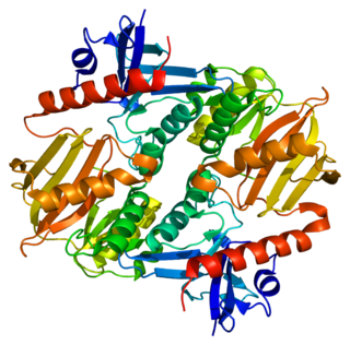
Synaptojanin is a protein involved in vesicle uncoating in neurons. This is an important regulatory lipid phosphatase. It dephosphorylates the D-5 position phosphate from phosphatidylinositol (3,4,5)-trisphosphate (PIP3) and Phosphatidylinositol (4,5)-bisphosphate(PIP2). It belongs to family of 5-phosphatases, which are structurally unrelated to D-3 inositol phosphatases like PTEN. Other members of the family of 5'phosphoinositide phosphatases include OCRL, SHIP1, SHIP2, INPP5J, INPP5E, INPP5B, INPP5A and SKIP.

Neurexins (NRXN) are a family of presynaptic cell adhesion proteins that have roles in connecting neurons at the synapse. They are located mostly on the presynaptic membrane and contain a single transmembrane domain. The extracellular domain interacts with proteins in the synaptic cleft, most notably neuroligin, while the intracellular cytoplasmic portion interacts with proteins associated with exocytosis. Neurexin and neuroligin "shake hands," resulting in the connection between the two neurons and the production of a synapse. Neurexins mediate signaling across the synapse, and influence the properties of neural networks by synapse specificity. Neurexins were discovered as receptors for α-latrotoxin, a vertebrate-specific toxin in black widow spider venom that binds to presynaptic receptors and induces massive neurotransmitter release. In humans, alterations in genes encoding neurexins are implicated in autism and other cognitive diseases, such as Tourette syndrome and schizophrenia.

Complexin (also known as synaphin) refers to a one of a small set of eukaryotic cytoplasmic neuronal proteins which binds to the SNARE protein complex (SNAREpin) with a high affinity. These are called synaphin 1 and 2. In the presence of Ca2+, the transport vesicle protein synaptotagmin displaces complexin, allowing the SNARE protein complex to bind the transport vesicle to the presynaptic membrane.

Sodium- and chloride-dependent glycine transporter 2, also known as glycine transporter 2 (GlyT2), is a protein that in humans is encoded by the SLC6A5 gene.

Synapsin-3 is a protein that in humans is encoded by the SYN3 gene.

Neuroligin (NLGN), a type I membrane protein, is a cell adhesion protein on the postsynaptic membrane that mediates the formation and maintenance of synapses between neurons. Neuroligins act as ligands for β-Neurexins, which are cell adhesion proteins located presynaptically. Neuroligin and β-neurexin "shake hands", resulting in the connection between two neurons and the production of a synapse. Neuroligins also affect the properties of neural networks by specifying synaptic functions, and they mediate signalling by recruiting and stabilizing key synaptic components. Neuroligins interact with other postsynaptic proteins to localize neurotransmitter receptors and channels in the postsynaptic density as the cell matures. Additionally, neuroligins are expressed in human peripheral tissues and have been found to play a role in angiogenesis. In humans, alterations in genes encoding neuroligins are implicated in autism and other cognitive disorders.
Axon terminals are distal terminations of the telodendria (branches) of an axon. An axon, also called a nerve fiber, is a long, slender projection of a nerve cell, or neuron, that conducts electrical impulses called action potentials away from the neuron's cell body, or soma, in order to transmit those impulses to other neurons, muscle cells or glands.
The ribbon synapse is a type of neuronal synapse characterized by the presence of an electron-dense structure, the synaptic ribbon, that holds vesicles close to the active zone. It is characterized by a tight vesicle-calcium channel coupling that promotes rapid neurotransmitter release and sustained signal transmission. Ribbon synapses undergo a cycle of exocytosis and endocytosis in response to graded changes of membrane potential. It has been proposed that most ribbon synapses undergo a special type of exocytosis based on coordinated multivesicular release. This interpretation has recently been questioned at the inner hair cell ribbon synapse, where it has been instead proposed that exocytosis is described by uniquantal release shaped by a flickering vesicle fusion pore.

Synapsin I, is the collective name for Synapsin Ia and Synapsin Ib, two nearly identical phosphoproteins that in humans are encoded by the SYN1 gene. In its phosphorylated form, Synapsin I may also be referred to as phosphosynaspin I. Synapsin I is the first of the proteins in the synapsin family of phosphoproteins in the synaptic vesicles present in the central and peripheral nervous systems. Synapsin Ia and Ib are close in length and almost the same in make up, however, Synapsin Ib stops short of the last segment of the C-terminal in the amino acid sequence found in Synapsin Ia.
Synapsin II is the collective name for synapsin IIa and synapsin IIb, two nearly identical phosphoproteins in the synapsin family that in humans are encoded by the SYN2 gene. Synapsins associate as endogenous substrates to the surface of synaptic vesicles and act as key modulators in neurotransmitter release across the presynaptic membrane of axonal neurons in the nervous system.

Thomas Christian Südhof, ForMemRS, is a German-American biochemist known for his study of synaptic transmission. Currently, he is a professor in the School of Medicine in the Department of Molecular and Cellular Physiology, and by courtesy in Neurology, and in Psychiatry and Behavioral Sciences at Stanford University.

The active zone or synaptic active zone is a term first used by Couteaux and Pecot-Dechavassinein in 1970 to define the site of neurotransmitter release. Two neurons make near contact through structures called synapses allowing them to communicate with each other. As shown in the adjacent diagram, a synapse consists of the presynaptic bouton of one neuron which stores vesicles containing neurotransmitter, and a second, postsynaptic neuron which bears receptors for the neurotransmitter, together with a gap between the two called the synaptic cleft. When an action potential reaches the presynaptic bouton, the contents of the vesicles are released into the synaptic cleft and the released neurotransmitter travels across the cleft to the postsynaptic neuron and activates the receptors on the postsynaptic membrane.
Neurotransmitters are released into a synapse in packaged vesicles called quanta. One quantum generates what is known as a miniature end plate potential (MEPP) which is the smallest amount of stimulation that one neuron can send to another neuron. Quantal release is the mechanism by which most traditional endogenous neurotransmitters are transmitted throughout the body. The aggregate sum of many MEPPs is known as an end plate potential (EPP). A normal end plate potential usually causes the postsynaptic neuron to reach its threshold of excitation and elicit an action potential. Electrical synapses do not use quantal neurotransmitter release and instead use gap junctions between neurons to send current flows between neurons. The goal of any synapse is to produce either an excitatory postsynaptic potential (EPSP) or an inhibitory postsynaptic potential (IPSP), which generate or repress the expression, respectively, of an action potential in the postsynaptic neuron. It is estimated that an action potential will trigger the release of approximately 20% of an axon terminal's neurotransmitter load.
