Synapsin 2

Last updated
SYN2
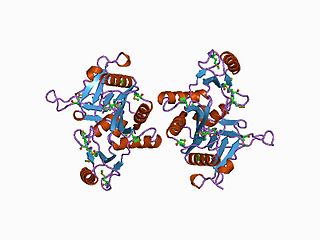
Protein SYN2 PDB 1i7l.png
Identifiers
Aliases SYN2 , SYNII, Synapsin 2, synapsin II
External IDs OMIM: 600755 MGI: 103020 HomoloGene: 49348 GeneCards: SYN2
Gene location (Human)
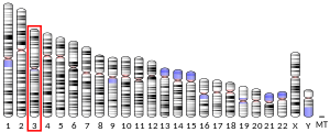
Ideogram human chromosome 3.svg
Chr. Chromosome 3 (human) [1]
Human chromosome 3 ideogram.svg
HSR 1996 II 3.5e.svg
Red rectangle 2x18.png
Band 3p25.2Start12,004,388 bp [1]
End12,192,032 bp [1]
RNA expression pattern
PBB GE SYN2 210247 at fs.png
More reference expression data
Orthologs
SpeciesHumanMouse
Entrez
Ensembl
UniProt
RefSeq (mRNA)

NM_003178
NM_133625

NM_001111015
NM_013681
NM_001326560

RefSeq (protein)

NP_003169
NP_598328

NP_001104485
NP_001313489
NP_038709

Location (UCSC) Chr 3: 12 – 12.19 Mb Chr 6: 115.13 – 115.28 Mb
PubMed search [3] [4]
Wikidata
View/Edit Human View/Edit Mouse

Synapsin II is the collective name for synapsin IIa and synapsin IIb, two nearly identical phosphoproteins in the synapsin family that in humans are encoded by the SYN2 gene. [5] [6] Synapsins associate as endogenous substrates to the surface of synaptic vesicles and act as key modulators in neurotransmitter release across the presynaptic membrane of axonal neurons in the nervous system.

Contents

Gene

Alternative splicing of the SYN2 gene results in two transcripts. The TIMP4 gene is located within an intron of this gene and is transcribed in the opposite direction. [6]

Protein

Synapsin II is a member of the synapsin family. Synapsins encode neuronal phosphoproteins which associate with the cytoplasmic surface of synaptic vesicles. Family members are characterized by common protein domains, and they are implicated in synaptogenesis and the modulation of neurotransmitter release, suggesting a potential role in several neuropsychiatric diseases. This member of the synapsin family encodes a neuron-specific phosphoprotein that selectively binds to small synaptic vesicles in the presynaptic nerve terminal. [6]

Synapsin II the collective name for two proteins, synapsin IIa and synapsin IIb, with synapsin IIa being the larger of the two isoforms. Their apparent molecular weights are 74,000 and 55,000 Da, per SDS gel electrophoresis. [7] Synapsin II along with synapsin I comprise approximately 9% of the proteins in highly purified samples of synaptic vesicles.

Structure

Synapsin II shares common domains within its amino acid sequence with other phosphoproteins in the synapsin family. [8] Sharing the same N-terminal, synapsin II diverges from synapsin I in its C-terminal domains. It is much shorter than synapsin I and is missing most of the elongated domains seen in synapsin I. Roughly 70% of the amino acid residues are common between the two synapsins, [7] which share common phosphorylation sites in the overlapping regions based on the homologous domains. Domain A of this neural protein contains phosphorylation sites for cAMP-dependent protein kinase and calcium/calmodulin-dependent protein kinase I, and domain B has two mitogen-activated protein kinase phosphorylation sites. At its B domain, between amino acids 43 and 121, synapsin II binds to a protein component in the cytosolic surface membrane of synaptic vesicles, organelles in neurons which carry neurotransmitters. [7]

Function

Synapsin II regulates synaptic function of neurons in the central and peripheral nervous system. [9] Synapsin IIa is the only synapsin isoform of the six synapsin isoforms (synapsin I-III each with isoforms A and B), which has been shown to significantly reverse synaptic depression and have a restorative effect on the density of synaptic vesicles within synapsinless neurons. Because of its restorative effect, synapsin IIa is believed to play a fundamental role in synaptic vesicle mobilization and reserve pool regulation in presynaptic nerve terminals. [10]

Lack of synapsins altogether in neurons, leads to behavioral alterations as well as epileptic-type seizures. The lack affects nervous signal transduction across excitatory and inhibitory synapses of neurons differently and is believed to be synapse-specific. Initial signal transduction appears to be unaffected by the lack of synapsins, but repeated stimulation of cultured synapsinless hippocampal neurons subsequently showed depressed responses at the excitatory synapse. At the inhibitory synapse, base signal transduction is reduced in neurons lacking pre-existing synapsins, but the reduced level of transduction is less affected by progressive stimulation. [11]

However, the restoration of synapsin IIa to neurons without pre-existing synapsins, can partially recover presumably lost signal transduction and slow the depression of synaptic response with progressive stimulation. Its isoform synapsin IIb may have a similar but weaker effect. Through fluorescence and staining, it has been demonstrated that synapsin IIa increases the number and density of glutamatergic synaptic vesicles in the nerve terminal of neural axons. The recovery of nervous signal transduction is attributed to the increase in density of synaptic vesicles, which carry neurotransmitters to the synaptic cleft, and the amount of synaptic vesicles in the reserve pool in the presence of synapsin IIa. [10] In turn, this is thought to increase the number of vesicles available for mobilization from the reserve pool to the ready-release pool. The reserve pool is the pool of synaptic vesicles which reside in the nerve terminal away from the presynaptic membrane of the axon, but are not in the ready to release or ready-release pool. Those vesicles in the ready-release pool reside very close to the presynaptic membrane and are primed to release neurotransmitters for nervous signal transduction.

Interactions

The synapsin II protein has been shown to interact with SYN1. [12]

Clinical significance

Mutations in the SYN2 gene may be associated with abnormal presynaptic function and schizophrenia. [6]

Related Research Articles

Chemical synapse

Chemical synapses are biological junctions through which neurons' signals can be sent to each other and to non-neuronal cells such as those in muscles or glands. Chemical synapses allow neurons to form circuits within the central nervous system. They are crucial to the biological computations that underlie perception and thought. They allow the nervous system to connect to and control other systems of the body.

Synaptic vesicle Neurotransmitters that are released at the synapse

In a neuron, synaptic vesicles store various neurotransmitters that are released at the synapse. The release is regulated by a voltage-dependent calcium channel. Vesicles are essential for propagating nerve impulses between neurons and are constantly recreated by the cell. The area in the axon that holds groups of vesicles is an axon terminal or "terminal bouton". Up to 130 vesicles can be released per bouton over a ten-minute period of stimulation at 0.2 Hz. In the visual cortex of the human brain, synaptic vesicles have an average diameter of 39.5 nanometers (nm) with a standard deviation of 5.1 nm.

Synaptophysin

Synaptophysin, also known as the major synaptic vesicle protein p38, is a protein that in humans is encoded by the SYP gene.
Synapsin

They are expressed in highest concentration in the nervous system.

Neurotransmission

Neurotransmission is the process by which signaling molecules called neurotransmitters are released by the axon terminal of a neuron, and bind to and react with the receptors on the dendrites of another neuron a short distance away. A similar process occurs in retrograde neurotransmission, where the dendrites of the postsynaptic neuron release retrograde neurotransmitters that signal through receptors that are located on the axon terminal of the presynaptic neuron, mainly at GABAergic and glutamatergic synapses.

Synaptotagmin

Synaptotagmins (SYTs) constitute a family of membrane-trafficking proteins that are characterized by an N-terminal transmembrane region (TMR), a variable linker, and two C-terminal C2 domains - C2A and C2B. There are 17 isoforms in the mammalian synaptotagmin family. There are several C2-domain containing protein families that are related to synaptotagmins, including transmembrane (Ferlins, Extended-Synaptotagmin (E-Syt) membrane proteins, and MCTPs) and soluble (RIMS1 and RIMS2, UNC13D, synaptotagmin-related proteins and B/K) proteins. The family includes synaptotagmin 1, a Ca2+ sensor in the membrane of the pre-synaptic axon terminal, coded by gene SYT1.

Synapse Junction between two neurons or a neuron and another cell

In the nervous system, a synapse is a structure that permits a neuron to pass an electrical or chemical signal to another neuron or to the target effector cell.

Complexin

Complexin (also known as synaphin) refers to a one of a small set of eukaryotic cytoplasmic neuronal proteins which binds to the SNARE protein complex (SNAREpin) with a high affinity. These are called synaphin 1 and 2. In the presence of Ca2+, the transport vesicle protein synaptotagmin displaces complexin, allowing the SNARE protein complex to bind the transport vesicle to the presynaptic membrane.

STX1A

Syntaxin-1A is a protein that in humans is encoded by the STX1A gene.

NRXN1

Neurexin-1-alpha is a protein that in humans is encoded by the NRXN1 gene.

APBA2

Amyloid beta A4 precursor protein-binding family A member 2 is a protein that in humans is encoded by the APBA2 gene.

RIMS1

Regulating synaptic membrane exocytosis protein 1 is a protein that in humans is encoded by the RIMS1 gene.

SYN3

Synapsin-3 is a protein that in humans is encoded by the SYN3 gene.

Neuroligin

Neuroligin (NLGN), a type I membrane protein, is a cell adhesion protein on the postsynaptic membrane that mediates the formation and maintenance of synapses between neurons. Neuroligins act as ligands for β-Neurexins, which are cell adhesion proteins located presynaptically. Neuroligin and β-neurexin "shake hands", resulting in the connection between two neurons and the production of a synapse. Neuroligins also affect the properties of neural networks by specifying synaptic functions, and they mediate signalling by recruiting and stabilizing key synaptic components. Neuroligins interact with other postsynaptic proteins to localize neurotransmitter receptors and channels in the postsynaptic density as the cell matures. Additionally, neuroligins are expressed in human peripheral tissues and have been found to play a role in angiogenesis. In humans, alterations in genes encoding neuroligins are implicated in autism and other cognitive disorders.

Axon terminal

Axon terminals are distal terminations of the telodendria (branches) of an axon. An axon, also called a nerve fiber, is a long, slender projection of a nerve cell, or neuron, that conducts electrical impulses called action potentials away from the neuron's cell body, or soma, in order to transmit those impulses to other neurons, muscle cells or glands.

The ribbon synapse is a type of neuronal synapse characterized by the presence of an electron-dense structure, the synaptic ribbon, that holds vesicles close to the active zone. It is characterized by a tight vesicle-calcium channel coupling that promotes rapid neurotransmitter release and sustained signal transmission. Ribbon synapses undergo a cycle of exocytosis and endocytosis in response to graded changes of membrane potential. It has been proposed that most ribbon synapses undergo a special type of exocytosis based on coordinated multivesicular release. This interpretation has recently been questioned at the inner hair cell ribbon synapse, where it has been instead proposed that exocytosis is described by uniquantal release shaped by a flickering vesicle fusion pore.

Synapsin I

Synapsin I, is the collective name for Synapsin Ia and Synapsin Ib, two nearly identical phosphoproteins that in humans are encoded by the SYN1 gene. In its phosphorylated form, Synapsin I may also be referred to as phosphosynaspin I. Synapsin I is the first of the proteins in the synapsin family of phosphoproteins in the synaptic vesicles present in the central and peripheral nervous systems. Synapsin Ia and Ib are close in length and almost the same in make up, however, Synapsin Ib stops short of the last segment of the C-terminal in the amino acid sequence found in Synapsin Ia.

Thomas C. Südhof

Thomas Christian Südhof, ForMemRS, is a German-American biochemist known for his study of synaptic transmission. Currently, he is a professor in the School of Medicine in the Department of Molecular and Cellular Physiology, and by courtesy in Neurology, and in Psychiatry and Behavioral Sciences at Stanford University.

Active zone

The active zone or synaptic active zone is a term first used by Couteaux and Pecot-Dechavassinein in 1970 to define the site of neurotransmitter release. Two neurons make near contact through structures called synapses allowing them to communicate with each other. As shown in the adjacent diagram, a synapse consists of the presynaptic bouton of one neuron which stores vesicles containing neurotransmitter, and a second, postsynaptic neuron which bears receptors for the neurotransmitter, together with a gap between the two called the synaptic cleft. When an action potential reaches the presynaptic bouton, the contents of the vesicles are released into the synaptic cleft and the released neurotransmitter travels across the cleft to the postsynaptic neuron and activates the receptors on the postsynaptic membrane.

Neurotransmitters are released into a synapse in packaged vesicles called quanta. One quantum generates what is known as a miniature end plate potential (MEPP) which is the smallest amount of stimulation that one neuron can send to another neuron. Quantal release is the mechanism by which most traditional endogenous neurotransmitters are transmitted throughout the body. The aggregate sum of many MEPPs is known as an end plate potential (EPP). A normal end plate potential usually causes the postsynaptic neuron to reach its threshold of excitation and elicit an action potential. Electrical synapses do not use quantal neurotransmitter release and instead use gap junctions between neurons to send current flows between neurons. The goal of any synapse is to produce either an excitatory postsynaptic potential (EPSP) or an inhibitory postsynaptic potential (IPSP), which generate or repress the expression, respectively, of an action potential in the postsynaptic neuron. It is estimated that an action potential will trigger the release of approximately 20% of an axon terminal's neurotransmitter load.

References

  1. 1 2 3 GRCh38: Ensembl release 89: ENSG00000157152 - Ensembl, May 2017
  2. 1 2 3 GRCm38: Ensembl release 89: ENSMUSG00000009394 - Ensembl, May 2017
  3. "Human PubMed Reference:". National Center for Biotechnology Information, U.S. National Library of Medicine.
  4. "Mouse PubMed Reference:". National Center for Biotechnology Information, U.S. National Library of Medicine.
  5. Li L, Chin LS, Greengard P, Copeland NG, Gilbert DJ, Jenkins NA (July 1995). "Localization of the synapsin II (SYN2) gene to human chromosome 3 and mouse chromosome 6". Genomics. 28 (2): 365–6. doi:10.1006/geno.1995.1162. PMID   8530057.
  6. 1 2 3 4 "Entrez Gene: SYN2 synapsin II".
  7. 1 2 3 Thiel G, Südhof TC, Greengard P (September 1990). "Synapsin II. Mapping of a domain in the NH2-terminal region which binds to small synaptic vesicles". J. Biol. Chem. 265 (27): 16527–33. PMID   2118908.
  8. Südhof TC, Czernik AJ, Kao HT, Takei K, Johnston PA, Horiuchi A, Kanazir SD, Wagner MA, Perin MS, De Camilli P (September 1989). "Synapsins: mosaics of shared and individual domains in a family of synaptic vesicle phosphoproteins". Science. 245 (4925): 1474–80. Bibcode:1989Sci...245.1474S. doi:10.1126/science.2506642. PMID   2506642.
  9. Greengard P, Valtorta F, Czernik AJ, Benfenati F (February 1993). "Synaptic vesicle phosphoproteins and regulation of synaptic function". Science. 259 (5096): 780–5. Bibcode:1993Sci...259..780G. CiteSeerX   10.1.1.336.2100 . doi:10.1126/science.8430330. PMID   8430330.
  10. 1 2 Gitler D, Cheng Q, Greengard P, Augustine GJ (October 2008). "Synapsin IIa controls the reserve pool of glutamatergic synaptic vesicles". J. Neurosci. 28 (43): 10835–43. doi:10.1523/JNEUROSCI.0924-08.2008. PMC   2605971 . PMID   18945891.
  11. Gitler D, Takagishi Y, Feng J, Ren Y, Rodriguiz RM, Wetsel WC, Greengard P, Augustine GJ (December 2004). "Different presynaptic roles of synapsins at excitatory and inhibitory synapses". J. Neurosci. 24 (50): 11368–80. doi:10.1523/JNEUROSCI.3795-04.2004. PMC   6730366 . PMID   15601943.
  12. Hosaka, M; Südhof T C (Jun 1999). "Homo- and heterodimerization of synapsins". J. Biol. Chem. 274 (24): 16747–53. doi: 10.1074/jbc.274.24.16747 . ISSN   0021-9258. PMID   10358015.

Further reading