Cytolysin

Last updated

Cytolysin refers to the substance secreted by microorganisms, plants or animals that is specifically toxic to individual cells, [1] [2] in many cases causing their dissolution through lysis. Cytolysins that have a specific action for certain cells are named accordingly. For instance, the cytolysins responsible for the destruction of red blood cells, thereby liberating hemoglobins, are named hemolysins , and so on. [3] Cytolysins may be involved in immunity as well as in venoms.

Contents

Hemolysin is also used by certain bacteria, such as Listeria monocytogenes , to disrupt the phagosome membrane of macrophages and escape into the cytoplasm of the cell.

History and background

The term "Cytolysin" or "Cytolytic toxin" was first introduced by Alan Bernheimer to describe membrane damaging toxins (MDTs) that have cytolytic effects to cells. [4] The first kind of cytolytic toxin discovered have hemolytic effects on erythrocytes of certain sensitive species, such as Human. For this reason "Hemolysin" was first used to describe any MDTs. In the 1960s certain MDTs were proved to be destructive on cells other than erythrocytes, such as leukocytes. The term "Cytolysin" is then introduced by Bernheimer to replace "Hemolysin". Cytolysins can destruct membranes without creating lysis to cells. [5] Therefore, "membrane damaging toxins" (MDTs) describes the essential actions of cytolysins. Cytolysins comprise more than 1/3 of all bacterial protein toxins. Bacterial protein toxins can be highly poisonous to human. For example, Botulinum is 3x10⁵ more toxic than snake venom to human and its toxic dose is only 0.8x10⁻⁸ mg. [6] A wide variety of gram-positive and gram-negative bacteria use cytolysin as their primary weapon for creating diseases, such as Enterococcus faecalis , [7] Staphylococcus and Clostridium perfringens .

A diverse range of studies has been done on cytolysins. Since the 1970s, more than 40 new cytolysins have been discovered and grouped into different families. [8] At genetic level, the genetic structures of about 70 Cytolysin proteins has been studied and published. [9] The detailed process of membrane damage has also been surveyed. Rossjohn et al. present the crystal structure of perfringolysin O, a thiol-activated cytolysin, which creates membrane holes on eukaryotic cells. A detailed model of membrane channel formation that reveals membrane insertion mechanism is constructed. [10] Shatursky et al. studied the membrane insertion mechanism of Perfringolysin O (PFO), a cholesterol-dependent pore-forming cytolysin produced by pathogenic Clostridium perfringens. Instead of using a single amphipathic β hairpin per polypeptide, PFO monomer contains two amphipathic β hairpins, each spans the whole membrane. Larry et al. focused on the membrane penetrating models of RTX toxins, a family of MDT secreted by many gram-negative bacteria. The insertion and transport process of the protein from RTX to target lipid membrane was revealed. [11]

Classification

The membrane-damaging cytolysins can be classified into three types based on their damaging mechanism:

Pore forming cytolysins

Pore structure representation 3.png
Porin-structure pore allows molecules of certain sizes to pass through.
Pore structure representation 2.png
Pores formed by membrane fusion could be made from only proteolipids.
Pore structure representation

Pore forming cytolysins (PFCs) comprise near 65% of all membrane-damaging cytolysins. [8] The first pore forming cytolysin is discovered by Manfred Mayer in 1972 of the C5-C9 insertion of erythrocytes. [12] PFCs can be produced by a wide variety of sources, such as bacteria, fungi and even plants. [13] The pathogenic process of PFCs normally involves forming channels or pores at the target cells' membranes. Note that the pores can have many structures. A porin-like structure allows molecules of certain sizes to pass through. Electric fields distribute unevenly across the pore and enable the selection molecules that can get through. [14] This type of structure is shown in staphylococcal α-hemolysin. [15] A pore can also be formed through membrane fusions. Controlled by Ca 2+, the membrane fusion of vesicles form water-filled pores from proteolipids. [16] Pore forming cytolysins such as perforin are used in cytotoxic killer T and NK cells to destroy infected cells.

pore forming process

Pore forming cytolysin 1.png
Cytolysins secreted by host cells (use bacteria as example)
Pore forming cytolysin 2.png
Cytolysins form cluster of oligomers on target-cell membranes.
Pore forming cytolysin 3.png
Cytolysins create channels (pores) on the target-cell membranes.
Pore forming process

A more complex pore formation process involves an oligomerization process of several PFC monomers. The pore forming process comprise three basic steps. The cytolysins are produced by certain microorganisms at first. Sometimes the producer organism needs to create a pore at its own membrane to release such cytolysins, like the case colicins produced by Escherichia coli . Cytolysins are released as protein monomers in a water-soluble state in this step. [17] Note that cytolysins are often toxic to its producing hosts as well. For example, colicins consume nucleic acids of cells by using several enzymes. [18] To prevent such toxicity, host cells produce immunity proteins for binding cytolysins before they do any damage inward. [8]

In the second step, cytolysins adhere to target cell membranes by matching the " receptors " on the membranes. Most receptors are proteins, but they can be other molecules as well, such as lipids or sugars. With the help of receptors, cytolysin monomers combine with each other and form clusters of oligomers. During this stage, cytolysins complete transition from water-soluble monomers state into oligomers state.

Finally, the formed cytolysin clusters penetrate target cells' membranes and form membrane pores. The size of these pores varies from 1–2 nm ( S. aureus α-toxin, E. coli α-hemolysin , Aeromonas aerolysin ) to 25–30 nm (streplysin O, pneumolysin ).

Depending on how the pores are formed, the pore forming cytolysins fall into two categories. Those forming pores with α-helices are named α-PFTs (Pore forming toxins). Those forming pores with β-barrel structures are named β-PFTs. Some of the common α-PFTs and β-PFTs are listed in the table below.

Common pore forming cytolysins
α-PFTsβ-PFTs
Colicin Ia, Pseudomonas aeruginosa extotoxin A, Actinia equina equinatoxin IIAerolysin, Clostrim septicum α-toxin, Staphylococcus aureus &alpha-hemolysin, Pseudomonas aeruginosa cytotoxin, anthrax protective antigen, cholesterol-dependent cytolysins.

Consequences of cytolysins

The lethal effects of pore-forming cytolysins are performed by causing influx and outflux disorder in a single cell. Pores that allow ions like Na+ to pass through created imbalance in the target cell which exceeds its ion-balancing capacity. Attacked cells therefore expand to lysis. [19] When target cell membranes are destructed, bacteria which produce the cytolysins can consume the intracellular elements of the cell, such as iron and cytokines. [8] Some enzymes that decompose target-cells' critical structures can enter the cells without obstructions.

Cholesterol-dependent cytolysin

One specific type of cytolysin is the cholesterol-dependent cytolysin (CDC). CDCs exist in many Gram-positive bacteria. The pore forming process of CDCs require the presence of cholesterols on target-cell membranes. The pore size created by CDC is large (25–30 nm) due to the oligomeric process of cytolysins. Note that cholesterol are not always necessary at during the adhering phase. For example, Intermedilysin requires only the presence of protein receptors when attaching to target cells and cholesterols are required at pore forming. [20] The formation of pores through CDCs involve an additional step than the steps analyzed above. The water-soluble monomers oligomerize to form an intermediate product named "pre-pore" complex and then a β-barrel is penetrated into the membrane. [20]

See also

Related Research Articles

<span class="mw-page-title-main">Exotoxin</span> Toxin from bacteria that destroys or disrupts cells

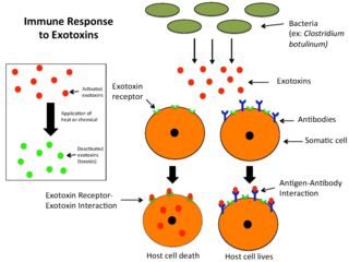
An exotoxin is a toxin secreted by bacteria. An exotoxin can cause damage to the host by destroying cells or disrupting normal cellular metabolism. They are highly potent and can cause major damage to the host. Exotoxins may be secreted, or, similar to endotoxins, may be released during lysis of the cell. Gram negative pathogens may secrete outer membrane vesicles containing lipopolysaccharide endotoxin and some virulence proteins in the bounding membrane along with some other toxins as intra-vesicular contents, thus adding a previously unforeseen dimension to the well-known eukaryote process of membrane vesicle trafficking, which is quite active at the host-pathogen interface.

<span class="mw-page-title-main">Porin (protein)</span> Group of transport proteins

Porins are beta barrel proteins that cross a cellular membrane and act as a pore, through which molecules can diffuse. Unlike other membrane transport proteins, porins are large enough to allow passive diffusion, i.e., they act as channels that are specific to different types of molecules. They are present in the outer membrane of gram-negative bacteria and some gram-positive mycobacteria, the outer membrane of mitochondria, and the outer chloroplast membrane.

Virulence factors are cellular structures, molecules and regulatory systems that enable microbial pathogens to achieve the following:

Adenylate cyclase toxin is a virulence factor produced by some members of the genus Bordetella. Together with the pertussis toxin it is the most important virulence factor of the causative agent of whooping cough, Bordetella pertussis. Bordetella bronchiseptica and Bordetella parapertussis, also able to cause pertussis-like symptoms, also produce adenylate cyclase toxin. It is a toxin secreted by the bacteria to influence the host immune system.

<span class="mw-page-title-main">Anthrax toxin</span> Tripartite protein complex secreted by virulent strains of Bacillus anthracis

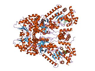
Anthrax toxin is a three-protein exotoxin secreted by virulent strains of the bacterium, Bacillus anthracis—the causative agent of anthrax. The toxin was first discovered by Harry Smith in 1954. Anthrax toxin is composed of a cell-binding protein, known as protective antigen (PA), and two enzyme components, called edema factor (EF) and lethal factor (LF). These three protein components act together to impart their physiological effects. Assembled complexes containing the toxin components are endocytosed. In the endosome, the enzymatic components of the toxin translocate into the cytoplasm of a target cell. Once in the cytosol, the enzymatic components of the toxin disrupts various immune cell functions, namely cellular signaling and cell migration. The toxin may even induce cell lysis, as is observed for macrophage cells. Anthrax toxin allows the bacteria to evade the immune system, proliferate, and ultimately kill the host animal. Research on anthrax toxin also provides insight into the generation of macromolecular assemblies, and on protein translocation, pore formation, endocytosis, and other biochemical processes.

Tetanolysin is a toxin produced by Clostridium tetani bacteria. Its function is unknown, but it is believed to contribute to the pathogenesis of tetanus. The other C. tetani toxin, tetanospasmin, is more definitively linked to tetanus. It is sensitive to oxygen.

<span class="mw-page-title-main">Colicin</span>

A colicin is a type of bacteriocin produced by and toxic to some strains of Escherichia coli. Colicins are released into the environment to reduce competition from other bacterial strains. Colicins bind to outer membrane receptors, using them to translocate to the cytoplasm or cytoplasmic membrane, where they exert their cytotoxic effect, including depolarisation of the cytoplasmic membrane, DNase activity, RNase activity, or inhibition of murein synthesis.

<span class="mw-page-title-main">Pore-forming toxin</span>

Pore-forming proteins are usually produced by bacteria, and include a number of protein exotoxins but may also be produced by other organisms such as earthworms, who produce lysenin. They are frequently cytotoxic, as they create unregulated pores in the membrane of targeted cells.

<span class="mw-page-title-main">Hemolysin</span> Molecule destroying the membrane of red blood cells

Hemolysins or haemolysins are lipids and proteins that cause lysis of red blood cells by disrupting the cell membrane. Although the lytic activity of some microbe-derived hemolysins on red blood cells may be of great importance for nutrient acquisition, many hemolysins produced by pathogens do not cause significant destruction of red blood cells during infection. However, hemolysins are often capable of lysing red blood cells in vitro.

Listeriolysin O (LLO) is a hemolysin produced by the bacterium Listeria monocytogenes, the pathogen responsible for causing listeriosis. The toxin may be considered a virulence factor, since it is crucial for the virulence of L. monocytogenes.

The Membrane Attack Complex/Perforin (MACPF) superfamily, sometimes referred to as the MACPF/CDC superfamily, is named after a domain that is common to the membrane attack complex (MAC) proteins of the complement system and perforin (PF). Members of this protein family are pore-forming toxins (PFTs). In eukaryotes, MACPF proteins play a role in immunity and development.

<i>Staphylococcus aureus</i> alpha toxin

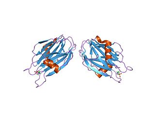
Alpha-toxin, also known as alpha-hemolysin (Hla), is the major cytotoxic agent released by bacterium Staphylococcus aureus and the first identified member of the pore forming beta-barrel toxin family. This toxin consists mostly of beta-sheets (68%) with only about 10% alpha-helices. The hly gene on the S. aureus chromosome encodes the 293 residue protein monomer, which forms heptameric units on the cellular membrane to form a complete beta-barrel pore. This structure allows the toxin to perform its major function, development of pores in the cellular membrane, eventually causing cell death.

Streptolysins are two hemolytic exotoxins from Streptococcus. Types include streptolysin O, which is oxygen-labile, and streptolysin S, which is oxygen-stable.

<span class="mw-page-title-main">Clostridium enterotoxin</span>

Clostridium enterotoxins are toxins produced by Clostridium species.

Microbial toxins are toxins produced by micro-organisms, including bacteria, fungi, protozoa, dinoflagellates, and viruses. Many microbial toxins promote infection and disease by directly damaging host tissues and by disabling the immune system. Endotoxins most commonly refer to the lipopolysaccharide (LPS) or lipooligosaccharide (LOS) that are in the outer plasma membrane of Gram-negative bacteria. The botulinum toxin, which is primarily produced by Clostridium botulinum and less frequently by other Clostridium species, is the most toxic substance known in the world. However, microbial toxins also have important uses in medical science and research. Currently, new methods of detecting bacterial toxins are being developed to better isolate and understand these toxin. Potential applications of toxin research include combating microbial virulence, the development of novel anticancer drugs and other medicines, and the use of toxins as tools in neurobiology and cellular biology.

The RTX toxin superfamily is a group of cytolysins and cytotoxins produced by bacteria. There are over 1000 known members with a variety of functions. The RTX family is defined by two common features: characteristic repeats in the toxin protein sequences, and extracellular secretion by the type I secretion systems (T1SS). The name RTX refers to the glycine and aspartate-rich repeats located at the C-terminus of the toxin proteins, which facilitate export by a dedicated T1SS encoded within the rtx operon.

The thiol-activated Cholesterol-dependent Cytolysin(CDC) family is a member of the MACPF superfamily. Cholesterol dependent cytolysins are a family of β-barrel pore-forming exotoxins that are secreted by gram-positive bacteria. CDCs are secreted as water-soluble monomers of 50-70 kDa, that when bound to the target cell, form a circular homo-oligomeric complex containing as many as 40 monomers. Through multiple conformational changes, the β-barrel transmembrane structure is formed and inserted into the target cell membrane. The presence of cholesterol in the target membrane is required for pore formation, though the presence of cholesterol is not required by all CDCs for binding. For example, intermedilysin secreted by Streptococcus intermedius will bind only to target membranes containing a specific protein receptor, independent of the presence of cholesterol, but cholesterol is required by intermedilysin for pore formation. While the lipid environment of cholesterol in the membrane can affect toxin binding, the exact molecular mechanism that cholesterol regulates the cytolytic activity of the CDC is not fully understood.

<span class="mw-page-title-main">Sea anemone cytotoxic protein</span>

In molecular biology, the sea anemone cytotoxic proteins are lethal pore-forming proteins, known collectively as actinoporins, a sub-class of cytolysins. There are several different groups of cytolysins based on their structure and function. This entry represents the most numerous group, the 20kDa highly basic peptides. These cytolysins form cation-selective pores in sphingomyelin-containing membranes. Examples include equinatoxins, sticholysins, magnificalysins, and tenebrosins, which exhibit pore-forming, haemolytic, cytotoxic, and heart stimulatory activities.

Nanosponges are a type of nanoparticle, often a synthesized carbon-containing polymer. They are porous in structure, pores being about 1–2 nanometers in size, and can therefore be targeted to absorb small amounts of matter or toxin. Nanosponges are often used in medicine as targeted drug delivery systems, detoxification methods, or as a way of damage control after an injury. They can also be used in environmental applications to clean up ecosystems by performing tasks like purifying water or metal deposits. Their small size allows them to move quickly through substances, like water or blood, efficiently finding and attacking unwanted matter. Nanosponges are often synthetically manufactured but oftentimes include natural materials to improve their efficiency when injected into the body. Nanosponges are superior to microsponges in application as the smaller size allows less disruption into the system in which it is implemented therefore imposing less risk of failed or detrimental effects. The prefix "nano" implies that items of this size are measured on a scale of meters.

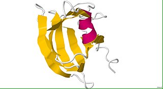
Lysenin is a pore-forming toxin (PFT) present in the coelomic fluid of the earthworm Eisenia fetida. Pore-forming toxins are a group of proteins that act as virulence factors of several pathogenic bacteria. Lysenin proteins are chiefly involved in the defense against eukaryotic and prokaryotic pathogens. Following the general mechanism of action of PFTs lysenin is segregated as a soluble monomer that binds specifically to a membrane receptor, sphingomyelin in the case of lysenin. After attaching to the membrane, the oligomerization begins, resulting in a nonamer on top of membrane, known as a prepore. After a conformational change, which could be triggered by a decrease of pH, the oligomer is inserted into the membrane in the so-called pore state.

References

  1. Computer Retrieval of Information on Scientific Projects (CRISP) - Thesaurus - Cytolysin Archived 2006-09-30 at the Wayback Machine
  2. "Cytolysin" entry from the American Heritage Medical Dictionary, on TheFreeDictionary.com (Retrieved on January 22, 2009)
  3. "Hemolysin" entry on TheFreeDictionary.com (Retrieved on January 22, 2009)
  4. Bernheimer A W (1970) Cytolytic toxins of bacteria, vol I. In: Ajl S, Kadis S, Montie TC (eds) Microbial toxins. Academic, New York, pp 183-212
  5. Thelestam, M. and Mollby, R. (1975) Infect. Immun, 11, 640-648.
  6. 1 2 Bacterial Protein Toxins
  7. Panthee, S; Paudel, A; Hamamoto, H; Ogasawara, AA; Iwasa, T; Blom, J; Sekimizu, K (24 March 2021). "Complete genome sequence and comparative genomic analysis of Enterococcus faecalis EF-2001, a probiotic bacterium". Genomics. 113 (3): 1534–1542. doi: 10.1016/j.ygeno.2021.03.021 . PMID   33771633.
  8. 1 2 3 4 Alouf, J. E. "Pore-forming bacterial protein toxins: an overview." Pore-forming toxins. Springer Berlin Heidelberg, 2001. 1-14.
  9. Kaper J, Hacker J (2000) Pathogenicity islands and other mobile virulence elements. ASM, Washington D.C.
  10. Rossjohn, Jamie, et al. "Structure of a cholesterol-binding, thiol-activated cytolysin and a model of its membrane form." Cell 89.5 (1997): 685-692.
  11. Lally, Edward T., et al. "The interaction between RTX toxins and target cells." Trends in Microbiology 7.9 (1999): 356-361.
  12. Mayer, Manfred M. "Mechanism of cytolysis by complement." Proceedings of the National Academy of Sciences 69.10 (1972): 2954-2958.
  13. Gilbert, R. J. C. "Pore-forming toxins." Cellular and Molecular Life Sciences 59.5 (2002): 832-844.
  14. Branden and Tooze, Introduction to Protein Structure, second edition
  15. Song L. Z., Hobaugh M. R., Shustak C., Cheley S., Bayley H. and Gouaux J. E. (1996) Structure of staphylococcal alpha-hemolysin, a heptamertic transmembrane pore. Science 274:1859–1866
  16. Peters, Christopher, et al. "Trans-complex formation by proteolipid channels in the terminal phase of membrane fusion." Nature 409.6820 (2001): 581-588.
  17. Lazdunski CJ, Baty 0, Geli V, Cavard 0, Morlon J, Lloubes R, Howard SP, Knibiehler M, Chartier M,Varenne S, et al. (1988) The membrane channel-forming colicin A:synthesis, secretion, structure, action and immunity. Biochim Biophys Acta 947:445-464
  18. James R, Kleanthous C, Moore GR (1996) The biology of E-colicins - paradigms and paradoxes. Microbiology UK 142:1569-1580
  19. Skals, Marianne, and Helle A. Praetorius. "Mechanisms of cytolysin-induced cell damage–a role for auto-and paracrine signalling." Acta Physiologica 209.2 (2013): 95-113.
  20. 1 2 Heuck, Alejandro P., Paul C. Moe, and Benjamin B. Johnson. "The cholesterol-dependent cytolysin family of gram-positive bacterial toxins." Cholesterol Binding and Cholesterol Transport Proteins:. Springer Netherlands, 2010. 551-577.