Hemolysin

Last updated
Leukocidin/Hemolysin toxin
7ahl.png
Staphylococcus aureus alpha toxin from S. aureus ( PDB: 7AHL ).
Identifiers
SymbolLeukocidin
Pfam PF07968
Pfam clan CL0636
InterPro IPR036435
Available protein structures:
Pfam   structures / ECOD  
PDB RCSB PDB; PDBe; PDBj
PDBsum structure summary

Hemolysins or haemolysins are lipids and proteins that cause lysis of red blood cells by disrupting the cell membrane. Although the lytic activity of some microbe-derived hemolysins on red blood cells may be of great importance for nutrient acquisition, many hemolysins produced by pathogens do not cause significant destruction of red blood cells during infection. However, hemolysins are often capable of lysing red blood cells in vitro.

Contents

While most hemolysins are protein compounds, some are lipid biosurfactants. [1]

Properties

Many bacteria produce hemolysins that can be detected in the laboratory. It is now believed that many clinically relevant fungi also produce hemolysins. [2] Hemolysins can be identified by their ability to lyse red blood cells in vitro.

Hemolysis by Streptococcus seen on a plate The process of hemolysis.png
Hemolysis by Streptococcus seen on a plate

Not only are the erythrocytes affected by hemolysins, but there are also some effects among other blood cells, such as leucocytes (white blood cells). Escherichia coli hemolysin is potentially cytotoxic to monocytes, lymphocytes and macrophages, leading them to autolysis and death.

Visualization of hemolysis (UK: haemolysis) of red blood cells in agar plates facilitates the categorization of Streptococcus.

Mechanism

One way hemolysin lyses erythrocytes is by forming pores in phospholipid bilayers. [3] [4] Other hemolysins lyse erythrocytes by hydrolyzing the phospholipids in the bilayer.

Pore formation

Many hemolysins are pore-forming toxins (PFT), which are able to cause the lysis of erythrocytes, leukocytes, and platelets by producing pores on the cytoplasmic membrane.

Hemolysin is normally secreted by the bacteria in a water-soluble way. These monomers diffuse to the target cells and are attached to them by specific receivers. After this is done, they oligomerize, creating ring-shaped heptamer complexes. [5]

Hemolysins can be secreted by many different kinds of bacteria such as Staphylococcus aureus, Escherichia coli or Vibrio parahemolyticus among other pathogens. We can take a look at the bacterium Staphylococcus aureus as a specific example of pore-forming hemolysin production. Staphylococcus aureus is a pathogen that causes many infectious diseases such as pneumonia and sepsis. It produces a ring-shaped complex called a staphylococcal alpha-hemolysin pore. In nature, Staphylococcus aureus secretes alpha-hemolysin monomers that bind to the outer membrane of susceptible cells. Upon binding, the monomers oligomerize to form a water-filled transmembrane channel that facilitates uncontrolled permeation of water, ions, and small organic molecules. Rapid discharge of vital molecules such as ATP, dissipation of the membrane potential and ion gradients, and irreversible osmotic swelling leading to the cell wall rupture (lysis) can cause death of the host cell.

This pore consists of seven alpha-hemolysin subunits, which represent the major cytotoxic agent that is freed by this kind of bacterium. These subunits attach to the target cells in the manner described before, and extend the lipid bilayer, forming the pore structures. These pores in the cellular membrane will eventually end up causing cell death, since it allows the exchange of monovalent ions that would cause the DNA fragmentation.

Enzymatic

Some hemolysins damage the erythrocyte membrane by cleaving the phospholipids in the membrane.

Staphylococcus aureus hemolysins

α-hemolysin

Alpha(a)-hemolysin of Staphylococcus aureus: macromolecular structure of the transmembrane pore. 7ahl opm.png
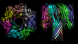
Alpha(α)-hemolysin of Staphylococcus aureus: macromolecular structure of the transmembrane pore.

Secreted by Staphylococcus aureus, this toxin binds with the outer membrane, with subsequent oligomerization of the toxin monomers to form water-filled channels. [6] These are responsible for osmotic phenomena, cell depolarization and loss of vital molecules (v.gr. ATP), leading to cell death. [7]

β-hemolysin

β-hemolysin (hlb; Q2FWP1 ) is a Phospholipase C toxin secreted by S. aureus. Upon investigating sheep erythrocytes, its toxic mechanism was discovered to be the hydrolysis of a specific membrane lipid, sphingomyelin, which accounts for 50% of the cell’s membrane. This degradation was followed by a noticeable rise of phosphoryl-choline due to the release of organic phosphorus from sphingomyelin and ultimately caused cell lysis. [8]

γ-Hemolysin

γ-Hemolysins are pore-forming toxins in the same family as α-Hemolysin. They are unique in that they come in two components, and hence are referred to as bi-component toxins (InterPro :  IPR003963 ). Compared to beta-hemolysin, it has a higher affinity for phosphocholines with short saturated acyl chains, especially if they have a conical form, whereas cylindrical lipids (e.g., sphingomyelin) hinder its activity. The lytic process, most commonly seen in leucocytes, is caused by pore formation induced by an oligomerized octamer that organizes in a ring structure. Once the prepore is formed, a more stable one ensues, named β-barrel. In this final part, the octamer binds with phosphatidylcholine. [9]

Structure

The structure of several hemolysins has been solved by X-ray crystallography in the soluble and pore-forming conformations. For example, α-hemolysin of Staphylococcus aureus forms a homo-heptameric β-barrel in biological membranes. [10] The Vibrio cholerae cytolysin [11] also forms a heptameric pore, however Staphylococcus aureus γ-hemolysin [12] forms a pore that is octameric.

The heptamer of α-hemolysin from Staphylococcus aureus has a mushroom-like shape and measures up to 100 Å in diameter and 100 Å in height. A membrane-spanning, solvent-accessible channel runs along the sevenfold axis and ranges from 14 Å to 46 Å in diameter. On the exterior of the 14-strand antiparallel β barrel there is a hydrophobic belt approximately 30 Å in width that provides a surface complementary to the nonpolar portion of the lipid bilayer. The interfaces are composed of both salt-links and hydrogen bonds, as well as hydrophobic interactions, and these contacts provide a molecular stability for the heptamer in SDS solutions even up to 65 °C. [13]

Role during infection

Hemolysins are thought to be responsible for many events in host cells. For example, iron may be a limiting factor in the growth of various pathogenic bacteria. [14] Since free iron may generate damaging free radicals, free iron is typically maintained at low concentrations within the body. Red blood cells are rich in iron-containing heme. Lysis of these cells releases heme into the surroundings, allowing the bacteria to take up the free iron. But hemolysin is related to bacteria not only in this way but also in some others.

As mentioned before, hemolysin is a potential virulence factor produced by microorganisms, which can put a human's health at risk. Despite causing some severe pathologies, many cases of hemolysis do not suppose a health hazard. But the fact that hemolysins (produced by pathogenic microorganisms during infections) are combined with other virulence factors may threaten a human's life to a greater extent.

The main consequence of hemolysis is hemolytic anemia, condition that involves the destruction of erythrocytes and their later removal from the bloodstream, earlier than expected in a normal situation. As the bone marrow cannot make erythrocytes fast enough to meet the body’s needs, oxygen does not arrive to body tissues properly. As a consequence, some symptoms may appear, such as fatigue, pain, arrhythmias, an enlarged heart or even heart failure, among others. [15]

Depending on the type of hemolysin and the microorganism that produces it, manifestation of symptoms and diseases may differ from one case to the other:

Both aerolysin and alpha-hemolysin are synthesized by extracellular bacteria, which infect specific tissue surfaces.

Hemolysins have proved to be a damaging factor for vital organs, through the activity of Staphylococcus aureus . S.aureus is a dangerous pathogen that may lead cells to necrotizing infections usually recognized by a massive inflammatory response leading to tissue damage or even tissue destruction. There is a clear example of this: the pneumonia produced by S.aureus. [16] In this case, it has been proven that alpha-hemolysin takes part in inducing necrotic pulmonary injury by the use of the NLRP3 inflammasome, which is responsible for inflammatory processes and of pyroptosis. Pneumonia caused by S.aureus is a common disease in some areas, which is the reason for the many studies in the field of immunology aimed at developing new farmacs to cure easily or prevent this kind of pneumonia. At the moment, apiegnin and beta-cyclodextrin are thought to alleviate S.aureus pneumonia, whereas the antibodies of anti alpha-hemlysin are thought to give protection. [17]

Further findings show that the main virulence factor of S. aureus, the pore-forming toxin α-hemolysin (Hla), is the secreted factor responsible for the activation of an alternative autophagic pathway. It has been demonstrated that this autophagic response is inhibited by artificially elevating the intracellular levels of cAMP. [18] This process is also mediated by the exchange factors RAPGEF3 and RAP2B.

Another interesting point is that pretreatment of leukocytes with doses of alpha-hemolysin at which nearly 80% of the cells survived decreased the ability of the cells to phagocytize bacteria and particles and to undergo chemotaxis. Premature activation of leukocytes and inhibition of phagocytosis and chemotaxis by alpha-hemolysin, if they occur in vivo, would greatly enhance the survival of an E. coli attack. [19]

Some hemolysins, such as listeriolysin O, allow bacteria to evade the immune system by escaping from phagosomes. Hemolysins may also mediate bacterial escape from host cells.

Regulation of gene expression

The regulation of gene expression of hemolysins (such as streptolysin S) is a system repressed in the presence of iron. [20] This ensures that hemolysin is produced only when needed. The regulation of the production of hemolysin in S.aureus(expression of hemolysin) is now possible due to in-vitro mutations that are related to serine/threonine kinase and phosphatase. [21]

Treatment

As hemolysins are produced by pathogenic organisms, the main treatment is the intake of antibiotics specific to the pathogen that have caused the infection. Moreover, some hemolysins may be neutralized by the action of anti-hemolysin antibodies, preventing a longer and more dangerous effect of hemolysis within the body.

When blood cells are being destroyed too fast, extra folic acid and iron supplements may be given or, in case of emergencies, a blood transfusion. In rare cases, the spleen must be removed because it filters blood and removes dead or damaged cells from the bloodstream, worsening the lack of erythrocytes. [22]

Applications

Medicine

Thermostable Direct Hemolysin (TDH; InterPro :  IPR005015 ) produced by Vibrio parahaemolyticus is now being studied in the field of oncology. It regulates cell proliferation in colon carcinoma cells. TDH induces Ca2+ influx from an extracellular environment accompanied by protein kinase C phosphorylation. Activated protein kinase C inhibits the tyrosine kinase activity of epidermal growth factor receptor (EGFR), the rational target of anti-colorectal cancer therapy. [23]

See also

Related Research Articles

<span class="mw-page-title-main">Hemolysis</span> Rupturing of red blood cells and release of their contents

Hemolysis or haemolysis, also known by several other names, is the rupturing (lysis) of red blood cells (erythrocytes) and the release of their contents (cytoplasm) into surrounding fluid. Hemolysis may occur in vivo or in vitro.

<i>Staphylococcus aureus</i> Species of Gram-positive bacterium

Staphylococcus aureus is a Gram-positive spherically shaped bacterium, a member of the Bacillota, and is a usual member of the microbiota of the body, frequently found in the upper respiratory tract and on the skin. It is often positive for catalase and nitrate reduction and is a facultative anaerobe that can grow without the need for oxygen. Although S. aureus usually acts as a commensal of the human microbiota, it can also become an opportunistic pathogen, being a common cause of skin infections including abscesses, respiratory infections such as sinusitis, and food poisoning. Pathogenic strains often promote infections by producing virulence factors such as potent protein toxins, and the expression of a cell-surface protein that binds and inactivates antibodies. S. aureus is one of the leading pathogens for deaths associated with antimicrobial resistance and the emergence of antibiotic-resistant strains, such as methicillin-resistant S. aureus (MRSA), is a worldwide problem in clinical medicine. Despite much research and development, no vaccine for S. aureus has been approved.

<span class="mw-page-title-main">Exotoxin</span> Toxin from bacteria that destroys or disrupts cells

An exotoxin is a toxin secreted by bacteria. An exotoxin can cause damage to the host by destroying cells or disrupting normal cellular metabolism. They are highly potent and can cause major damage to the host. Exotoxins may be secreted, or, similar to endotoxins, may be released during lysis of the cell. Gram negative pathogens may secrete outer membrane vesicles containing lipopolysaccharide endotoxin and some virulence proteins in the bounding membrane along with some other toxins as intra-vesicular contents, thus adding a previously unforeseen dimension to the well-known eukaryote process of membrane vesicle trafficking, which is quite active at the host–pathogen interface.

<span class="mw-page-title-main">Enterotoxin</span> Toxin from a microorganism affecting the intestines

An enterotoxin is a protein exotoxin released by a microorganism that targets the intestines.

<span class="mw-page-title-main">Antimicrobial peptides</span> Class of peptides that have antimicrobial activity

Antimicrobial peptides (AMPs), also called host defence peptides (HDPs) are part of the innate immune response found among all classes of life. Fundamental differences exist between prokaryotic and eukaryotic cells that may represent targets for antimicrobial peptides. These peptides are potent, broad spectrum antibiotics which demonstrate potential as novel therapeutic agents. Antimicrobial peptides have been demonstrated to kill Gram negative and Gram positive bacteria, enveloped viruses, fungi and even transformed or cancerous cells. Unlike the majority of conventional antibiotics it appears that antimicrobial peptides frequently destabilize biological membranes, can form transmembrane channels, and may also have the ability to enhance immunity by functioning as immunomodulators.

<span class="mw-page-title-main">Hemolysis (microbiology)</span>

Hemolysis is the breakdown of red blood cells. The ability of bacterial colonies to induce hemolysis when grown on blood agar is used to classify certain microorganisms. This is particularly useful in classifying streptococcal species. A substance that causes hemolysis is a hemolysin.

Virulence factors are cellular structures, molecules and regulatory systems that enable microbial pathogens to achieve the following:

Adenylate cyclase toxin is a virulence factor produced by some members of the genus Bordetella. Together with the pertussis toxin it is the most important virulence factor of the causative agent of whooping cough, Bordetella pertussis. Bordetella bronchiseptica and Bordetella parapertussis, also able to cause pertussis-like symptoms, also produce adenylate cyclase toxin. It is a toxin secreted by the bacteria to influence the host immune system.

<span class="mw-page-title-main">Panton–Valentine leukocidin</span>

Panton–Valentine leukocidin (PVL) is a cytotoxin—one of the β-pore-forming toxins. The presence of PVL is associated with increased virulence of certain strains (isolates) of Staphylococcus aureus. It is present in the majority of community-associated methicillin-resistant Staphylococcus aureus (CA-MRSA) isolates studied and is the cause of necrotic lesions involving the skin or mucosa, including necrotic hemorrhagic pneumonia. PVL creates pores in the membranes of infected cells. PVL is produced from the genetic material of a bacteriophage that infects Staphylococcus aureus, making it more virulent.

Cytolysin refers to the substance secreted by microorganisms, plants or animals that is specifically toxic to individual cells, in many cases causing their dissolution through lysis. Cytolysins that have a specific action for certain cells are named accordingly. For instance, the cytolysins responsible for the destruction of red blood cells, thereby liberating hemoglobins, are named hemolysins, and so on. Cytolysins may be involved in immunity as well as in venoms.

<span class="mw-page-title-main">Pore-forming toxin</span>

Pore-forming proteins are usually produced by bacteria, and include a number of protein exotoxins but may also be produced by other organisms such as apple snails that produce perivitellin-2 or earthworms, who produce lysenin. They are frequently cytotoxic, as they create unregulated pores in the membrane of targeted cells.

RNAIII is a stable 514 nt regulatory RNA transcribed by the P3 promoter of the Staphylococcus aureus quorum-sensing agr system ). It is the major effector of the agr regulon, which controls the expression of many S. aureus genes encoding exoproteins and cell wall associated proteins plus others encoding regulatory proteins The RNAIII transcript also encodes the 26 amino acid δ-haemolysin peptide (Hld). RNAIII contains many stem loops, most of which match the Shine-Dalgarno sequence involved in translation initiation of the regulated genes. Some of these interactions are inhibitory, others stimulatory; among the former is the regulatory protein Rot. In vitro, RNAIII is expressed post exponentially, inhibiting translation of the surface proteins, notably protein A, while stimulating that of the exoproteins, many of which are tissue-degrading enzymes or cytolysins. Among the latter is the important virulence factor, α-hemolysin (Hla), whose translation RNAIII activates by preventing the formation of an inhibitory foldback loop in the hla mRNA leader.

<i>Staphylococcus aureus</i> alpha toxin

Alpha-toxin, also known as alpha-hemolysin (Hla), is the major cytotoxic agent released by bacterium Staphylococcus aureus and the first identified member of the pore forming beta-barrel toxin family. This toxin consists mostly of beta-sheets (68%) with only about 10% alpha-helices. The hly gene on the S. aureus chromosome encodes the 293 residue protein monomer, which forms heptameric units on the cellular membrane to form a complete beta-barrel pore. This structure allows the toxin to perform its major function, development of pores in the cellular membrane, eventually causing cell death.

'Staphylococcus aureus delta toxin is a toxin produced by Staphylococcus aureus. It has a wide spectrum of cytolytic activity.

Microbial toxins are toxins produced by micro-organisms, including bacteria, fungi, protozoa, dinoflagellates, and viruses. Many microbial toxins promote infection and disease by directly damaging host tissues and by disabling the immune system. Endotoxins most commonly refer to the lipopolysaccharide (LPS) or lipooligosaccharide (LOS) that are in the outer plasma membrane of Gram-negative bacteria. The botulinum toxin, which is primarily produced by Clostridium botulinum and less frequently by other Clostridium species, is the most toxic substance known in the world. However, microbial toxins also have important uses in medical science and research. Currently, new methods of detecting bacterial toxins are being developed to better isolate and understand these toxin. Potential applications of toxin research include combating microbial virulence, the development of novel anticancer drugs and other medicines, and the use of toxins as tools in neurobiology and cellular biology.

The RTX toxin superfamily is a group of cytolysins and cytotoxins produced by bacteria. There are over 1000 known members with a variety of functions. The RTX family is defined by two common features: characteristic repeats in the toxin protein sequences, and extracellular secretion by the type I secretion systems (T1SS). The name RTX refers to the glycine and aspartate-rich repeats located at the C-terminus of the toxin proteins, which facilitate export by a dedicated T1SS encoded within the rtx operon.

Clostridium perfringens beta toxin is one of the four major lethal protein toxins produced by Clostridium perfringens Type B and Type C strains. It is a necrotizing agent and it induces hypertension by release of catecholamine. It has been shown to cause necrotic enteritis in mammals and induces necrotizing intestinal lesions in the rabbit ileal loop model. C. perfringens beta toxin is susceptible to breakdown by proteolytic enzymes, particularly trypsin. Beta toxin is therefore highly lethal to infant mammals because of trypsin inhibitors present in the colostrum.

The thiol-activated Cholesterol-dependent Cytolysin(CDC) family is a member of the MACPF superfamily. Cholesterol dependent cytolysins are a family of β-barrel pore-forming exotoxins that are secreted by gram-positive bacteria. CDCs are secreted as water-soluble monomers of 50-70 kDa, that when bound to the target cell, form a circular homo-oligomeric complex containing as many as 40 monomers. Through multiple conformational changes, the β-barrel transmembrane structure is formed and inserted into the target cell membrane. The presence of cholesterol in the target membrane is required for pore formation, though the presence of cholesterol is not required by all CDCs for binding. For example, intermedilysin secreted by Streptococcus intermedius will bind only to target membranes containing a specific protein receptor, independent of the presence of cholesterol, but cholesterol is required by intermedilysin for pore formation. While the lipid environment of cholesterol in the membrane can affect toxin binding, the exact molecular mechanism that cholesterol regulates the cytolytic activity of the CDC is not fully understood.

Actinobacillus pleuropneumoniae, is a Gram-negative, facultative anaerobic, respiratory pathogen found in pigs. It was first reported in 1957, and was formally declared to be the causative agent of porcine pleuropneumonia in 1964. It was reclassified in 1983 after DNA studies showed it was more closely related to A. lignieresii.

Staphylococcus pseudintermedius is a gram positive coccus bacteria of the genus Staphylococcus found worldwide. It is primarily a pathogen for domestic animals, but has been known to affect humans as well. S. pseudintermedius is an opportunistic pathogen that secretes immune modulating virulence factors, has many adhesion factors, and the potential to create biofilms, all of which help to determine the pathogenicity of the bacterium. Diagnoses of Staphylococcus pseudintermedius have traditionally been made using cytology, plating, and biochemical tests. More recently, molecular technologies like MALDI-TOF, DNA hybridization and PCR have become preferred over biochemical tests for their more rapid and accurate identifications. This includes the identification and diagnosis of antibiotic resistant strains.

References

  1. Stipcevic T, Piljac T, Isseroff RR (November 2005). "Di-rhamnolipid from Pseudomonas aeruginosa displays differential effects on human keratinocyte and fibroblast cultures". J. Dermatol. Sci. 40 (2): 141–3. doi:10.1016/j.jdermsci.2005.08.005. PMC   1592130 . PMID   16199139.
  2. Vesper SJ, Vesper MJ (2004). Possible role of fungal hemolysins in sick building syndrome. Adv. Appl. Microbiol. Advances in Applied Microbiology. Vol. 55. pp. 191–213. doi:10.1016/S0065-2164(04)55007-4. ISBN   9780120026579. PMID   15350795.
  3. Chalmeau J, Monina N, Shin J, Vieu C, Noireaux V (January 2011). "α-Hemolysin pore formation into a supported phospholipid bilayer using cell-free expression". Biochim. Biophys. Acta. 1808 (1): 271–8. doi: 10.1016/j.bbamem.2010.07.027 . PMID   20692229.
  4. Bhakdi S, Mackman N, Menestrina G, Gray L, Hugo F, Seeger W, Holland IB (June 1988). "The hemolysin of Escherichia coli". Eur. J. Epidemiol. 4 (2): 135–43. doi:10.1007/BF00144740. PMID   3042445. S2CID   8237698.
  5. Thompson JR, Cronin B, Bayley H, Wallace MI (December 2011). "Rapid assembly of a multimeric membrane protein pore". Biophys. J. 101 (11): 2679–83. Bibcode:2011BpJ...101.2679T. doi:10.1016/j.bpj.2011.09.054. PMC   3297801 . PMID   22261056.
  6. Krasil’nikov O.V.; Ternovsky, VI.; Tashmukhamedov, BA. Properties of conductivity channels induced in phospholipid bilayer membanes by alpha-staphylotoxin. //Biofizika (Moscow), — 1981.—V. 26, — N.2, —P. 271—276.
  7. McGillivray DJ, Heinrich F, Valincius G, Ignatjev I, Vanderah DJ, Lösche M, Kasianowicz JJ. "Membrane Association of α-Hemolysin: Proteins Functionally Reconstituted in tBLMs". Carnegie Mellon University.
  8. Maheswaran SK, Lindorfer RK (November 1967). "Staphylococcal beta-hemolysin. II. Phospholipase C activity of purified beta-hemolysin". J. Bacteriol. 94 (5): 1313–9. doi:10.1128/JB.94.5.1313-1319.1967. PMC   276826 . PMID   4964474.
  9. Dalla Serra M, Coraiola M, Viero G, Comai M, Potrich C, Ferreras M, Baba-Moussa L, Colin DA, Menestrina G, Bhakdi S, Prévost G (2005). "Staphylococcus aureus bicomponent gamma-hemolysins, HlgA, HlgB, and HlgC, can form mixed pores containing all components". J Chem Inf Model. 45 (6): 1539–45. doi:10.1021/ci050175y. PMID   16309251.
  10. Song L, Hobaugh MR, Shustak C, Cheley S, Bayley H, Gouaux JE (December 1996). "Structure of staphylococcal alpha-hemolysin, a heptameric transmembrane pore". Science. 274 (5294): 1859–66. Bibcode:1996Sci...274.1859S. doi:10.1126/science.274.5294.1859. PMID   8943190. S2CID   45663016.
  11. PDB: 3o44 ; De S, Olson R (May 2011). "Crystal structure of the Vibrio cholerae cytolysin heptamer reveals common features among disparate pore-forming toxins". Proc. Natl. Acad. Sci. U.S.A. 108 (18): 7385–90. Bibcode:2011PNAS..108.7385D. doi: 10.1073/pnas.1017442108 . PMC   3088620 . PMID   21502531.
  12. PDB: 3b07 ; Yamashita K, Kawai Y, Tanaka Y, Hirano N, Kaneko J, Tomita N, Ohta M, Kamio Y, Yao M, Tanaka I (October 2011). "Crystal structure of the octameric pore of staphylococcal γ-hemolysin reveals the β-barrel pore formation mechanism by two components". Proc. Natl. Acad. Sci. U.S.A. 108 (42): 17314–9. Bibcode:2011PNAS..10817314Y. doi: 10.1073/pnas.1110402108 . PMC   3198349 . PMID   21969538.
  13. Gouaux E (1998). "α-Hemolysin from Staphylococcus aureus: an archetype of β-barrel, channel-forming toxins". J. Struct. Biol. 121 (2): 110–22. doi: 10.1006/jsbi.1998.3959 . PMID   9615434.
  14. Sritharan M (July 2006). "Iron and bacterial virulence". Indian J Med Microbiol. 24 (3): 163–4. doi: 10.1016/S0255-0857(21)02343-4 . PMID   16912433.
  15. "What Is Hemolytic Anemia? - NHLBI, NIH". United States National Institutes of Health. 2011-04-01. Retrieved 2012-11-24.
  16. Kebaier C, Chamberland RR, Allen IC, Gao X, Broglie PM, Hall JD, Jania C, Doerschuk CM, Tilley SL, Duncan JA (March 2012). "Staphylococcus aureus α-hemolysin mediates virulence in a murine model of severe pneumonia through activation of the NLRP3 inflammasome". J. Infect. Dis. 205 (5): 807–17. doi:10.1093/infdis/jir846. PMC   3274379 . PMID   22279123.
  17. Dong J, Qiu J, Wang J, Li H, Dai X, Zhang Y, Wang X, Tan W, Niu X, Deng X, Zhao S (October 2012). "Apigenin alleviates the symptoms of Staphylococcus aureus pneumonia by inhibiting the production of alpha-hemolysin". FEMS Microbiol. Lett. 338 (2): 124–31. doi: 10.1111/1574-6968.12040 . PMID   23113475.
  18. Mestre MB, Colombo MI (October 2012). "Staphylococcus aureus promotes autophagy by decreasing intracellular cAMP levels". Autophagy. 8 (12): 1865–7. doi:10.4161/auto.22161. PMC   3541307 . PMID   23047465.
  19. Cavalieri SJ, Snyder IS (September 1982). "Effect of Escherichia coli alpha-hemolysin on human peripheral leukocyte function in vitro". Infect. Immun. 37 (3): 966–74. doi:10.1128/IAI.37.3.966-974.1982. PMC   347633 . PMID   6752033.
  20. Griffiths BB, McClain O (1988). "The role of iron in the growth and hemolysin (Streptolysin S) production in Streptococcus pyogenes". J. Basic Microbiol. 28 (7): 427–36. doi:10.1002/jobm.3620280703. PMID   3065477. S2CID   23325588.
  21. Burnside K, Lembo A, de Los Reyes M, Iliuk A, Binhtran NT, Connelly JE, Lin WJ, Schmidt BZ, Richardson AR, Fang FC, Tao WA, Rajagopal L (2010). "Regulation of hemolysin expression and virulence of Staphylococcus aureus by a serine/threonine kinase and phosphatase". PLOS ONE. 5 (6): e11071. Bibcode:2010PLoSO...511071B. doi: 10.1371/journal.pone.0011071 . PMC   2884019 . PMID   20552019.
  22. Ragle BE, Bubeck Wardenburg J (July 2009). "Anti-alpha-hemolysin monoclonal antibodies mediate protection against Staphylococcus aureus pneumonia". Infect. Immun. 77 (7): 2712–8. doi:10.1128/IAI.00115-09. PMC   2708543 . PMID   19380475.
  23. Karmakar P, Chakrabarti MK (July 2012). "Thermostable direct hemolysin diminishes tyrosine phosphorylation of epidermal growth factor receptor through protein kinase C dependent mechanism". Biochim. Biophys. Acta. 1820 (7): 1073–80. doi:10.1016/j.bbagen.2012.04.011. PMID   22543197.