Streptococcus

Last updated

Contents

Streptococcus
Streptococci.jpg
Scientific classification OOjs UI icon edit-ltr.svg
Domain: Bacteria
Phylum: Bacillota
Class: Bacilli
Order: Lactobacillales
Family: Streptococcaceae
Genus: Streptococcus
Rosenbach, 1884
Species [1]

Streptococcus is a genus of gram-positive coccus (pl.: cocci) or spherical bacteria that belongs to the family Streptococcaceae, within the order Lactobacillales (lactic acid bacteria), in the phylum Bacillota. [2] Cell division in streptococci occurs along a single axis, so as they grow, they tend to form pairs or chains that may appear bent or twisted. This differs from staphylococci, which divide along multiple axes, thereby generating irregular, grape-like clusters of cells. Most streptococci are oxidase-negative and catalase-negative, and many are facultative anaerobes (capable of growth both aerobically and anaerobically).

The term was coined in 1877 by Viennese surgeon Albert Theodor Billroth (1829–1894), [3] by combining the prefix "strepto-" (from Ancient Greek : στρεπτός , romanized: streptós, lit. 'easily twisted, pliant' [4] ), together with the suffix "-coccus" (from Modern Latin : coccus, from Ancient Greek: κόκκος, romanized: kókkos, lit.'grain, seed, berry'. [5] ) In 1984, many bacteria formerly grouped in the genus Streptococcus were separated out into the genera Enterococcus and Lactococcus . [6] Currently, over 50 species are recognised in this genus. This genus has been found to be part of the salivary microbiome. [7]

Pathogenesis and classification

In addition to streptococcal pharyngitis (strep throat), certain Streptococcus species are responsible for many cases of pink eye, [8] meningitis, bacterial pneumonia, endocarditis, erysipelas, and necrotizing fasciitis (the 'flesh-eating' bacterial infections). However, many streptococcal species are not pathogenic, and form part of the commensal human microbiota of the mouth, skin, intestine, and upper respiratory tract. Streptococci are also a necessary ingredient in producing Emmentaler ("Swiss") cheese. [9]

Species of Streptococcus are classified based on their hemolytic properties. [10] Alpha-hemolytic species cause oxidization of iron in hemoglobin molecules within red blood cells, giving it a greenish color on blood agar. Beta-hemolytic species cause complete rupture of red blood cells. On blood agar, this appears as wide areas clear of blood cells surrounding bacterial colonies. Gamma-hemolytic species cause no hemolysis. [11]

Beta-hemolytic streptococci are further classified by Lancefield grouping, a serotype classification (that is, describing specific carbohydrates present on the bacterial cell wall). [6] The 21 described serotypes are named Lancefield groups A to W (excluding I and J). This system of classification was developed by Rebecca Lancefield, a scientist at Rockefeller University. [12]

In the medical setting, the most important groups are the alpha-hemolytic streptococci S. pneumoniae and Streptococcusviridans group, and the beta-hemolytic streptococci of Lancefield groups A and B (also known as "group A strep" and "group B strep").

Table: Medically relevant streptococci (not all are alpha-hemolytic) [10]

SpeciesHostDisease
S. pyogenes human pharyngitis, cellulitis, erysipelas
S. agalactiae human, cattle neonatal meningitis and sepsis
S. dysgalactiae human, animals endocarditis, bacteremia, pneumonia, meningitis, respiratory infections
S. gallolyticus human, animalsbiliary or urinary tract infections, endocarditis
S. anginosus human, animalssubcutaneous/organ abscesses, meningitis, respiratory infections
S. sanguinis humanendocarditis, dental caries
S. suis swinemeningitis
S. mitis humanendocarditis
S. mutans humandental caries
S. pneumoniae human pneumonia

Alpha-hemolytic

When alpha-hemolysis (α-hemolysis) is present, the agar under the colony will appear dark and greenish due to the conversion of hemoglobin to green biliverdin. Streptococcus pneumoniae and a group of oral streptococci (Streptococcus viridans or viridans streptococci) display alpha-hemolysis. Alpha-hemolysis is also termed incomplete hemolysis or partial hemolysis because the cell membranes of the red blood cells are left intact. This is also sometimes called green hemolysis because of the color change in the agar.[ citation needed ]

Pneumococci

  • S. pneumoniae (sometimes called pneumococcus), is a leading cause of bacterial pneumonia and occasional etiology of otitis media, sinusitis, meningitis, and peritonitis. Inflammation is thought to be the major cause of how pneumococci cause disease, hence the tendency of diagnoses associated with them to involve inflammation. They possess no Lancefield antigens. [2]

The viridans group: alpha-hemolytic

Beta-hemolytic

Beta-hemolysis (β-hemolysis), sometimes called complete hemolysis, is a complete lysis of red cells in the media around and under the colonies: the area appears lightened (yellow) and transparent. Streptolysin, an exotoxin, is the enzyme produced by the bacteria which causes the complete lysis of red blood cells. There are two types of streptolysin: Streptolysin O (SLO) and streptolysin S (SLS). Streptolysin O is an oxygen-sensitive cytotoxin, secreted by most group A Streptococcus (GAS), and interacts with cholesterol in the membrane of eukaryotic cells (mainly red and white blood cells, macrophages, and platelets), and usually results in beta-hemolysis under the surface of blood agar. Streptolysin S is an oxygen-stable cytotoxin also produced by most GAS strains which results in clearing on the surface of blood agar. SLS affects immune cells, including polymorphonuclear leukocytes and lymphocytes, and is thought to prevent the host immune system from clearing infection. Streptococcus pyogenes, or GAS, displays beta hemolysis.

Some weakly beta-hemolytic species cause intense hemolysis when grown together with a strain of Staphylococcus. This is called the CAMP test. Streptococcus agalactiae displays this property. Clostridium perfringens can be identified presumptively with this test. Listeria monocytogenes is also positive on sheep's blood agar.

Alpha-hemolytic S. viridans (right) and beta-hemolytic S. pyogenes (left) streptococci growing on blood agar Alpha and Beta haemolytic streptococci.jpg
Alpha-hemolytic S. viridans (right) and beta-hemolytic S. pyogenes (left) streptococci growing on blood agar

Group A

Group A S. pyogenes is the causative agent in a wide range of group A streptococcal infections (GAS). These infections may be noninvasive or invasive. The noninvasive infections tend to be more common and less severe. The most common of these infections include streptococcal pharyngitis (strep throat) and impetigo. [13] Scarlet fever is another example of Group A noninvasive infection.

The invasive infections caused by group A beta-hemolytic streptococci tend to be more severe and less common. This occurs when the bacterium is able to infect areas where it is not usually found, such as the blood and the organs. [14] The diseases that may be caused include streptococcal toxic shock syndrome, necrotizing fasciitis, pneumonia, and bacteremia. [13] Globally, GAS has been estimated to cause more than 500,000 deaths every year, making it one of the world's leading pathogens. [13]

Additional complications may be caused by GAS, namely acute rheumatic fever and acute glomerulonephritis. Rheumatic fever, a disease that affects the joints, kidneys, and heart valves, is a consequence of untreated strep A infection caused not by the bacterium itself, instead due to the antibodies created by the immune system to fight off the infection cross-reacting with other proteins in the body. This "cross-reaction" causes the body to essentially attack itself and leads to the damage above. A similar autoimmune mechanism initiated by Group A beta-hemolytic streptococcal (GABHS) infection is hypothesized to cause pediatric autoimmune neuropsychiatric disorders associated with streptococcal infections (PANDAS), wherein autoimmune antibodies affect the basal ganglia, causing rapid onset of psychiatric, motor, sleep, and other symptoms in pediatric patients.

GAS infection is generally diagnosed with a rapid strep test or by culture.

Group B

S. agalactiae , or group B streptococcus, GBS, causes pneumonia and meningitis in newborns and the elderly, with occasional systemic bacteremia. Importantly, Streptococcus agalactiae is the most common cause of meningitis in infants from one month to three months old. They can also colonize the intestines and the female reproductive tract, increasing the risk for premature rupture of membranes during pregnancy, and transmission of the organism to the infant. The American College of Obstetricians and Gynecologists, American Academy of Pediatrics, and the Centers for Disease Control recommend all pregnant women between 35 and 37 weeks gestation to be tested for GBS. Women who test positive should be given prophylactic antibiotics during labor, which will usually prevent transmission to the infant. [15]

The United Kingdom has chosen to adopt a risk factor-based protocol, rather than the culture-based protocol followed in the US. [16] Current guidelines state that if one or more of the following risk factors is present, then the woman should be treated with intrapartum antibiotics:

  • GBS bacteriuria during this pregnancy
  • History of GBS disease in a previous infant
  • Intrapartum fever (≥38 °C)
  • Preterm labour (<37 weeks)
  • Prolonged rupture of membranes (>18 hours)

This protocol results in the administration of intrapartum antibiotics to 15–20% of pregnant women and prevention of 65–70% of cases of early onset GBS sepsis. [17]

Group C

This group includes S. equi, which causes strangles in horses, [18] and S. zooepidemicus S. equi is a clonal descendant or biovar of the ancestral S. zooepidemicus — which causes infections in several species of mammals, including cattle and horses. S. dysgalactiae subsp. dysgalactiae [19] is also a member of group C, beta-haemolytic streptococci that can cause pharyngitis and other pyogenic infections similar to group A streptococci.

Group D (enterococci)

Many former group D streptococci have been reclassified and placed in the genus Enterococcus (including E. faecalis, E. faecium, E. durans, and E. avium). [20] For example, Streptococcus faecalis is now Enterococcus faecalis . E. faecalis is sometimes alpha-hemolytic and E. faecium is sometimes beta hemolytic. [21]

The remaining nonenterococcal group D strains include Streptococcus gallolyticus , Streptococcus bovis , Streptococcus equinus and Streptococcus suis .

Nonhemolytic streptococci rarely cause illness. However, weakly hemolytic group D beta-hemolytic streptococci and Listeria monocytogenes (which is actually a gram-positive bacillus) should not be confused with nonhemolytic streptococci.

Group F streptococci

Group F streptococci were first described in 1934 by Long and Bliss amongst the "minute haemolytic streptococci". [22] They are also known as Streptococcus anginosus (according to the Lancefield classification system) or as members of the S. milleri group (according to the European system).

Group G streptococci

These streptococci are usually, but not exclusively, beta-hemolytic. Streptococcus dysgalactiae subsp. canis [19] is the predominant subspecies encountered. It is a particularly common GGS in humans, although it is typically found on animals. S. phocae is a GGS subspecies that has been found in marine mammals and marine fish species. In marine mammals it has been mainly associated with meningoencephalitis, sepsis, and endocarditis, but is also associated with many other pathologies. Its environmental reservoir and means of transmission in marine mammals is not well characterized.

Group H streptococci

Group H streptococci cause infections in medium-sized canines. Group H streptococci rarely cause human illness unless a human has direct contact with the mouth of a canine. One of the most common ways this can be spread is human-to-canine, mouth-to-mouth contact. However, the canine may lick the human's hand and infection can be spread, as well. [23]

Clinical identification

Example of a workup algorithm of possible bacterial infection in cases with no specifically requested targets (non-bacteria, mycobacteria etc.), with most common situations and agents seen in a New England setting. Main Streptococcus groups are included as "Strep." at bottom left. Diagnostic algorithm of possible bacterial infection.png
Example of a workup algorithm of possible bacterial infection in cases with no specifically requested targets (non-bacteria, mycobacteria etc.), with most common situations and agents seen in a New England setting. Main Streptococcus groups are included as "Strep." at bottom left.

In clinical practice, the most common groups of Streptococcus can be distinguished by simple bench tests, such as the PYR test for group A streptococcus. There are also latex agglutination kits which can distinguish each of the main groups seen in clinical practice.

Molecular taxonomy and phylogenetics

Phylogenetic tree of Streptococcus species, based on data from PATRIC. 16S groups are indicated by brackets and their key members are highlighted in red. Streptococcus phylogenetic tree.png
Phylogenetic tree of Streptococcus species, based on data from PATRIC. 16S groups are indicated by brackets and their key members are highlighted in red.

Streptococci have been divided into six groups on the basis of their 16S rDNA sequences: S. anginosus, S. gallolyticus, S. mitis, S. mutans, S. pyogenes and S. salivarius. [25] The 16S groups have been confirmed by whole genome sequencing (see figure). The important pathogens S. pneumoniae and S. pyogenes belong to the S. mitis and S. pyogenes groups, respectively, [26] while the causative agent of dental caries, Streptococcus mutans , is basal to the Streptococcus group.

A conceptual diagram of Streptococcus subclade taxonomy based on phylogenetic trees and the conserved signature indels (CSIs) that are specifically shared by groups of streptococci. The number of CSIs identified for each group is shown. Streptococcus subclades.png
A conceptual diagram of Streptococcus subclade taxonomy based on phylogenetic trees and the conserved signature indels (CSIs) that are specifically shared by groups of streptococci. The number of CSIs identified for each group is shown.

Recent technological advances have resulted in an increase of available genome sequences for Streptococcus species, allowing for more robust and reliable phylogenetic and comparative genomic analyses to be conducted. [27] In 2018, the evolutionary relationships within Streptococcus was re-examined by Patel and Gupta through the analysis of comprehensive phylogenetic trees constructed based on four different datasets of proteins and the identification of 134 highly specific molecular signatures (in the form of conserved signature indels) that are exclusively shared by the entire genus or its distinct subclades. [27]

The results revealed the presence of two main clades at the highest level within Streptococcus, termed the "Mitis-Suis" and "Pyogenes-Equinus-Mutans" clades. [27] The "Mitis-Suis" main clade comprises the Suis subclade and the Mitis clade, which encompasses the Angiosus, Pneumoniae, Gordonii and Parasanguinis subclades. The second main clade, the "Pyogenes-Equinus-Mutans", includes the Pyogenes, Mutans, Salivarius, Equinus, Sobrinus, Halotolerans, Porci, Entericus and Orisratti subclades. In total, 14 distinct subclades have been identified within the genus Streptococcus, each supported by reliable branching patterns in phylogenetic trees and by the presence of multiple conserved signature indels in different proteins that are distinctive characteristics of the members of these 14 clades. [27] A summary diagram showing the overall relationships among the Streptococcus based on these studies is depicted in a figure on this page.

Genomics

Common and species-specific genes among Streptococcus sanguinis, S. mutans, and S. pneumoniae. Modified after Xu et al. (2007) Streptococcus gene overlap.png
Common and species-specific genes among Streptococcus sanguinis , S. mutans , and S. pneumoniae . Modified after Xu et al. (2007)

The genomes of hundreds of species have been sequenced. [29] Most Streptococcus genomes are 1.8 to 2.3 Mb in size and encode 1,700 to 2,300 proteins. Some important genomes are listed in the table. [30] The four species shown in the table (S. pyogenes, S. agalactiae, S. pneumoniae, and S. mutans) have an average pairwise protein sequence identity of about 70%. [30]

featureS. pyogenesS. agalactiaeS. pneumoniaeS. mutans
base pairs 1,852,4422,211,4882,160,8372,030,921
ORFs 1792211822361963
prophages yesnonono

Bacteriophage

Bacteriophages have been described for many species of Streptococcus. 18 prophages have been described in S. pneumoniae that range in size from 38 to 41 kb in size, encoding from 42 to 66 genes each. [31] Some of the first Streptococcus phages discovered were Dp-1 [32] [33] and ω1 (alias ω-1). [34] [35] [36] In 1981 the Cp (Complutense phage 1, officially Streptococcus virus Cp1, Picovirinae ) family was discovered with Cp-1 as its first member. [37] Dp-1 and Cp-1 infect both S. pneumoniae and S. mitis . [38] However, the host ranges of most Streptococcus phages have not been investigated systematically.

Natural genetic transformation

Natural genetic transformation involves the transfer of DNA from one bacterium to another through the surrounding medium. Transformation is a complex process dependent on expression of numerous genes. To be capable of transformation a bacterium must enter a special physiologic state referred to as competence. S. pneumoniae, S. mitis and S. oralis can become competent, and as a result actively acquire homologous DNA for transformation by a predatory fratricidal mechanism [39] This fratricidal mechanism mainly exploits non-competent siblings present in the same niche [40] Among highly competent isolates of S. pneumoniae, Li et al. [41] showed that nasal colonization fitness and virulence (lung infectivity) depend on an intact competence system. Competence may allow the streptococcal pathogen to use external homologous DNA for recombinational repair of DNA damages caused by the hosts oxidative attack. [42]

See also

Related Research Articles

<span class="mw-page-title-main">Group A streptococcal infection</span> Medical condition

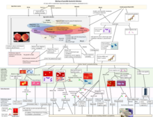
Group A streptococcal infections are a number of infections with Streptococcus pyogenes, a group A streptococcus (GAS). S. pyogenes is a species of beta-hemolytic Gram-positive bacteria that is responsible for a wide range of infections that are mostly common and fairly mild. If the bacteria enter the bloodstream an infection can become severe and life-threatening, and is called an invasive GAS (iGAS).

<i>Streptococcus pyogenes</i> Species of bacterium

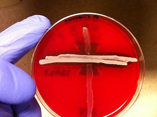
Streptococcus pyogenes is a species of Gram-positive, aerotolerant bacteria in the genus Streptococcus. These bacteria are extracellular, and made up of non-motile and non-sporing cocci that tend to link in chains. They are clinically important for humans, as they are an infrequent, but usually pathogenic, part of the skin microbiota that can cause Group A streptococcal infection. S. pyogenes is the predominant species harboring the Lancefield group A antigen, and is often called group A Streptococcus (GAS). However, both Streptococcus dysgalactiae and the Streptococcus anginosus group can possess group A antigen as well. Group A streptococci, when grown on blood agar, typically produce small (2–3 mm) zones of beta-hemolysis, a complete destruction of red blood cells. The name group A (beta-hemolytic) Streptococcus is thus also used.

<span class="mw-page-title-main">Scarlet fever</span> Infectious disease caused by Streptococcus pyogenes

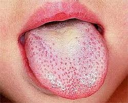
Scarlet fever, also known as scarlatina, is an infectious disease caused by Streptococcus pyogenes, a Group A streptococcus (GAS). It most commonly affects children between five and 15 years of age. The signs and symptoms include a sore throat, fever, headache, swollen lymph nodes, and a characteristic rash. The face is flushed and the rash is red and blanching. It typically feels like sandpaper and the tongue may be red and bumpy. The rash occurs as a result of capillary damage by exotoxins produced by S.pyogenes. On darker-pigmented skin the rash may be hard to discern.

<span class="mw-page-title-main">Viridans streptococci</span> Species of bacterium

The viridans streptococci are a large group of commensal streptococcal Gram-positive bacteria species that are α-hemolytic, producing a green coloration on blood agar plates, although some species in this group are actually γ-hemolytic, meaning they produce no change on blood agar. The pseudo-taxonomic term "Streptococcus viridans" is often used to refer to this group of species, but writers who do not like to use the pseudotaxonomic term prefer the terms viridans streptococci, viridans group streptococci (VGS), or viridans streptococcal species.

<span class="mw-page-title-main">Hemolysis (microbiology)</span> Breakdown of red blood cells

Hemolysis is the breakdown of red blood cells. The ability of bacterial colonies to induce hemolysis when grown on blood agar is used to classify certain microorganisms. This is particularly useful in classifying streptococcal species. A substance that causes hemolysis is a hemolysin.

<span class="mw-page-title-main">Rebecca Lancefield</span> 20th-century American microbiologist

Rebecca Craighill Lancefield was a prominent American microbiologist. She joined the Rockefeller Institute for Medical Research in New York in 1918, and was associated with that institute throughout her long and outstanding career. Her bibliography comprises more than 50 publications published over 60 years.

<i>Streptococcus oralis</i> Species of bacterium

Streptococcus oralis is a Gram positive viridans streptococcus of the Streptococcus mitis group. S. oralis is one of the pioneer species associated with eubiotic dental pellicle biofilms, and can be found in high numbers on most oral surfaces. It has been, however, found to be an opportunistic pathogen as well.

<i>Arcanobacterium haemolyticum</i> Species of bacterium

Arcanobacterium haemolyticum is a species of bacteria classified as a gram-positive bacillus. It is catalase-negative, facultative anaerobic, beta-hemolytic, and not motile. It has been known to cause head and neck infections, pharyngitis, and sinusitis.

Anti-streptolysin O is the antibody made against streptolysin O, an immunogenic, oxygen-labile streptococcal hemolytic exotoxin produced by most strains of group A and many strains of groups C and G Streptococcus bacteria. The "O" in the name stands for oxygen-labile; the other related toxin being oxygen-stable streptolysin-S. The main function of streptolysin O is to cause hemolysis —in particular, beta-hemolysis.

Streptolysins are two homogenous exotoxins from Streptococcus pyogenes. Types include streptolysin O, which is oxygen-labile, and streptolysin S, which is oxygen-stable.

<span class="mw-page-title-main">CAMP test</span> Microbiological method for identification

The CAMP test (Christie–Atkins–Munch-Peterson) is a test to identify group B β-hemolytic streptococci based on their formation of a substance that enlarges the area of hemolysis formed by the β-hemolysin elaborated from Staphylococcus aureus.

<i>Streptococcus anginosus</i> Species of bacterium

Streptococcus anginosus is a species of Streptococcus. This species, Streptococcus intermedius, and Streptococcus constellatus constitute the anginosus group, which is sometimes also referred to as the milleri group after the previously assumed but later refuted idea of a single species Streptococcus milleri. Phylogenetic relatedness of S. anginosus, S. constellatus, and S. intermedius has been confirmed by rRNA sequence analysis.

<i>Streptococcus dysgalactiae</i> Species of bacterium

Streptococcus dysgalactiae is a gram positive, beta-haemolytic, coccal bacterium belonging to the family Streptococcaceae. It is capable of infecting both humans and animals, but is most frequently encountered as a commensal of the alimentary tract, genital tract, or less commonly, as a part of the skin flora. The clinical manifestations in human disease range from superficial skin-infections and tonsillitis, to severe necrotising fasciitis and bacteraemia. The incidence of invasive disease has been reported to be rising. Several different animal species are susceptible to infection by S. dysgalactiae, but bovine mastitis and infectious arthritis in lambs have been most frequently reported.

<span class="mw-page-title-main">Streptococcal intertrigo</span> Medical condition

Streptococcal intertrigo is a skin condition that is secondary to a streptococcal bacterial infection. It is often seen in infants and young children and can be characterized by a fiery-red color of the skin, foul odor with an absence of satellite lesions, and skin softening in the neck, armpits or folds of the groin. Newborn children and infants commonly develop intertrigo because of physical features such as deep skin folds, short neck, and flexed posture. Prompt diagnosis by a medical professional and treatment with topical and/or oral antibiotics can effectively relieve symptoms.

<i>Streptococcus iniae</i> Species of bacterium

Streptococcus iniae is a species of Gram-positive, sphere-shaped bacterium belonging to the genus Streptococcus. Since its isolation from an Amazon freshwater dolphin in the 1970s, S. iniae has emerged as a leading fish pathogen in aquaculture operations worldwide, resulting in over US$100M in annual losses. Since its discovery, S. iniae infections have been reported in at least 27 species of cultured or wild fish from around the world. Freshwater and saltwater fish including tilapia, red drum, hybrid striped bass, and rainbow trout are among those susceptible to infection by S. iniae. Infections in fish manifest as meningoencephalitis, skin lesions, and septicemia.

Perianal cellulitis, also known as perianitis or perianal streptococcal dermatitis, is a bacterial infection affecting the lower layers of the skin (cellulitis) around the anus. It presents as bright redness in the skin and can be accompanied by pain, difficulty defecating, itching, and bleeding. This disease is considered a complicated skin and soft tissue infection (cSSTI) because of the involvement of the deeper soft tissues.

Bacteriophage T12 is a bacteriophage that infects Streptococcus pyogenes bacteria. It is a proposed species of the family Siphoviridae in the order Caudovirales also known as tailed viruses. It converts a harmless strain of bacteria into a virulent strain. It carries the speA gene which codes for erythrogenic toxin A. speA is also known as streptococcal pyogenic exotoxin A, scarlet fever toxin A, or even scarlatinal toxin. Note that the name of the gene "speA" is italicized; the name of the toxin "speA" is not italicized. Erythrogenic toxin A converts a harmless, non-virulent strain of Streptococcus pyogenes to a virulent strain through lysogeny, a life cycle which is characterized by the ability of the genome to become a part of the host cell and be stably maintained there for generations. Phages with a lysogenic life cycle are also called temperate phages. Bacteriophage T12, proposed member of family Siphoviridae including related speA-carrying bacteriophages, is also a prototypic phage for all the speA-carrying phages of Streptococcus pyogenes, meaning that its genome is the prototype for the genomes of all such phages of S. pyogenes. It is the main suspect as the cause of scarlet fever, an infectious disease that affects small children.

<span class="mw-page-title-main">Lancefield grouping</span> System for classifying streptococci bacteria

Lancefield grouping is a system of classification that classifies catalase-negative Gram-positive cocci based on the carbohydrate composition of bacterial antigens found on their cell walls. The system, created by Rebecca Lancefield, was historically used to organize the various members of the family Streptococcaceae, which includes the genera Lactococcus and Streptococcus, but now is largely superfluous due to explosive growth in the number of streptococcal species identified since the 1970s. However, it has retained some clinical usefulness even after the taxonomic changes, and as of 2018, Lancefield designations are still often used to communicate medical microbiological test results.

<span class="mw-page-title-main">Colonial morphology</span> Examination of microbial colonies

In microbiology, colonial morphology refers to the visual appearance of bacterial or fungal colonies on an agar plate. Examining colonial morphology is the first step in the identification of an unknown microbe. The systematic assessment of the colonies' appearance, focusing on aspects like size, shape, colour, opacity, and consistency, provides clues to the identity of the organism, allowing microbiologists to select appropriate tests to provide a definitive identification.

Streptococcosis is a set of diseases varying from mild to fatal side effects of the infection which originate from the bacterial group streptococcus, particularly in animals. Some of the areas of infection include wounds, body tissue, and respiratory areas. Research within horses, dogs, cats, wound injuries and swine infections have been done to document specific side effects from streptococcosis. Streptococcosis can occur to both humans and animals, the most common including horses, guinea pigs, dogs, cats and fish; while uncommon animals infected include monkeys, cattle, sheep, goats, ferrets and poultry. The wide range of diseases is due to the variability of streptococcus strains which thus creates multiple species for the diseases to occur.

References

  1. 1 2 3 4 5 6 7 8 9 10 11 12 13 14 15 16 17 18 19 20 21 22 23 24 25 26 27 28 29 30 31 32 33 34 35 36 37 38 39 40 41 42 43 44 45 46 47 48 49 50 51 52 53 54 55 56 57 58 59 60 61 62 63 64 65 66 67 68 69 70 71 72 Parte AC. "Streptococcus". LPSN .
  2. 1 2 3 Ryan KJ, Sherris JC, eds. (1994). Sherris Medical Microbiology (3rd ed.). Appleton & Lange. pp.  266–7. ISBN   0-8385-8541-8.
  3. "streptococcus". Online Etymology Dictionary. Retrieved 25 July 2018.
  4. στρεπτός  in Liddell, Henry George ; Scott, Robert (1940) A Greek–English Lexicon, revised and augmented throughout by Jones, Sir Henry Stuart , with the assistance of McKenzie, Roderick. Oxford: Clarendon Press. In the Perseus Digital Library , Tufts University.
  5. κόκκος  in Liddell and Scott
  6. 1 2 Facklam R (October 2002). "What happened to the streptococci: overview of taxonomic and nomenclature changes". Clinical Microbiology Reviews. 15 (4): 613–630. doi:10.1128/CMR.15.4.613-630.2002. PMC   126867 . PMID   12364372.
  7. Wang K, Lu W, Tu Q, Ge Y, He J, Zhou Y, et al. (March 2016). "Preliminary analysis of salivary microbiome and their potential roles in oral lichen planus". Scientific Reports. 6 (1): 22943. Bibcode:2016NatSR...622943W. doi:10.1038/srep22943. PMC   4785528 . PMID   26961389.
  8. "How to Get Rid of Pinkeye, Symptoms, Treatment, Causes & Pictures".
  9. "Streptococcus | Center for Academic Research and Training in Anthropogeny (CARTA)". carta.anthropogeny.org. Retrieved 2022-07-23.
  10. 1 2 Patterson MJ (1996). Baron S; et al. (eds.). Streptococcus. In: Baron's Medical Microbiology (4th ed.). Univ of Texas Medical Branch. ISBN   978-0-9631172-1-2. (via NCBI Bookshelf).
  11. Sharma S, Khanna G, Gangane SD (2019-07-13). Textbook of Pathology and Genetics for Nurses E-Book. Elsevier Health Sciences. ISBN   978-81-312-5538-4.
  12. Carroll KC (August 2019). Munson E (ed.). "Biographical Feature: Rebecca Lancefield, Ph.D". Journal of Clinical Microbiology. 57 (8). doi:10.1128/JCM.00728-19. PMC   6663886 . PMID   31142605.
  13. 1 2 3 Cohen-Poradosu R, Kasper DL (October 2007). "Group A streptococcus epidemiology and vaccine implications". Clinical Infectious Diseases. 45 (7): 863–865. doi: 10.1086/521263 . PMID   17806050.
  14. "Streptococcal Infections (Invasive Group A Strep)". New York City Department of Health and Mental Hygiene. Archived from the original on 6 November 2012. Retrieved 21 November 2012.
  15. Schrag S, Gorwitz R, Fultz-Butts K, Schuchat A (August 2002). "Prevention of perinatal group B streptococcal disease. Revised guidelines from CDC". MMWR. Recommendations and Reports. 51 (RR-11): 1–22. PMID   12211284.
  16. "Prevention of Early-onset Neonatal Group B Streptococcal Disease: Green-top Guideline No. 36". BJOG. 124 (12): e280–e305. November 2017. doi: 10.1111/1471-0528.14821 . PMID   28901693.
  17. Norwitz ER, Schorge JO (2013). Obstetrics and Gynecology at a Glance (4th ed.). Chichester: John Wiley & Sons, Ltd. ISBN   978-1118341735.
  18. Harrington DJ, Sutcliffe IC, Chanter N (April 2002). "The molecular basis of Streptococcus equi infection and disease". Microbes and Infection. 4 (4): 501–510. doi: 10.1016/S1286-4579(02)01565-4 . PMID   11932201.
  19. 1 2 Haslam DB, St Geme III JW (2023). "122 - Groups C and G Streptococci". In Long SS, Prober CG, Fischer M, Kimberlin D (eds.). Principles and Practice of Pediatric Infectious Diseases (Sixth ed.). Elsevier. pp. 752–753. doi:10.1016/B978-0-323-75608-2.00122-1. ISBN   978-0-323-75608-2. Note that according to the same source, the subspecies equisimilis is a grouping of large S. dysgalactiae colonies, whether they are members of Group C or Group G.
  20. Köhler W (June 2007). "The present state of species within the genera Streptococcus and Enterococcus". International Journal of Medical Microbiology. 297 (3): 133–150. doi:10.1016/j.ijmm.2006.11.008. PMID   17400023.
  21. Holt et al. (1994). Bergey's Manual of Determinative Bacteriology (9th ed.). Lippincott Williams & Wilkins. ISBN   0-683-00603-7
  22. Whitworth JM (November 1990). "Lancefield group F and related streptococci". Journal of Medical Microbiology. 33 (3): 135–151. doi: 10.1099/00222615-33-3-135 . PMID   2250284.[ permanent dead link ]
  23. "Bacterial Infection (Streptococcus) in Dogs". petmd.com. Retrieved 12 December 2014.
  24. "Bacteria-Firmicutes-Bacilli-Lactobacillales-Streptococcaceae-Streptococcus". PATRIC, University of Chicago. Retrieved 12 December 2014.
  25. Kawamura Y, Hou XG, Sultana F, Miura H, Ezaki T (April 1995). "Determination of 16S rRNA sequences of Streptococcus mitis and Streptococcus gordonii and phylogenetic relationships among members of the genus Streptococcus". International Journal of Systematic Bacteriology. 45 (2): 406–408. doi: 10.1099/00207713-45-2-406 . PMID   7537076.
  26. Liu, D., Molecular Detection of Human Bacterial Pathogens (Boca Raton: CRC Press, 2011), p. 324.
  27. 1 2 3 4 5 Patel S, Gupta RS (December 2018). "Robust demarcation of fourteen different species groups within the genus Streptococcus based on genome-based phylogenies and molecular signatures". Infection, Genetics and Evolution. 66: 130–151. doi:10.1016/j.meegid.2018.09.020. PMID   30248475. S2CID   52813184.
  28. Xu P, Alves JM, Kitten T, Brown A, Chen Z, Ozaki LS, et al. (April 2007). "Genome of the opportunistic pathogen Streptococcus sanguinis". Journal of Bacteriology. 189 (8): 3166–3175. doi:10.1128/JB.01808-06. PMC   1855836 . PMID   17277061.
  29. "Streptococcus". PATRIC. Blacksburg, VA: Virginia Bioinformatics Institute. Archived from the original on 2013-03-10.
  30. 1 2 Ferretti JJ, Ajdic D, McShan WM (May 2004). "Comparative genomics of streptococcal species". The Indian Journal of Medical Research. 119 (Suppl): 1–6. PMID   15232152.
  31. McShan, W. Michael; Nguyen, Scott V. (2016), Ferretti, Joseph J.; Stevens, Dennis L.; Fischetti, Vincent A. (eds.), "The Bacteriophages of Streptococcus pyogenes", Streptococcus pyogenes: Basic Biology to Clinical Manifestations, Oklahoma City (OK): University of Oklahoma Health Sciences Center, PMID   26866212 , retrieved 2024-02-07
  32. McDonnell M, Ronda C, Tomasz A (1975) "Diplophage": a bacteriophage of Diplococcus pneumoniae. Virology 63:577–582
  33. NCBI: Streptococcus phage Dp-1 (species)
  34. Tiraby JG, Tiraby E, Fox MS (Dec 1975) Pneumococcal bacteriophages. Virology 68:566–569. doi:10.1016/0042-6822(75)90300-1. PMID   844
  35. López R (September 2004). "Streptococcus pneumoniae and its bacteriophages: one long argument". International Microbiology. 7 (3): 163–171. PMID   15492930. PDF via web archive (9 Aug 2017)
  36. Rubens López, Ernesto García: Recent trends on the molecular biology of pneumococcal capsules, lytic enzymes, and bacteriophage, Oxford Academic FEMS Microbiology Reviews. Volume 28, Issue 5. 1 Nov 2004, pp. 554—580, doi:10.1016/j.femsre.2004.05.002 (Free Fulltext)
  37. Ronda C, López R, García E (November 1981). "Isolation and characterization of a new bacteriophage, Cp-1, infecting Streptococcus pneumoniae". Journal of Virology. 40 (2): 551–559. doi:10.1128/JVI.40.2.551-559.1981. PMC   256658 . PMID   6275103.
  38. Ouennane S, Leprohon P, Moineau S (2015). "Diverse virulent pneumophages infect Streptococcus mitis". PLOS ONE. 10 (2): e0118807. Bibcode:2015PLoSO..1018807O. doi: 10.1371/journal.pone.0118807 . PMC   4334900 . PMID   25692983.
  39. Johnsborg O, Eldholm V, Bjørnstad ML, Håvarstein LS (July 2008). "A predatory mechanism dramatically increases the efficiency of lateral gene transfer in Streptococcus pneumoniae and related commensal species". Molecular Microbiology. 69 (1): 245–253. doi: 10.1111/j.1365-2958.2008.06288.x . PMID   18485065. S2CID   30923996.
  40. Claverys JP, Håvarstein LS (March 2007). "Cannibalism and fratricide: mechanisms and raisons d'être". Nature Reviews. Microbiology. 5 (3): 219–229. doi:10.1038/nrmicro1613. PMID   17277796. S2CID   35433490.
  41. Li G, Liang Z, Wang X, Yang Y, Shao Z, Li M, et al. (June 2016). "Addiction of Hypertransformable Pneumococcal Isolates to Natural Transformation for In Vivo Fitness and Virulence". Infection and Immunity. 84 (6): 1887–1901. doi:10.1128/IAI.00097-16. PMC   4907133 . PMID   27068094.
  42. Michod RE, Bernstein H, Nedelcu AM (May 2008). "Adaptive value of sex in microbial pathogens". Infection, Genetics and Evolution. 8 (3): 267–285. doi:10.1016/j.meegid.2008.01.002. PMID   18295550.