Tularemia

Last updated
Tularemia
Other namesTularaemia, Pahvant Valley plague, [1] rabbit fever, [1] deer fly fever, Ohara's fever [2]
Tularemia lesion.jpg
A tularemia lesion on the back of the right hand
Specialty Infectious disease
Symptoms Fever, skin ulcer, large lymph nodes [3]
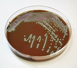
Causesbacterium Francisella tularensis (spread by ticks, deer flies, contact with infected animals) [4]
Diagnostic method Blood tests, microbial culture [5]
Prevention Insect repellent, wearing long pants, rapidly removing ticks, not disturbing dead animals [6]
Medication Streptomycin, Gentamicin, doxycycline, ciprofloxacin [5]
Prognosis Generally good with treatment [4]
Frequency~200 cases per year (US) [7]

Tularemia, also known as rabbit fever, is an infectious disease caused by the bacterium Francisella tularensis . [4] Symptoms may include fever, skin ulcers, and enlarged lymph nodes. [3] Occasionally, a form that results in pneumonia or a throat infection may occur. [3]

Contents

The bacterium is typically spread by ticks, deer flies, or contact with infected animals. [4] It may also be spread by drinking contaminated water or breathing in contaminated dust. [4] It does not spread directly between people. [8] Diagnosis is by blood tests or cultures of the infected site. [5] [9]

Prevention is by using insect repellent, wearing long pants, rapidly removing ticks, and not disturbing dead animals. [6] Treatment is typically with the antibiotic streptomycin. [9] Gentamicin, doxycycline, or ciprofloxacin may also be used. [5]

Between the 1970s and 2015, around 200 cases were reported in the United States a year. [7] Males are affected more often than females. [7] It occurs most frequently in the young and the middle aged. [7] In the United States, most cases occur in the summer. [7] The disease is named after Tulare County, California, where the disease was discovered in 1911. [10] A number of other animals, such as rabbits, may also be infected. [4]

Signs and symptoms

Depending on the site of infection, tularemia has six characteristic clinical variants: ulceroglandular (the most common type representing 75% of all forms), glandular, oropharyngeal, pneumonic, oculoglandular, and typhoidal. [11]

The incubation period for tularemia is 1 to 14 days; most human infections become apparent after three to five days. [12] In most susceptible mammals, the clinical signs include fever, lethargy, loss of appetite, signs of sepsis, and possibly death. Nonhuman mammals rarely develop the skin lesions seen in people. Subclinical infections are common, and animals often develop specific antibodies to the organism. Fever is moderate or very high, and tularemia bacilli can be isolated from blood cultures at this stage. The face and eyes redden and become inflamed. Inflammation spreads to the lymph nodes, which enlarge and may suppurate (mimicking bubonic plague). Lymph node involvement is accompanied by a high fever. [13]

Cause

Tularemia is caused by the bacteria Francisella tularensis which is typically spread by ticks, deer flies, and contact with infected animals. [4]

Bacteria

Chocolate agar culture showing Francisella tularensis colonies Chocolate agar 1.jpg
Chocolate agar culture showing Francisella tularensis colonies
Another culture of Francisella tularensis Francisella tularensis 01.jpg
Another culture of Francisella tularensis

The bacteria can penetrate into the body through damaged skin, mucous membranes, and inhalation. Humans are most often infected by tick/deer fly bite or through handling an infected animal. Ingesting infected water, soil, or food can also cause infection. Hunters are at a higher risk for this disease because of the potential of inhaling the bacteria during the skinning process. It has been contracted from inhaling particles from an infected rabbit ground up in a lawnmower (see below). Tularemia is not spread directly from person to person. [14] Humans can also be infected through bioterrorism attempts. [15]

Francisella tularensis can live both within and outside the cells of the animal it infects, meaning it is a facultative intracellular bacterium. [16] It primarily infects macrophages, a type of white blood cell, and thus is able to evade the immune system. The course of disease involves the spread of the organism to multiple organ systems, including the lungs, liver, spleen, and lymphatic system. The course of disease is different depending on the route of exposure. Mortality in untreated (before the antibiotic era) patients has been as high as 50% in the pneumoniac and typhoidal forms of the disease, which however account for less than 10% of cases. [17]

Spread

The most common way the disease is spread is via arthropod vectors. Ticks involved include Amblyomma , Dermacentor , Haemaphysalis , and Ixodes . [18] Rodents, rabbits, and hares often serve as reservoir hosts, [19] but waterborne infection accounts for 5–10% of all tularemia in the United States. [20] Tularemia can also be transmitted by biting flies, particularly the deer fly Chrysops discalis . Individual flies can remain infectious for 14 days and ticks for over two years.[ citation needed ] Tularemia may also be spread by direct contact with contaminated animals or material, by ingestion of poorly cooked flesh of infected animals or contaminated water, or by inhalation of contaminated dust. [21]

Diagnosis

Pathology

In lymph node biopsies, the typical histopathologic pattern is characterized by geographic areas of necrosis with neutrophils and necrotizing granulomas. The pattern is non specific and similar to other infectious lymphadenopathies. [22]

The laboratorial isolation of F. tularensis requires special media such as buffered charcoal yeast extract agar. It cannot be isolated in the routine culture media because of the need for sulfhydryl group donors (such as cysteine). The microbiologist must be informed when tularemia is suspected not only to include the special media for appropriate isolation, but also to ensure that safety precautions are taken to avoid contamination of laboratory personnel. Serological tests (detection of antibodies in the serum of the patients) are available and widely used. Cross reactivity with Brucella can confuse interpretation of the results, so diagnosis should not rely only on serology. Molecular methods such as PCR are available in reference laboratories.[ citation needed ]

Prevention

There are no safe, available, approved vaccines against tularemia. However, vaccination research and development continues, with live attenuated vaccines being the most thoroughly researched and most likely candidate for approval. [23] Sub-unit vaccine candidates, such as killed-whole cell vaccines, are also under investigation, however research has not reached a state of public use. [23]

Optimal preventative practices include limiting direct exposure when handling potentially infected animals by wearing gloves and face masks (importantly when skinning deceased animals). [24]

Treatment

If infection occurs or is suspected, treatment is generally with the antibiotics streptomycin or gentamicin. [24] Doxycycline was previously used. [25] Gentamicin may be easier to obtain than streptomycin. [25] There is also tentative evidence to support the use of quinolone antibiotics. [25]

Prognosis

Since the discovery of antibiotics, the rate of death associated with tularemia has decreased from 60% to less than 4%. [24]

Epidemiology

Tularemia is most common in the Northern Hemisphere, including North America and parts of Europe and Asia. [24] It occurs between 30° and 71° north latitude. [24]

In the United States, although records show that tularemia was never particularly common, incidence rates continued to drop over the course of the 20th century. Between 1990 and 2000, the rate dropped to less than 1 per one million, meaning the disease is extremely rare in the United States today. [26]

In Europe, tularemia is generally rare, though outbreaks with hundreds of cases occur every few years in neighboring Finland and Sweden. [27] In Sweden over a period from 1984 to 2012 a total of 4,830 cases of tularemia occurred (most of the infections were acquired within the country). About 1.86 cases per 100,000 persons occur each year with higher rates in those between 55 and 70. [28]

Outbreaks

In the 14th century BC, Tularemia spread throughout the Hittite Empire, known as the Hittite plague, and was the first recorded use of biological warfare.

From May to October 2000, an outbreak of tularemia in Martha's Vineyard, Massachusetts, resulted in one fatality, and brought the interest of the United States Centers for Disease Control and Prevention (CDC) as a potential investigative ground for aerosolised Francisella tularensis. For a time, Martha's Vineyard was identified as the only place in the world where documented cases of tularemia resulted from lawn mowing. [29] However, in May 2015 [30] a resident of Lafayette, Colorado, died from aerosolised F. tularensis, which was also connected to lawn mowing, highlighting this new vector of risk.

An outbreak of tularemia occurred in Kosovo in 1999–2000. [31]

In 2004, three researchers at Boston Medical Center, in Massachusetts, were accidentally infected with F. tularensis, after apparently failing to follow safety procedures. [32]

In 2005, small amounts of F. tularensis were detected in the National Mall area of Washington, D.C., the morning after an antiwar demonstration on September 24, 2005. Biohazard sensors were triggered at six locations surrounding the Mall. While thousands of people were potentially exposed, no infections were reported. The detected bacteria likely originated from a natural source, not from a bioterror attempt. [33]

In 2005, an outbreak occurred in Germany amongst participants in a hare hunt. About 27 people came into contact with contaminated blood and meat after the hunt. Ten of the exposed, aged 11 to 73, developed tularemia. One of these died due to complications caused by chronic heart disease. [34]

Tularemia is endemic in the Gori region of the Eurasian country of Georgia. The last outbreak was in 2006. [35] The disease is also endemic on the uninhabited Pakri Islands off the northern coast of Estonia. Used for bombing practice by Soviet forces, chemical and bacteriological weapons may have been dropped on these islands. [36]

In July 2007, an outbreak was reported in the Spanish autonomous region of Castile and León and traced to the plague of voles infesting the region. Another outbreak had taken place ten years before in the same area. [37]

In January 2011, researchers searching for brucellosis among feral pig populations in Texas discovered widespread tularemia infection or evidence of past infection in feral hog populations of at least two Texas counties, even though tularemia is not normally associated with pigs at all. Precautions were recommended for those who hunt, dress, or prepare feral hogs. Since feral hogs roam over large distances, concern exists that tularemia may spread or already be present in feral hogs over a wide geographic area. [38]

In November 2011, it was found in Tasmania. Reports claimed it to be the first in the Southern Hemisphere. [39] However, the causative organism was documented to have been isolated from a foot wound in the Northern Territory in 2003. [40]

In 2014, at least five cases of tularemia were reported in Colorado and at least three more cases in early 2015, including one death as a result of lawn mowing, as noted above. [30] In the summer of 2015, a popular hiking area just north of Boulder was identified as a site of animal infection and signs were posted to warn hikers.[ citation needed ]

History

The tularemia bacterium was first isolated by G.W. McCoy of the United States Public Health Service plague lab and reported in 1912. [41] [42] Scientists determined tularemia could be dangerous to humans; a human being may catch the infection after contacting an infected animal. The ailment soon became associated with hunters, cooks and agricultural workers. [43]

Biological weapon

The Centers for Disease Control and Prevention (CDC) regard F. tularensis as a viable biological warfare agent, and it has been included in the biological warfare programs of the United States, Soviet Union and Japan at various times. [44] A former Soviet biological weapons scientist, Ken Alibek, has alleged that an outbreak of tularemia among German soldiers shortly before the Battle of Stalingrad was due to the release of F. tularensis by Soviet forces. Others who have studied the pathogen "propose that an outbreak resulting from natural causes is more likely". [45] [46] In the United States, practical research into using rabbit fever as a biological warfare agent took place in 1954 at Pine Bluff Arsenal, Arkansas, an extension of the Fort Detrick program. [47] It was viewed as an attractive agent because:[ citation needed ]

The Schu S4 strain was standardized as "Agent UL" for use in the United States M143 bursting spherical bomblet. It was a lethal biological warfare agent with an anticipated fatality rate of 40–60%. The rate-of-action was around three days, with a duration-of-action of one to three weeks (treated) and two to three months (untreated), with frequent relapses. UL was streptomycin resistant. The aerobiological stability of UL was a major concern, being sensitive to sunlight, and losing virulence over time after release. When the 425 strain was standardized as "agent JT" (an incapacitant rather than lethal agent), the Schu S4 strain's symbol was changed again to SR.[ citation needed ]

Both wet and dry types of F. tularensis (identified by the codes TT and ZZ) were examined during the "Red Cloud" tests, which took place from November 1966 to February 1967 in the Tanana Valley, Alaska. [48]

Other animals

Cats and dogs can acquire the disease from the bite of a tick or flea that has fed on an infected host, such as a rabbit or rodent. For treatment of infected cats, antibiotics are the preferred treatment, including tetracycline, chloramphenicol or streptomycin. Long treatment courses may be necessary as relapses are common. [49]

Related Research Articles

<span class="mw-page-title-main">Bioterrorism</span> Terrorism involving biological agents

Bioterrorism is terrorism involving the intentional release or dissemination of biological agents. These agents include bacteria, viruses, insects, fungi, and/or their toxins, and may be in a naturally occurring or a human-modified form, in much the same way as in biological warfare. Further, modern agribusiness is vulnerable to anti-agricultural attacks by terrorists, and such attacks can seriously damage economy as well as consumer confidence. The latter destructive activity is called agrobioterrorism and is a subtype of agro-terrorism.

<span class="mw-page-title-main">Plague (disease)</span> Disease caused by Yersinia pestis bacterium

Plague is an infectious disease caused by the bacterium Yersinia pestis. Symptoms include fever, weakness and headache. Usually this begins one to seven days after exposure. There are three forms of plague, each affecting a different part of the body and causing associated symptoms. Pneumonic plague infects the lungs, causing shortness of breath, coughing and chest pain; bubonic plague affects the lymph nodes, making them swell; and septicemic plague infects the blood and can cause tissues to turn black and die.

<span class="mw-page-title-main">Anthrax</span> Infection caused by Bacillus anthracis bacteria

Anthrax is an infection caused by the bacterium Bacillus anthracis. Infection typically occurs by contact with the skin, inhalation, or intestinal absorption. Symptom onset occurs between one day and more than two months after the infection is contracted. The skin form presents with a small blister with surrounding swelling that often turns into a painless ulcer with a black center. The inhalation form presents with fever, chest pain and shortness of breath. The intestinal form presents with diarrhea, abdominal pains, nausea and vomiting.

<span class="mw-page-title-main">Q fever</span> Coxiella burnetii infection

Q fever or query fever is a disease caused by infection with Coxiella burnetii, a bacterium that affects humans and other animals. This organism is uncommon, but may be found in cattle, sheep, goats, and other domestic mammals, including cats and dogs. The infection results from inhalation of a spore-like small-cell variant, and from contact with the milk, urine, feces, vaginal mucus, or semen of infected animals. Rarely, the disease is tick-borne. The incubation period can range from 9 to 40 days. Humans are vulnerable to Q fever, and infection can result from even a few organisms. The bacterium is an obligate intracellular pathogenic parasite.

<span class="mw-page-title-main">Shigellosis</span> Medical condition

Shigellosis is an infection of the intestines caused by Shigella bacteria. Symptoms generally start one to two days after exposure and include diarrhea, fever, abdominal pain, and feeling the need to pass stools even when the bowels are empty. The diarrhea may be bloody. Symptoms typically last five to seven days and it may take several months before bowel habits return entirely to normal. Complications can include reactive arthritis, sepsis, seizures, and hemolytic uremic syndrome.

<span class="mw-page-title-main">Whooping cough</span> Human disease caused by the bacteria Bordetella pertussis

Whooping cough, also known as pertussis or the 100-day cough, is a highly contagious, vaccine-preventable bacterial disease. Initial symptoms are usually similar to those of the common cold with a runny nose, fever, and mild cough, but these are followed by two or three months of severe coughing fits. Following a fit of coughing, a high-pitched whoop sound or gasp may occur as the person breathes in. The violent coughing may last for 10 or more weeks, hence the phrase "100-day cough". The cough may be so hard that it causes vomiting, rib fractures, and fatigue. Children less than one year old may have little or no cough and instead have periods where they cannot breathe. The incubation period is usually seven to ten days. Disease may occur in those who have been vaccinated, but symptoms are typically milder.

Brucellosis is a zoonosis caused by ingestion of unpasteurized milk from infected animals, or close contact with their secretions. It is also known as undulant fever, Malta fever, and Mediterranean fever.

<span class="mw-page-title-main">Glanders</span> Horse disease that can be transmitted to humans

Glanders is a contagious zoonotic infectious disease that occurs primarily in horses, mules, and donkeys. It can be contracted by other animals, such as dogs, cats, pigs, goats, and humans. It is caused by infection with the bacterium Burkholderia mallei.

<span class="mw-page-title-main">Campylobacteriosis</span> Medical condition

Campylobacteriosis is among the most common infections caused by a bacterium in humans, often as a foodborne illness. It is caused by the Campylobacter bacterium, most commonly C. jejuni. It produces an inflammatory, sometimes bloody, diarrhea or dysentery syndrome, and usually cramps, fever and pain.

<i>Francisella</i> Genus of bacteria

Francisella is a genus of Gram-negative bacteria. They are small coccobacillary or rod-shaped, nonmotile organisms, which are also facultative intracellular parasites of macrophages. Strict aerobes, Francisella colonies bear a morphological resemblance to those of the genus Brucella. Some Francisella species are pathogenic bacteria but some others are endosymbionts of ticks. Ticks do not use any other food source than vertebrate blood and therefore ingest high levels of protein, iron and salt, but few vitamins. To overcome these nutritional deficiencies, ticks have evolved obligate interactions with nutritional endosymbionts, including Francisella endosymbionts. Their experimental elimination typically results in decreased tick survival, molting, fecundity and egg viability, as well as in physical abnormalities, which all are fully restored with an oral supplement of B vitamins. The genome sequencing of Francisella endosymbionts confirmed that they consistently produce three B vitamin types, biotin (vitamin B7), riboflavin (B2) and folate (B9). Francisella endosymbionts are often misidentified as Francisella tularensis; however, Francisella endosymbionts lack virulence genes and cannot infect humans.

<i>Francisella tularensis</i> Species of bacterium

Francisella tularensis is a pathogenic species of Gram-negative coccobacillus, an aerobic bacterium. It is nonspore-forming, nonmotile, and the causative agent of tularemia, the pneumonic form of which is often lethal without treatment. It is a fastidious, facultative intracellular bacterium, which requires cysteine for growth. Due to its low infectious dose, ease of spread by aerosol, and high virulence, F. tularensis is classified as a Tier 1 Select Agent by the U.S. government, along with other potential agents of bioterrorism such as Yersinia pestis, Bacillus anthracis, and Ebola virus. When found in nature, Francisella tularensis can survive for several weeks at low temperatures in animal carcasses, soil, and water. In the laboratory, F. tularensis appears as small rods, and is grown best at 35–37 °C.

<i>Burkholderia mallei</i> Species of bacterium

Burkholderia mallei is a Gram-negative, bipolar, aerobic bacterium, a human and animal pathogen of genus Burkholderia causing glanders; the Latin name of this disease (malleus) gave its name to the species causing it. It is closely related to B. pseudomallei, and by multilocus sequence typing it is a subspecies of B. pseudomallei.B. mallei evolved from B. pseudomallei by selective reduction and deletions from the B. pseudomallei genome. Unlike B. pseudomallei and other genus members, B. mallei is nonmotile; its shape is coccobacillary measuring some 1.5–3.0 μm in length and 0.5–1.0 μm in diameter with rounded ends.

<span class="mw-page-title-main">Meningococcal disease</span> Often life-threatening bacterial infection

Meningococcal disease describes infections caused by the bacterium Neisseria meningitidis. It has a high mortality rate if untreated but is vaccine-preventable. While best known as a cause of meningitis, it can also result in sepsis, which is an even more damaging and dangerous condition. Meningitis and meningococcemia are major causes of illness, death, and disability in both developed and under-developed countries.

<span class="mw-page-title-main">Ehrlichiosis</span> Medical condition

Ehrlichiosis is a tick-borne bacterial infection, caused by bacteria of the family Anaplasmataceae, genera Ehrlichia and Anaplasma. These obligate intracellular bacteria infect and kill white blood cells.

<span class="mw-page-title-main">Bubonic plague</span> Human and animal disease

Bubonic plague is one of three types of plague caused by the bacterium Yersinia pestis. One to seven days after exposure to the bacteria, flu-like symptoms develop. These symptoms include fever, headaches, and vomiting, as well as swollen and painful lymph nodes occurring in the area closest to where the bacteria entered the skin. Acral necrosis, the dark discoloration of skin, is another symptom. Occasionally, swollen lymph nodes, known as "buboes", may break open.

<span class="mw-page-title-main">African tick bite fever</span> Medical condition

African tick bite fever (ATBF) is a bacterial infection spread by the bite of a tick. Symptoms may include fever, headache, muscle pain, and a rash. At the site of the bite there is typically a red skin sore with a dark center. The onset of symptoms usually occurs 4–10 days after the bite. Complications are rare but may include joint inflammation. Some people do not develop symptoms.

Pathogenic <i>Escherichia coli</i> Strains of E. coli that can cause disease

Escherichia coli is a gram-negative, rod-shaped bacterium that is commonly found in the lower intestine of warm-blooded organisms (endotherms). Most E. coli strains are harmless, but pathogenic varieties cause serious food poisoning, septic shock, meningitis, or urinary tract infections in humans. Unlike normal flora E. coli, the pathogenic varieties produce toxins and other virulence factors that enable them to reside in parts of the body normally not inhabited by E. coli, and to damage host cells. These pathogenic traits are encoded by virulence genes carried only by the pathogens.

Francisella novicida is a bacterium of the Francisellaceae family, which consist of Gram-negative pathogenic bacteria. These bacteria vary from small cocci to rod-shaped, and are most known for their intracellular parasitic capabilities. In this family, six species have been identified; however, the species F. novicida is under intense scrutiny. Though some believe it should be classified with its own species designation, others argue it should be reclassified as a subspecies under F. tularensis. If it were to be classified as a subspecies, F. novicida would join the other known subspecies including F. t. tularensis and F. t. holarctica. Biochemical assays for identifying F. tularensis subtypes and strains are not ideal because the results are often non-definitive and subject to variation, therefore these assays should only be considered as supplementary tests for identification of Francisella species and subspecies. Several strains of F. novicida or F. novicida-like bacteria have been described, and these strains may be resolved by PCR-based methods.

<span class="mw-page-title-main">Legionnaires' disease</span> Form of atypical pneumonia

Legionnaires' disease is a form of atypical pneumonia caused by any species of Legionella bacteria, quite often Legionella pneumophila. Signs and symptoms include cough, shortness of breath, high fever, muscle pains, and headaches. Nausea, vomiting, and diarrhea may also occur. This often begins 2–10 days after exposure.

<span class="mw-page-title-main">Hittite plague</span> 14th century BC epidemic of tularemia

The Hittite Plague or Hand of Nergal was an epidemic, possibly of tularemia, which occurred in the mid-to-late 14th century BC.

References

  1. 1 2 Rapini, Ronald P.; Bolognia, Jean L.; Jorizzo, Joseph L. (2007). Dermatology: 2-Volume Set. St. Louis: Mosby. ISBN   978-1-4160-2999-1.
  2. James, William D.; Berger, Timothy G. (2006). Andrews' Diseases of the Skin: clinical Dermatology. Saunders Elsevier. p. 286. ISBN   978-0-7216-2921-6.
  3. 1 2 3 "Signs and Symptoms Tularemia". CDC. October 2015. Retrieved 8 November 2017.
  4. 1 2 3 4 5 6 7 "Tularemia". CDC. October 2015.
  5. 1 2 3 4 "Diagnosis and Treatment Tularemia". CDC. October 2015. Retrieved 8 November 2017.
  6. 1 2 "Prevention Tularemia". www.cdc.gov. October 2015. Retrieved 8 November 2017.
  7. 1 2 3 4 5 "Statistics Tularemia". CDC. November 2016. Retrieved 8 November 2017.
  8. "Transmission Tularemia". CDC. October 2015. Retrieved 8 November 2017.
  9. 1 2 "Clinicians Tularemia". www.cdc.gov. September 2016. Retrieved 8 November 2017.
  10. Hagan, William Arthur; Bruner, Dorsey William; Timoney, John Francis (1988). Hagan and Bruner's Microbiology and Infectious Diseases of Domestic Animals: With Reference to Etiology, Epizootiology, Pathogenesis, Immunity, Diagnosis, and Antimicrobial Susceptibility. Cornell University Press. p. 132. ISBN   978-0801418969.
  11. Plourde PJ, Embree J, Friesen F, Lindsay G, Williams T; Embree; Friesen; Lindsay; Williams (June 1992). "Glandular tularemia with typhoidal features in a Manitoba child". CMAJ. 146 (11): 1953–5. PMC   1490377 . PMID   1596844.{{cite journal}}: CS1 maint: multiple names: authors list (link)
  12. Office international des épizooties. (2000). Manual of standards for diagnostic tests and vaccines: lists A and B diseases of mammals, birds and bees. Paris, France: Office international des épizooties. pp. 494–6, 1394. ISBN   978-92-9044-510-4.
  13. Peace or Pestilence? Biological Warfare and How to Avoid It (1949), New York City: McGraw-Hill.
  14. "WHO Guidelines on Tularemia" Published 2007
  15. "Tularemia | CDC". www.cdc.gov. Retrieved 2017-03-12.
  16. Kinkead, LC; Allen, LA (September 2016). "Multifaceted effects of Francisella tularensis on human neutrophil function and lifespan". Immunological Reviews. 273 (1): 266–81. doi:10.1111/imr.12445. PMC   5000853 . PMID   27558340.
  17. "Tularemia: Current, comprehensive information on pathogenesis, microbiology, epidemiology, diagnosis, treatment, and prophylaxis". CIDRAP. Archived from the original on 2009-02-01. Retrieved 2008-09-29.
  18. George W. Beran; James H. Steele (22 October 1994). Handbook of Zoonoses: Bacterial, rickettsial, chlamydial, and mycotic. CRC Press. pp. 117–. ISBN   978-0-8493-3205-0 . Retrieved 28 October 2010.
  19. Mörner T (December 1992). "The ecology of tularaemia". Rev. Sci. Tech. 11 (4): 1123–30. doi: 10.20506/rst.11.4.657 . PMID   1305858.
  20. Jellison WL, Owen C, Bell JF, Kohls GM (1961). "Tularemia and animal populations". Wildl Dis. 17: 1–22.
  21. "Tularemia Transmission". Centers for Disease Control and Prevention. October 26, 2015. Retrieved 2017-10-06.
  22. Rosado FG, Stratton CW, Mosse CA Clinicopathologic correlation of epidemiologic and histopathologic features of pediatric bacterial lymphadenitis. Arch Pathol Lab Med. 2011 Nov;135(11):1490-3. http://www.archivesofpathology.org/doi/pdf/10.5858/arpa.2010-0581-OA
  23. 1 2 Putzova, D; Senitkova, I; Stulik, J (19 May 2016). "Tularemia vaccines". Folia Microbiologica. 61 (6): 495–504. doi:10.1007/s12223-016-0461-z. ISSN   1874-9356. PMID   27194547. S2CID   1227719.
  24. 1 2 3 4 5 Penn, R.L. (2014). Francisella tularensis (Tularemia) In J. E. Bennett, R. Dolin, & M. J. Blaser (Eds.), Mandell, Douglas, and Bennett's Principles and Practice of Infectious Diseases (8th ed.). Philadelphia, PA: Churchill Livingstone. pp. 2590–2602. ISBN   978-1-4557-4801-3.
  25. 1 2 3 Hepburn, MJ; Simpson, AJ (April 2008). "Tularemia: current diagnosis and treatment options" (PDF). Expert Review of Anti-infective Therapy. 6 (2): 231–40. doi:10.1586/14787210.6.2.231. PMID   18380605. S2CID   9036831. Archived (PDF) from the original on 2013-12-19. Retrieved 2013-06-30.
  26. Hayes E, Marshall S, Dennis D, et al. (March 2002). "Tularemia--United States, 1990-2000". MMWR. 51 (JULIOes=181–4): 181–4. PMID   11900351.
  27. Rossow, Heidi (2015-10-09). Epidemiology of tularemia in Finland. University of Helsinki. hdl:10138/156555.
  28. Desvars A, Furberg M, Hjertqvist M, et al. (January 2015). "Epidemiology and ecology of tularemia in Sweden, 1984–2012". Emerg Infect Dis. 21 (1): 32–39. doi:10.3201/eid2101.140916. PMC   4285262 . PMID   25529978.
  29. Feldman KA, Enscore RE, Lathrop SL, et al. (November 2001). "An outbreak of primary pneumonic tularemia on Martha's Vineyard". New England Journal of Medicine . 345 (22): 1601–6. doi: 10.1056/NEJMoa011374 . PMID   11757506.
  30. 1 2 Byars, Mitchell (May 28, 2015). "Lafayette resident contracts tularemia after mowing lawn, dies of other medical complications". Daily Camera . Retrieved 2018-06-19.
  31. Tularemia Outbreak Investigation in Kosovo: Case Control and Environmental Studies. Emerg Infect Dis. -Reintjes R, Dedushaj I, Gjini A, Jorgensen TR, Cotter B, Lieftucht A, et al. - Retrieved 3 Jan 2012
  32. Smith S (2005-03-29). "City tells BU to bolster safety of its medical labs". Boston Globe . Retrieved 2007-05-09.
  33. Dvorak P (2005-10-02). "Health Officials Vigilant for Illness After Sensors Detect Bacteria on Mall: Agent Found as Protests Drew Thousands of Visitors". Washington Post . p. C13. Retrieved 2007-05-08. A week after six bioterrorism sensors detected the presence of a dangerous bacterium on the Mall, health officials said there are no reports that any of the thousands of people in the nation's capital Sept. 24 have tularemia, the illness that results from exposure to the bacteria.
  34. Epidemiologisches Bulletin (pdf) des Robert Koch-Instituts Nr. 50 16. Dezember 2005
  35. According to staff at Georgia's National Center for Disease Control, an outbreak of tularemia occurred in the village of Zemo Rene east of Gori in December 2005 and January 2006. Twenty-six persons tested positive for the bacteria, and 45 tested positive for antibodies. No cases were fatal. The source was deemed to be a water spring. Previous outbreaks were in Tamarasheni (2005) and Ruisi (1997 and 1998).
  36. "Human Impact on Groundwater Management in Northern Estonia." (PDF). Archived from the original (PDF) on 2014-04-07. Retrieved 2014-04-05.
  37. "Diagnóstico de un brote de tularemia en Castilla-León" (PDF). Archived from the original (PDF) on March 9, 2009.
  38. Davis, John (January 25, 2011). "Researchers Warn of Tularemia In Area Feral Hogs". Texas Tech Today. Texas Tech University. Archived from the original on 2013-06-02. Retrieved 2018-06-19.
  39. Biological war disease found in Tasmania Australian Broadcasting Corporation - Retrieved 4 Nov 2011.
  40. Whipp MJ; Davis JM; Lum G; et al. (2003). "Characterization of a novicida-like subspecies of Francisella tularensis isolated in Australia". Journal of Medical Microbiology . 52 (Pt 9): 839–42. doi:10.1099/jmm.0.05245-0. PMID   12909664.
  41. Tärnvik, A.; Berglund, L. (February 1, 2003). "Tularaemia". European Respiratory Journal. 21 (2): 361–373. doi: 10.1183/09031936.03.00088903 . PMID   12608453. S2CID   219200617 via erj.ersjournals.com.
  42. McCoy GW, Chapin CW. Bacterium tularense, the cause of a plaguelike disease of rodents. Public Health Bull 1912;53:17–23.
  43. "Archived copy". Archived from the original on 2007-06-13. Retrieved 2007-06-04.{{cite web}}: CS1 maint: archived copy as title (link)
  44. Dennis DT, Inglesby TV, Henderson DA, et al. (June 2001). "Tularemia as a biological weapon: medical and public health management". JAMA. 285 (21): 2763–73. doi:10.1001/jama.285.21.2763. PMID   11386933.
  45. Croddy E, Krcalova S (October 2001). "Tularemia, Biological Warfare, and the Battle for Stalingrad (1942-1943)". Military Medicine . 166 (10): 837–838. doi: 10.1093/milmed/166.10.837 . PMID   11603230. Archived from the original on 2012-04-02.
  46. Sjöstedt A (June 2007). "Tularemia: history, epidemiology, pathogen physiology, and clinical manifestations". Annals of the New York Academy of Sciences . 1105 (1): 1–29. Bibcode:2007NYASA1105....1S. doi:10.1196/annals.1409.009. PMID   17395726. S2CID   26000749.
  47. Kanti Ghosh, Tushar, Prelas, Mark, Viswanath, Dabir: Science and Technology of Terrorism and Counterterrorism. CRC Press, 2002. Page 97. ISBN   0-8247-0870-9
  48. "Fact Sheet - Red Cloud Archived 2009-03-09 at the Wayback Machine ", Office of the Assistant Secretary of Defense (Health Affairs), Deployment Health Support Directorate.
  49. Eldredge, Debra M.; Carlson, Delbert G.; Carlson, Liisa D.; Giffin, James M. (2008). Cat Owner's Home Veterinary Handbook. Howell Book House.