Burkholderia mallei | |
---|---|
![]() | |
Scientific classification ![]() | |
Domain: | Bacteria |
Phylum: | Pseudomonadota |
Class: | Betaproteobacteria |
Order: | Burkholderiales |
Family: | Burkholderiaceae |
Genus: | Burkholderia |
Species: | B. mallei |
Binomial name | |
Burkholderia mallei (Zopf 1885) Yabuuchi et al. 1993 | |
Synonyms | |
Bacillus malleiZopf 1885 Contents
|
Burkholderia mallei is a Gram-negative, bipolar[ clarification needed ], aerobic bacterium, a human and animal pathogen of genus Burkholderia causing glanders; the Latin name of this disease (malleus) gave its name to the species causing it. It is closely related to B. pseudomallei, and by multilocus sequence typing it is a subspecies of B. pseudomallei. [1] B. mallei evolved from B. pseudomallei by selective reduction and deletions from the B. pseudomallei genome. [2] Unlike B. pseudomallei and other genus members, B. mallei is nonmotile; its shape is coccobacillary measuring some 1.5–3.0 μm in length and 0.5–1.0 μm in diameter with rounded ends.
Wilhelm Schütz and Friedrich Löffler first isolated B. mallei in 1882. [3] It was isolated from an infected liver and spleen of a horse. [4] This bacterium is also one of the first to be identified containing a type VI secretion system which is important for its pathogenicity. [5] In 1885, the German Botanist and Bacteriologist, Wilhelm Zopf (1846–1909) gave the pathogen its binomial name, after analyzing samples of the bacterium. He further refined his observations with the pathogen in 1886. [6]
Most organisms within the Burkholderiaceae live in soil; however, B. mallei does not. Because B. mallei is an obligate mammalian pathogen, it must infect a host mammal to live and to be transmitted from one host to another. [7]
B. mallei is very closely related to B. pseudomallei, being 99% identical in conserved genes when compared to B. pseudomallei. B. malllei has about 1.4 Mb less DNA than B. pseudomallei. [4] B. mallei may have actually evolved from a strain of B. pseudomallei after the latter had infected an animal. The bacterium would have lost the genes that were not necessary for living in an animal host. This suggestion has found support from studies that compare strains of B. mallei to B. pseudomallei and indicate that their two respective genomes are very similar. The genes that allowed the bacterium to survive in a soil environment, like genes that gave B. mallei the capacity to protect against bactericidals, antibiotics, and antifungals, were likely deleted. Thus, the reason that B. mallei is not found outside of a host is because it lacks the genes necessary for survival in the soil. Genome comparisons also seem to indicate that the B. mallei is still evolving and adapting to an intracellular lifestyle. [8]
The genome of B. mallei was sequenced in the United States by The Institute of Genomic Research. The size of the genome is smaller than that of B. pseudomallei. The B. mallei sequence revealed a chromosome of 3.5 mega base pairs (Mb) and a 2.3 Mb "megaplasmid". Many insertion sequences and phase-variable genes were also found. [3] The genome for B. mallei is made up of two circular chromosomes. Chromosome 1 is where genes relating to metabolism, capsule formation, and lipopolysaccharide biosynthesis are located. B. mallei has a polysaccharide capsule which indicates its potential as a pathogen. Chromosome 2 is where most of the information regarding secretion systems and virulence-associated genes are located. [4] Multilocus sequence typing has revealed that B. mallei most likely evolved from a B. pseudomallei clone reduction. About 1000 B. pseudomellei genes are absent or varying in the B. mallei genome. B. mallei's genome also has a large amount of insertion sequences. [8]
Burkholderia mallei was first called "Bacillus mallei" and was in the genus Pseudomonas until the early 1990s. It is now part of the genus Burkholderia. [3]
No standardised system exists for differentiating between B. mallei and B. pseudomallei. The methods that have been used to differentiate and identify one strain from the other include ribotyping, pulsed-field gel electrophoresis, multilocus enzyme electrophoresis, random amplified polymorphic DNA analysis, and multilocus sequence typing. [3] Comparing the DNA of B. mallei and B. pseudomallei must be done at the 23S rDNA level, however, since no identifiable difference is found between the two species at the 16S rDNA level. [9]
Both B. mallei and B. pseudomallei can be cultured in a laboratory; nutrient agar can be used to grow the bacteria. When grown in culture, B. mallei grows in smooth, grey, translucent colonies. In a period of 18 hours at 37 °C, a B. mallei colony can grow to about 0.5–1.0 mm in diameter. B. mallei culture growth on MacConkey agar is variable. Many microbiologists are unfamiliar with B. mallei and as a result it has frequently been misidentified as a Pseudomonas species or as a contaminant in a culture. [3]
The bacterium is susceptible to numerous disinfectants including benzalkonium chloride, iodine, mercuric chloride, potassium permanganate, 1% sodium hypochlorite, and ethanol. The micro-organism can also be destroyed by heating or ultraviolet light. Antibiotics such as streptomycin, amikacin, tetracycline, doxycycline, carbapenems, ceftazidime, amoxicillin/clavulanic acid, piperacillin, chloramphenicol, and sulfathiazole have been reported to be effective against the bacteria in vitro . B. mallei, like B. pseudomallei, is also resistant to a number of antibiotics including aminoglycosides, polymyxins, and beta-lactams. No vaccine is currently available for humans or animals to protect against B. mallei infection. [3] An animal model that will predict immune responses necessary to create immunity to the bacterium is needed before a vaccine can be developed. Mice are fairly close to humans in their susceptibility to B. mallei and would be the ideal choice of animal for creating a model for the vaccine. [4]
Burkholderia mallei is responsible for causing glanders disease, which historically mostly affected animals, such as horses, mules, and donkeys, and rarely humans. Horses are considered the natural host for B. mallei infection and are highly susceptible to it. [4] B. mallei infects and gains access to the cell of its host through lysis of the entry vacuole. B. mallei has bacterial protein-dependent, actin-based motility once inside the cell. It is also able to initiate host cell fusion that results in multinucleated giant cells (MNGCs). The consequence of MNGCs has yet to be determined, but it may allow the bacteria to spread to different cells, evade responses by the infected host's immune system, or allow the bacteria to remain in the host longer. B. mallei is able to survive inside host cells through its capabilities in disrupting the bacteria-killing functions of the cell. It leaves the vacuoles early, which allows for efficient replication of the bacteria inside the cell. Leaving the cell early also keeps the bacteria from being destroyed by lysosomal defensins and other pathogen-killing agents. MNGCs may help protect the bacteria from immune responses. [10] B. mallei's ability to live within the host cell makes developing a vaccine against it difficult and complex. The vaccine would need to create a cell-mediated immune response, as well as a humoral response to the bacteria in to be effective in protecting against B. mallei. In regards to a vaccine against B. mallei, the closeness of B. mallei to B. pseudomallei may make it possible that a vaccine developed for either type would be effective against the other. [7]
Horses chronically infected with B. mallei with glanders disease typically experience mucus-containing nasal discharge, lung lesions, and nodules around the liver or spleen. Acute infection in horses results in a high fever, loss of fat or muscle, erosion of the surface of the nasal septum, hemorrhaging or mucus discharge. The bacterium mostly affects the lungs and airways. [4] Human infection with B. mallei is rare, although it occasionally occurs among laboratory workers dealing with the bacteria or those who are frequently near infected animals. [3] The bacteria usually infect a person through their eyes, nose, mouth, or cuts in the skin. Once people are infected, they develop a fever and rigors. Eventually, they get pneumonia, pustules, and abscesses, which prove fatal within a week to 10 days if left untreated by antibiotics. The way someone is infected by the bacteria also affects the type of symptoms that will result. If the bacteria enter through the skin, a local skin infection can result, while inhaling B. mallei can cause septicemic or pulmonary, muscular, hepatic, or splenous infections. B. mallei infection has a fatality rate of 95% if left untreated, and a 50% fatality rate in individuals treated with antibiotics. [4]
In the first days of B. mallei infection, neutrophils, macrophages, and T cells go to the spleen in great quantities. The early cellular response to B. mallei infection involves Gr-1+ (antigen) cells, and implies their importance to immunity against this bacterial infection. T cells (nitric oxide) are actually more involved in combating B. mallei in the later stages of its infection of a host. [11]
Lipopolysaccharide isolated from B. mallei demonstrated significantly lower biological activity as compared to the LPS from Escherichia coli , in agreement with the lower degree of acylation of its lipid A: the major forms of B. mallei lipid A were penta- and tetraacylated, whereas classical lipid A from E. coli was hexaacylated. In addition, lipid A from B. mallei contains 4-amino-4-deoxyarabinose residue in almost half of the molecules, which would partially neutralize the negative charge of the phosphate groups necessary for the interaction with the positively charged amino acids of TLR4. At the same time, lipid A acyl chains in B. mallei were on the average longer (14–16 carbon atoms) than those in E. coli (14 carbon atoms), yet LPS from B. mallei appeared to be a weaker activator. B. mallei may employ LPS with low biological activity to evade proper recognition by the TLR4/MD-2 complex of innate immune system, dampening the host immune response and increasing the risk of bacterial dissemination. [12]
Burkholderia mallei has been eradicated in the United States and most Western countries, but still affects animals in Africa, Asia, the Middle East, Central America, and South America. [4] Many Western countries were able to eliminate the disease through glanders control programs and laws requiring notification of cases of infection to health departments and the destruction of any animal affected with B. mallei. [13]
Burkholderia mallei and B. pseudomallei have a history of being on a list of potential biological warfare agents. The Centers for Disease Control and Prevention classifies B. mallei as a category B critical biological agent. [7] As a result, research regarding B. mallei may only be done in biosafety level 3 facilities in the US and internationally. Though it is so highly infective and a potential biological weapon, little research has been conducted on this bacterium. [4] B. mallei and B. pseudomallei under the policy of Institutional Oversight of Life Sciences Dual Use Research of Concern would be subject to oversight to ensure the responsible investigation of these agents. [14]
In March 2000, one of the first cases since the 1940s of glanders in the United States occurred in a young microbiologist working for the U.S. Army Medical Research Institute for Infectious Diseases. The researcher had type 1 diabetes and had been working with B. mallei for about two years, but he did not always wear gloves while conducting his research. The researcher experienced enlargement of the lymph nodes and a fever which lasted for 10 days even with antibiotic treatment. In the following weeks, the researcher experienced fatigue, rigors, night sweats, and loss of weight. The next month, his symptoms seemed to disappear after treatment with clarithromycin, but after the medication was stopped, the symptoms reappeared. After conducting multiple tests on cultures from the researcher's blood and a biopsied portion of a liver abscess, the bacterium was identified as B. mallei. Once it was established what infected the researcher, another course of antibiotics was given (imipenem and doxycycline) with 6 months of treatment. After a year, the researcher made a full recovery.
This incident also showed how a cut or skin abrasion is not absolutely necessary to contract the disease, as the researcher had no recollection of any cut or accident while working in the laboratory. The case was significant as it showed the difficulty that microbiology laboratories have in identifying bioweapon agents and the potential consequences if measures are not taken to prepare for an actual biological attack. [13]
Burkholderia mallei was intentionally used to infect animals and humans during World War I. The Germans used B. mallei to infect animals that were being sent from neutral countries to the Allies with glanders. [3] The Germans' plans for biological warfare started in 1915 on the East Coast of the United States; they intended to infect and kill the livestock that were being sent to the Allies and facilitate the transfer of the disease to humans. The East Coast was where many animals were being assembled for shipment to the Allies fighting in Europe. The Germans also targeted Romania, Norway, and Spain's animal supplies with cultures of glanders. The German biological sabotage eventually spread to Argentina, where agents would rely on bacterial cultures from Spain to infect the cattle, horses, and mules that Argentina was supplying to the Allies. The German use of microbes as weapons is one of the only documented attacks of intentionally using biological weapons against neutral countries. [15]
The Japanese used B. mallei in their biological warfare research units. The most notable and notorious unit, Unit 731, used the bacterium to conduct experiments on live human subjects. However, the Japanese did not end up creating a biological weapon out of B. mallei. They did actually use B. mallei to test its effectiveness in contaminating water supplies, and the results of these tests were successful.
The Russians' biological weapons program also took an interest in B. mallei and conducted field tests with it. Some of the researchers from the program were actually infected and killed by it during the course of their research. It has been suggested that the Soviet Union eventually used B. mallei during their war in Afghanistan against the mujahideen . [3]
A human pathogen is a pathogen that causes disease in humans.
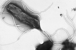
Helicobacter pylori, previously known as Campylobacter pylori, is a gram-negative, flagellated, helical bacterium. Mutants can have a rod or curved rod shape, and these are less effective. Its helical body is thought to have evolved in order to penetrate the mucous lining of the stomach, helped by its flagella, and thereby establish infection. The bacterium was first identified as the causal agent of gastric ulcers in 1983 by the Australian doctors Barry Marshall and Robin Warren.
Melioidosis is an infectious disease caused by a gram-negative bacterium called Burkholderia pseudomallei. Most people exposed to B. pseudomallei experience no symptoms; however, those who do experience symptoms have signs and symptoms that range from mild, such as fever and skin changes, to severe with pneumonia, abscesses, and septic shock that could cause death. Approximately 10% of people with melioidosis develop symptoms that last longer than two months, termed "chronic melioidosis".
Glanders is a contagious zoonotic infectious disease that occurs primarily in horses, mules, and donkeys. It can be contracted by other animals, such as dogs, cats, pigs, goats, and humans. It is caused by infection with the bacterium Burkholderia mallei.
Rickettsia prowazekii is a species of gram-negative, alphaproteobacteria, obligate intracellular parasitic, aerobic bacillus bacteria that is the etiologic agent of epidemic typhus, transmitted in the feces of lice. In North America, the main reservoir for R. prowazekii is the flying squirrel. R. prowazekii is often surrounded by a protein microcapsular layer and slime layer; the natural life cycle of the bacterium generally involves a vertebrate and an invertebrate host, usually an arthropod, typically the human body louse. A form of R. prowazekii that exists in the feces of arthropods remains stably infective for months. R. prowazekii also appears to be the closest semi-free-living relative of mitochondria, based on genome sequencing.
Rickettsia rickettsii is a Gram-negative, intracellular, coccobacillus bacterium that was first discovered in 1902. Having a reduced genome, the bacterium harvests nutrients from its host cell to carry out respiration, making it an organoheterotroph. Maintenance of its genome is carried out through vertical gene transfer where specialization of the bacterium allows it to shuttle host sugars directly into its TCA cycle.
Burkholderia is a genus of Pseudomonadota whose pathogenic members include the Burkholderia cepacia complex, which attacks humans and Burkholderia mallei, responsible for glanders, a disease that occurs mostly in horses and related animals; Burkholderia pseudomallei, causative agent of melioidosis; and Burkholderia cepacia, an important pathogen of pulmonary infections in people with cystic fibrosis (CF). Burkholderia species is also found in marine environments. S.I. Paul et al. (2021) isolated and characterized Burkholderia cepacia from marine sponges of the Saint Martin's Island of the Bay of Bengal, Bangladesh.
Corynebacterium diphtheriae is a Gram-positive pathogenic bacterium that causes diphtheria. It is also known as the Klebs–Löffler bacillus because it was discovered in 1884 by German bacteriologists Edwin Klebs (1834–1912) and Friedrich Löffler (1852–1915). The bacteria are usually harmless unless they are infected by a bacteriophage that carries a gene that gives rise to a toxin. This toxin causes the disease. Diphtheria is caused by the adhesion and infiltration of the bacteria into the mucosal layers of the body, primarily affecting the respiratory tract and the subsequent release of an exotoxin. The toxin has a localized effect on skin lesions, as well as a metastatic, proteolytic effects on other organ systems in severe infections. Originally a major cause of childhood mortality, diphtheria has been almost entirely eradicated due to the vigorous administration of the diphtheria vaccination in the 1910s.
Francisella tularensis is a pathogenic species of Gram-negative coccobacillus, an aerobic bacterium. It is nonspore-forming, nonmotile, and the causative agent of tularemia, the pneumonic form of which is often lethal without treatment. It is a fastidious, facultative intracellular bacterium, which requires cysteine for growth. Due to its low infectious dose, ease of spread by aerosol, and high virulence, F. tularensis is classified as a Tier 1 Select Agent by the U.S. government, along with other potential agents of bioterrorism such as Yersinia pestis, Bacillus anthracis, and Ebola virus. When found in nature, Francisella tularensis can survive for several weeks at low temperatures in animal carcasses, soil, and water. In the laboratory, F. tularensis appears as small rods, and is grown best at 35–37 °C.
Neisseria meningitidis, often referred to as the meningococcus, is a Gram-negative bacterium that can cause meningitis and other forms of meningococcal disease such as meningococcemia, a life-threatening sepsis. The bacterium is referred to as a coccus because it is round, and more specifically a diplococcus because of its tendency to form pairs.
Burkholderia pseudomallei is a Gram-negative, bipolar, aerobic, motile rod-shaped bacterium. It is a soil-dwelling bacterium endemic in tropical and subtropical regions worldwide, particularly in Thailand and northern Australia. It was reported in 2008 that there had been an expansion of the affected regions due to significant natural disasters, and it could be found in Southern China, Hong Kong, and countries in America. B. pseudomallei, amongst other pathogens, has been found in monkeys imported into the United States from Asia for laboratory use, posing a risk that the pathogen could be introduced into the country.
Chlamydia felis is a Gram-negative, obligate intracellular bacterial pathogen that infects cats. It is endemic among domestic cats worldwide, primarily causing inflammation of feline conjunctiva, rhinitis and respiratory problems. C. felis can be recovered from the stomach and reproductive tract. Zoonotic infection of humans with C. felis has been reported. Strains FP Pring and FP Cello have an extrachromosomal plasmid, whereas the FP Baker strain does not. FP Cello produces lethal disease in mice, whereas the FP Baker does not. An attenuated FP Baker strain, and an attenuated 905 strain, are used as live vaccines for cats.
Chromobacterium violaceum is a Gram-negative, facultative anaerobic, non-sporing coccobacillus. It is motile with the help of a single flagellum which is located at the pole of the coccobacillus. Usually, there are one or two more lateral flagella as well. It is part of the normal flora of water and soil of tropical and sub-tropical regions of the world. It produces a natural antibiotic called violacein, which may be useful for the treatment of colon and other cancers. It grows readily on nutrient agar, producing distinctive smooth low convex colonies with a characteristic striking dark violet metallic sheen. Some strains of the bacteria which do not produce this pigment have also been reported. It has the ability to break down tarballs.
Burkholderia thailandensis is a nonfermenting motile, Gram-negative bacillus that occurs naturally in soil. It is closely related to Burkholderia pseudomallei, but unlike B. pseudomallei, it only rarely causes disease in humans or animals. The lethal inoculum is approximately 1000 times higher than for B. pseudomallei. It is usually distinguished from B. pseudomallei by its ability to assimilate arabinose. Other differences between these species include lipopolysaccharide composition, colony morphology, and differences in metabolism.
Pathogenic bacteria are bacteria that can cause disease. This article focuses on the bacteria that are pathogenic to humans. Most species of bacteria are harmless and are often beneficial but others can cause infectious diseases. The number of these pathogenic species in humans is estimated to be fewer than a hundred. By contrast, several thousand species are part of the gut flora present in the digestive tract.
Leptospira interrogans is a species of obligate aerobic spirochaete bacteria shaped like a corkscrew with hooked and spiral ends. L. interrogans is mainly found in warmer tropical regions. The bacteria can live for weeks to months in the ground or water. Leptospira is one of the genera of the spirochaete phylum that causes severe mammalian infections. This species is pathogenic to some wild and domestic animals, including pet dogs. It can also spread to humans through abrasions on the skin, where infection can cause flu-like symptoms with kidney and liver damage. Human infections are commonly spread by contact with contaminated water or soil, often through the urine of both wild and domestic animals. Some individuals are more susceptible to serious infection, including farmers and veterinarians who work with animals.
Pathogenomics is a field which uses high-throughput screening technology and bioinformatics to study encoded microbe resistance, as well as virulence factors (VFs), which enable a microorganism to infect a host and possibly cause disease. This includes studying genomes of pathogens which cannot be cultured outside of a host. In the past, researchers and medical professionals found it difficult to study and understand pathogenic traits of infectious organisms. With newer technology, pathogen genomes can be identified and sequenced in a much shorter time and at a lower cost, thus improving the ability to diagnose, treat, and even predict and prevent pathogenic infections and disease. It has also allowed researchers to better understand genome evolution events - gene loss, gain, duplication, rearrangement - and how those events impact pathogen resistance and ability to cause disease. This influx of information has created a need for bioinformatics tools and databases to analyze and make the vast amounts of data accessible to researchers, and it has raised ethical questions about the wisdom of reconstructing previously extinct and deadly pathogens in order to better understand virulence.
Pathema was one of the eight bioinformatics resource centers funded by the National Institute of Allergy and Infectious Diseases (NIAID), a component of the National Institute of Health (NIH), which is an agency of the United States Department of Health and Human Services.
Escherichia coli is a gram-negative, rod-shaped bacterium that is commonly found in the lower intestine of warm-blooded organisms (endotherms). Most E. coli strains are harmless, but pathogenic varieties cause serious food poisoning, septic shock, meningitis, or urinary tract infections in humans. Unlike normal flora E. coli, the pathogenic varieties produce toxins and other virulence factors that enable them to reside in parts of the body normally not inhabited by E. coli, and to damage host cells. These pathogenic traits are encoded by virulence genes carried only by the pathogens.
In biology, a pathogen, in the oldest and broadest sense, is any organism or agent that can produce disease. A pathogen may also be referred to as an infectious agent, or simply a germ.