Mycoplasma

Last updated

Contents

Mycoplasma
M. haemofelis IP2011.jpg
Mycoplasma haemofelis
Scientific classification OOjs UI icon edit-ltr.svg
Domain: Bacteria
Phylum: Mycoplasmatota
Class: Mollicutes
Order: Mycoplasmatales
Family: Mycoplasmataceae
Genus: Mycoplasma
J.Nowak 1929
Type species
Mycoplasma mycoides
(Borrel et al. 1910) Freundt 1955 (Approved Lists 1980)
Species

See text

Synonyms
  • " Asterococcus " Borrel et al. 1910 non Scherffel 1908 non Borkhsenius 1960
  • "Asteromyces" Wroblewski 1931 non Moreau & Moreau ex Hennebert 1962
  • "Borrelomyces" Turner 1935
  • "Bovimyces" Sabin 1941
  • HaemobartonellaTyzzer & Weinman 1939
  • " Pleuropneumonia " Tulasne & Brisou 1955
Mycoplasmosis
Specialty Infectious disease

Mycoplasma is a genus of bacteria that, like the other members of the class Mollicutes , lack a cell wall around their cell membranes. [1] Peptidoglycan (murein) is absent. This characteristic makes them naturally resistant to antibiotics that target cell wall synthesis (like the beta-lactam antibiotics). They can be parasitic or saprotrophic. Several species are pathogenic in humans, including M. pneumoniae , which is an important cause of "walking" pneumonia and other respiratory disorders, and M. genitalium , which is believed to be involved in pelvic inflammatory diseases. Mycoplasma species (like the other species of the class Mollicutes) are among the smallest organisms yet discovered, [2] can survive without oxygen, and come in various shapes. For example, M. genitalium is flask-shaped (about 300 x 600 nm), while M. pneumoniae is more elongated (about 100 x 1000 nm), many Mycoplasma species are coccoid. Hundreds of Mycoplasma species infect animals. [3]

In casual speech, the name "mycoplasma" (plural mycoplasmas or mycoplasms) generally refers to all members of the class Mollicutes. In formal scientific classification, the designation Mycoplasma refers exclusively to the genus, a member of the Mycoplasmataceae, the only family in the order Mycoplasmatales (see "scientific classification").

Etymology

The term "mycoplasma", from the Greek μύκης, mykes (fungus) and πλάσμα, plasma (formed), was first used by Albert Bernhard Frank in 1889 to describe an altered state of plant cell cytoplasm resulting from infiltration by fungus-like microorganisms. [4] [5] Julian Nowak later proposed the name mycoplasma for certain filamentous microorganisms imagined to have both cellular and acellular stages in their lifecycles, which could explain how they were visible with a microscope, but passed through filters impermeable to other bacteria. [6] Later, the name for these mycoplasmas was pleuropneumonia-like organisms (PPLO), broadly referring to organisms similar in colonial morphology and filterability to the causative agent (a Mycoplasma species) of contagious bovine pleuropneumonia. [7] At present, all these organisms are classified as Mollicutes, and the term Mycoplasma solely refers to the genus.[ citation needed ]

Species that infect humans

Species of Mycoplasma, other than those listed below, have been recovered from humans, but are assumed to have been contracted from a non-human host. The following species use humans as the primary host:[ citation needed ]

Pathophysiology

Mycoplasma species have been isolated from women with bacterial vaginosis. [3] M. genitalium is found in women with pelvic inflammatory disease. [9] In addition, infection is associated with increased risk of cervicitis, infertility, preterm birth and spontaneous abortion. [10] Mycoplasma genitalium has developed resistance to some antibiotics. [11] Mycoplasma species are associated with infant respiratory distress syndrome, bronchopulmonary dysplasia, and intraventricular hemorrhage in preterm infants. [3]

Characteristics

Over 100 species have been included in the genus Mycoplasma, a member of the class Mollicutes. They are parasites or commensals of humans, animals, and plants. The genus Mycoplasma uses vertebrate and arthropod hosts. [12] Dietary nitrogen availability has been shown to alter codon bias and genome evolution in Mycoplasma and Phytoplasma. [13]

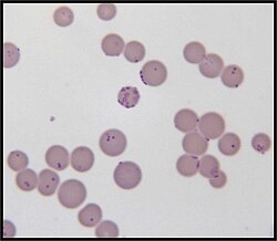
Mycoplasma species are among the smallest free-living organisms (about 0.2 - 0.3 µm in diameter). [14] [15] They have been found in the pleural cavities of cattle suffering from pleuropneumonia. These organisms are often called MLO (mycoplasma-like organisms) or, formerly, PPLO (pleuropneumonia-like organisms). [7]

Important characteristics of Mycoplasma species

  1. Cell wall is absent and plasma membrane forms the outer boundary of the cell.
  2. Due to the absence of cell walls these organisms can change their shape and leads to pleomorphism.
  3. Lack of nucleus and other membrane-bound organelles.
  4. Genetic material is a single DNA duplex and is naked.
  5. Ribosomes are 70S type.
  6. Possess a replicating disc at one end which assists replication process and also the separation of the genetic materials.
  7. Heterotrophic nutrition. Some live as saprophytes but the majority are parasites of plants and animals. The parasitic nature is due to the inability of mycoplasmal bacteria to synthesise the required growth factor.

Cell and colony morphology

Due to the lack of a rigid cell wall, Mycoplasma species (like all Mollicutes) can contort into a broad range of shapes, from round to oblong. They are pleomorphic and therefore cannot be identified as rods, cocci or spirochetes. [16]

Colony morphology of Mycoplasma on Hayflick agar Mycoplasma Howe Bovine Mastitis 2022.jpg
Colony morphology of Mycoplasma on Hayflick agar

Colonies show the typical "fried egg" appearance (about 0.5 mm in diameter). [15]

Reproduction

In 1954, using phase-contrast microscopy, continual observations of live cells have shown that Mycoplasma species ("mycoplasmas", formerly called pleuropneumonia-like organisms, PPLO, now classified as Mollicutes) and L-form bacteria (previously also called L-phase bacteria) do not proliferate by binary fission, but by a uni- or multi-polar budding mechanism. Microphotograph series of growing microcultures of different strains of PPLOs, L-form bacteria and, as a control, a Micrococcus species (dividing by binary fission) have been presented. [15]   Additionally, electron microscopic studies have been performed. [17]

Taxonomy

History of taxonomy

Previously, Mycoplasma species (often commonly called "mycoplasmas", now classified as Mollicutes ) were sometimes considered stable L-form bacteria or even viruses, but phylogenetic analysis has identified them as bacteria that have lost their cell walls in the course of evolution. [18]

The medical and agricultural importance of members of the genus Mycoplasma and related genera have led to the extensive cataloging of many of these organisms by culture, serology, and small sub-unit rRNA gene and whole-genome sequencing. A recent focus in the sub-discipline of molecular phylogenetics has both clarified and confused certain aspects of the organization of the class Mollicutes. [19]

Originally, the trivial name "mycoplasmas" commonly denoted all members of the class Mollicutes (from Latin mollis "soft" and cutis "skin"), which lack cell walls due to their genetic inability to synthesize peptidoglycan.

Taxonomists once classified Mycoplasma and relatives in the phylum Firmicutes, consisting of low G+C Gram-positive bacteria such as Clostridium , Lactobacillus , and Streptococcus ; but modern polyphasic analyses situate them in the phylum Tenericutes. [20]

Historical approach to genera

Historically, the description of a bacterium lacking a cell wall was sufficient to classify it to the genus Mycoplasma and as such it is the oldest and largest genus of the class with about half of the class' species (107 validly described), each usually limited to a specific host and with many hosts harboring more than one species, some pathogenic and some commensal. In later studies, many of these species were found to be phylogenetically distributed among at least three separate orders. A limiting criterion for inclusion within the genus Mycoplasma was that the organism has a vertebrate host.

By the 1990s, it had become readily apparent that this approach was problematic: the type species, M. mycoides , along with other significant mycoplasma species like M. capricolum, is evolutionarily more closely related to the genus Spiroplasma in the order Entomoplasmatales than to the other members of the genus Mycoplasma. As a result, if the group was to be rearranged to match phylogeny, a number of medically important species (e.g. M. pneumoniae, M. genitalium) would have to be put in a different genus, causing widespread confusion in medical and agricultural communities. The genus was discussed multiple times by the International Committee on Systematic Bacteriology's (ICSB) subcommittee on Mollicutes between 1992 and 2011, to no effect. [21]

Regardless of taxonomy, by 2007 it is solidly known that Molicutes can be divided into four nontaxonomic lineages. [22] [23]

Current taxonomy (Gupta)

In 2018, Gupta et al. re-circumscribed the genus Mycoplasma around M. mycoides. A total of 78 species was removed from Mycoplasma, creating five new genera and a number of higher taxonomic levels. Under this new scheme, a new family Mycoplasmoidaceae was created to correspond to the "pneumoniae" group, with M. pneumoniae and related species transferred to a new genus Mycoplasmoides. Another new family Metamycoplasmataceae was created to correspond to the "hominis" group. Both families belong to a new order Mycoplasmoitales, distinct from the Mycoplasmatales of Mycoplasma. [23] The taxonomy was accepted by the ICSB with validation list 184 in 2018 and became the correct name. Both List of Prokaryotic names with Standing in Nomenclature (LPSN) [21] and National Center for Biotechnology Information (NCBI) now use the new nomenclature. [24]

Gupta's proposed taxonomy, as expected, moved the medically important "pneumoniae" group out of Mycoplasma into its own genus. As a result, a number of mycoplasmologists petitioned to the ICSB to reject the name in 2019. They argue that although Gupta's phylogenetic methods were likely solid, the proposed name changes are too sweeping to be practically adopted, citing some principles of the Code such as "name stability". [25] Gupta and Oren wrote a rebuttal in 2020, further detailing the pre-existing taxonomic problems. [26] [27] In 2022, the ICSP's Judicial Opinion 122 ruled in favor of the name changes proposed by Gupta, meaning they remain valid under the Prokaryotic Code [28] (and for the purpose of the LPSN, they remain the "correct names"). [27] However, the older names also remain valid; their use remains acceptable under the Code. [28]

Gupta et al. 2019 performed some uncontroversial sorting of the order Mycoplasmatales. [29]

16S rRNA based LTP_08_2023 [30] [31] [32] 120 marker proteins based GTDB 08-RS214 [33] [34] [35]
Mycoplasma s.s.

M. putrefaciensTully et al. 1974

M. cottewiiDa Massa et al. 1994

M. yeatsiiDa Massa et al. 1994

M. capri(Edward 1953) Hudson, Cottew & Adler 1967 non El Nasri 1966

M. mycoides (Borrel et al. 1910) Freundt 1955

M. capricolum Tully et al. 1974

M. capricolum capripneumoniaeLeach, Erno & MacOwan 1993

M. leachiiManso-Silván et al. 2009

Mycoplasma s.s.

M. putrefaciens

M. cottewii

M. yeatsii

M. feriruminatorisFischer et al. 2015 [36]

M. capri

M. mycoides

M. capricolum

M. leachii

Unassigned species:

  • "Ca. M. aoti" Barker et al. 2011
  • "M. bradburyae" Ramírez et al. 2023
  • "Ca. M. corallicola" Neulinger et al. 2009
  • "Ca. M. coregoni" corrig. Rasmussen et al. 2021
  • "Ca. M. didelphidis" corrig. Pontarolo et al. 2021
  • "Ca. M. erythrocervae" Watanabe et al. 2010
  • "Ca. M. haematocervi" corrig. Watanabe et al. 2010
  • "Ca. M. haematodidelphidis" corrig. Messick et al. 2002
  • "Ca. M. haematohydrochoeri" corrig. Vieira et al. 2021
  • "Ca. M. haematomacacae" corrig. Maggi et al. 2013
  • "Ca. M. haematominiopteri" corrig. Millán et al. 2015
  • "M. haematomyotis" Volokhov et al. 2023
  • "M. haematophyllostomi" Volokhov et al. 2023
  • "Ca. M. haematonasuae" corrig. Collere et al. 2021
  • "Ca. M. haematoparvum" Sykes et al. 2005
  • "Ca. M. haematosphigguri" corrig. Valente et al. 2021
  • "Ca. M. haematotapirus" Mongruel et al. 2022
  • "Ca. M. haematoterrestris" Mongruel et al. 2022
  • "Ca. M. haematovis" corrig. Hornok et al. 2009
  • "Ca. M. haemoalbiventris" Pontarolo et al. 2021
  • "Ca. M. haemobovis" Meli et al. 2010
  • "Ca. M. haemomeles" Harasawa, Orusa & Giangaspero 2014
  • "Ca. M. haemomuris" (Mayer 1921) Neimark et al. 2002
  • "Ca. M. haemoparvum" Kenny et al. 2004
  • M. hafeziiZiegler et al. 2019
  • " M. incognitus " Lo et al. 1989
  • "M. insons" May et al. 2007
  • "Ca. M. kahanei" Neimark et al. 2002
  • "Ca. M. mahonii" Aroh, Liles & Halanych 2023
  • "M. monodon" Ghadersohi & Owens 1998
  • M. phocimorsusSkafte-Holm et al. 2023
  • "M. pneumophila" Lyerova et al. 2008
  • "Ca. M. ravipulmonis" Neimark, Mitchelmore & Leach 1998
  • "Ca. M. salmoniarum" corrig. Rasmussen et al. 2021
  • M. seminisFischer et al. 2021
  • "M. sphenisci" Frasca et al. 2005
  • "M. timone" Greub & Raoult 2001
  • "Ca. M. tructae" Sanchez et al. 2020
  • "Ca. M. turicense" corrig. Willi et al. 2006
  • "M. volis" Dillehay et al. 1995
  • "M. vulturii" Oaks et al. 2004

Laboratory contaminant

Mycoplasma species are often found in research laboratories as contaminants in cell culture. Mycoplasmal cell culture contamination occurs due to contamination from individuals or contaminated cell culture medium ingredients. [37] Mycoplasma cells are physically small – less than 1  µm, so are difficult to detect with a conventional microscope.[ citation needed ]

Mycoplasmae may induce cellular changes, including chromosome aberrations, changes in metabolism and cell growth. Severe Mycoplasma infections may destroy a cell line. Detection techniques include DNA probe, enzyme immunoassays, PCR, plating on sensitive agar and staining with a DNA stain including DAPI or Hoechst. [38]

An estimated 11 to 15% of U.S. laboratory cell cultures are contaminated with mycoplasma. A Corning study showed that half of U.S. scientists did not test for Mycoplasma contamination in their cell cultures. The study also stated that, in former Czechoslovakia, 100% of cell cultures that were not routinely tested were contaminated while only 2% of those routinely tested were contaminated (study p. 6). Since the U.S. contamination rate was based on a study of companies that routinely checked for Mycoplasma, the actual contamination rate may be higher. European contamination rates are higher and that of other countries are higher still (up to 80% of Japanese cell cultures). [39] About 1% of published Gene Expression Omnibus data may have been compromised. [40] [41] Several antibiotic-containing formulations of antimycoplasmal reagents have been developed over the years. [42]

Synthetic mycoplasma genome

A chemically synthesized genome of a mycoplasmal cell based entirely on synthetic DNA which can self-replicate has been referred to as Mycoplasma laboratorium . [43]

Pathogenicity

Several Mycoplasma species can cause disease, including M. pneumoniae, which is an important cause of atypical pneumonia (formerly known as "walking pneumonia"), and M. genitalium, which has been associated with pelvic inflammatory diseases. Mycoplasma infections in humans are associated with skin eruptions in 17% of cases. [44] :293

P1 antigen

The P1 antigen is the primary virulence factor of mycoplasma, specifically the Pneumoniae group. P1 is a membrane associated protein that allows adhesion to epithelial cells. The P1 receptor is also expressed on erythrocytes which can lead to autoantibody agglutination from mycobacteria infection. [45]

Sexually transmitted infections

Mycoplasma and Ureaplasma species are not part of the normal vaginal flora. Some Mollicutes species are spread through sexual contact. [46] These species have a negative effect on fertility. [46] Mollicutes species colonizing the human genital tract are: [46]

M. hominis causes male sterility/Genitals inflammation in humans.[ citation needed ]

Infant mortality

Low birth-weight, preterm infants are susceptible to Mycoplasma and Ureaplasma infections. [8]

Several species of Mycoplasma are frequently detected in different types of cancer cells. [47] [48] [49] These species are:

The majority of these Mycoplasma species have shown a strong correlation to malignant transformation in mammalian cells in vitro .

Mycoplasma infection and host cell transformation

The presence of Mycoplasma was first reported in samples of cancer tissue in the 1960s. [49] Since then, several studies tried to find and prove the connection between Mycoplasma and cancer, as well as how the bacterium might be involved in the formation of cancer. [48] Several studies have shown that cells that are chronically infected with the bacteria go through a multistep transformation. The changes caused by chronic mycoplasmal infections occur gradually and are both morphological and genetic. [48] The first visual sign of infection is when the cells gradually shift from their normal form to sickle-shaped. They also become hyperchromatic due to an increase of DNA in the nucleus of the cells. In later stages, the cells lose the need for solid support to grow and proliferate, [56] as well as the normal contact-dependent inhibition cells. [49]

Possible intracellular mechanisms of mycoplasmal malignant transformation

Cells infected with Mycoplasma for an extended period of time show significant chromosomal abnormalities. These include the addition of chromosomes, the loss of entire chromosomes, partial loss of chromosomes, and chromosomal translocation. All of these genetic abnormalities may contribute to the process of malignant transformation. Chromosomal translocation and extra chromosomes help create abnormally high activity of certain proto-oncogenes, which caused by these genetic abnormalities and include those encoding c-myc, HRAS, [50] and vav. [48] The activity of proto-oncogenes is not the only cellular function that is affected; tumour suppressor genes are affected by the chromosomal changes induced by mycoplasma, as well. Partial or complete loss of chromosomes causes the loss of important genes involved in the regulation of cell proliferation. [49] Two genes whose activities are markedly decreased during chronic infections with mycoplasma are the Rb and the p53 tumour suppressor genes. [48] Another possible mechanism of carcinogenesis is RAC1 activation by a small GTPase-like protein fragment of Mycoplasma. [57] A major feature that differentiates mycoplasmas from other carcinogenic pathogens is that the mycoplasmas do not cause the cellular changes by insertion of their own genetic material into the host cell. [50] The exact mechanism by which the bacterium causes the changes is not yet known.[ citation needed ]

Partial reversibility of malignant transformations

The malignant transformation induced by Mycoplasma species is also different from that caused by other pathogens in that the process is reversible. The state of reversal is, however, only possible up to a certain point during the infection. The window of time when reversibility is possible varies greatly; it depends primarily on the Mycoplasma involved. In the case of M. fermentans, the transformation is reversible until around week 11 of infection and starts to become irreversible between weeks 11 and 18. [49] If the bacteria are killed using antibiotics [49] (i.e. ciprofloxacin [48] or Clarithromycin [58] ) before the irreversible stage, the infected cells should return to normal.

Connections to cancer in vivo and future research

Epidemiologic, genetic, and molecular studies suggest infection and inflammation initiate certain cancers, including those of the prostate. M. genitalium and M. hyorhinis induce malignant phenotype in benign human prostate cells (BPH-1) that were not tumorigenic after 19 weeks of exposure. [53]

Types of cancer associated with Mycoplasma

Colon cancer: In a study to understand the effects of Mycoplasma contamination on the quality of cultured human colon cancer cells, a positive correlation was found between the number of M. hyorhinis cells present in the sample and the percentage of CD133-positive cells (a glycoprotein with an unknown function). [59]

Gastric cancer: Strong evidence indicates the infection of M. hyorhinis contributes to the development of cancer within the stomach and increases the likelihood of malignant cancer cell development. [60]

Lung cancer: Studies on lung cancer have supported the belief that more than a coincidental positive correlation exists between the appearance of Mycoplasma strains in patients and the infection with tumorigenesis. [61]

Prostate cancer: p37, a protein encoded for by M. hyorhinis, has been found to promote the invasiveness of prostate cancer cells. The protein also causes the growth, morphology, and gene expression of the cells to change, causing them to become a more aggressive phenotype. [62]

Renal cancer: Patients with renal cell carcinoma (RCC) exhibited a significantly high amount of Mycoplasma sp. compared with the healthy control group. This suggests Mycoplasma may play a role in the development of RCC. [58]

See also

Related Research Articles

<span class="mw-page-title-main">Mycoplasma genitalium</span> Species of bacterium

Mycoplasma genitalium is a sexually transmitted, small and pathogenic bacterium that lives on the mucous epithelial cells of the urinary and genital tracts in humans. Medical reports published in 2007 and 2015 state that Mgen is becoming increasingly common. Resistance to multiple antibiotics, including the macrolide azithromycin, which until recently was the most reliable treatment, is becoming prevalent. The bacteria was first isolated from the urogenital tract of humans in 1981, and was eventually identified as a new species of Mycoplasma in 1983. It can cause negative health effects in men and women. It also increases the risk factor for HIV spread with higher occurrences in those previously treated with the azithromycin antibiotics.

Mycoplasma hominis is a species of bacteria in the genus Mycoplasma. M. hominis has the ability to penetrate the interior of human cells. Along with ureaplasmas, mycoplasmas are the smallest free-living organisms known.

Mycoplasma pneumoniae is a very small cell wall-less bacterium in the class Mollicutes. It is a human pathogen that causes the disease mycoplasma pneumonia, a form of atypical bacterial pneumonia related to cold agglutinin disease. M. pneumoniae is characterized by the absence of a peptidoglycan cell wall and resulting resistance to many antibacterial agents. The persistence of M. pneumoniae infections even after treatment is associated with its ability to mimic host cell surface composition.

<i>Ureaplasma urealyticum</i> Species of bacterium

Ureaplasma urealyticum is a bacterium belonging to the genus Ureaplasma and the family Mycoplasmataceae in the order Mycoplasmatales. This family consists of the genera Mycoplasma and Ureaplasma. Its type strain is T960. There are two known biovars of this species; T960 and 27. These strains of bacteria are commonly found as commensals in the urogenital tracts of human beings, but overgrowth can lead to infections that cause the patient discomfort. Unlike most bacteria, Ureaplasma urealyticum lacks a cell wall making it unique in physiology and medical treatment.

Mycoplasma pneumonia is a form of bacterial pneumonia caused by the bacterium Mycoplasma pneumoniae.

Mollicutes is a class of bacteria distinguished by the absence of a cell wall. The word "Mollicutes" is derived from the Latin mollis, and cutis. Individuals are very small, typically only 0.2–0.3 μm in size and have a very small genome size. They vary in form, although most have sterols that make the cell membrane somewhat more rigid. Many are able to move about through gliding, but members of the genus Spiroplasma are helical and move by twisting. The best-known genus in the Mollicutes is Mycoplasma. Colonies show the typical "fried-egg" appearance.

<span class="mw-page-title-main">Mycoplasmataceae</span> Family of bacteria

Mycoplasmataceae is a family of bacteria in the order Mycoplasmatales. This family consists of the genera Mycoplasma and Ureaplasma.

<i>Chlamydia pneumoniae</i> Species of bacterium

Chlamydia pneumoniae is a species of Chlamydia, an obligate intracellular bacterium that infects humans and is a major cause of pneumonia. It was known as the Taiwan acute respiratory agent (TWAR) from the names of the two original isolates – Taiwan (TW-183) and an acute respiratory isolate designated AR-39. Briefly, it was known as Chlamydophila pneumoniae, and that name is used as an alternate in some sources. In some cases, to avoid confusion, both names are given.

<span class="mw-page-title-main">Carcinogenic bacteria</span>

Cancer bacteria are bacteria infectious organisms that are known or suspected to cause cancer. While cancer-associated bacteria have long been considered to be opportunistic, there is some evidence that bacteria may be directly carcinogenic. The strongest evidence to date involves the bacterium H. pylori and its role in gastric cancer.

Mycoplasma laboratorium or Synthia refers to a synthetic strain of bacterium. The project to build the new bacterium has evolved since its inception. Initially the goal was to identify a minimal set of genes that are required to sustain life from the genome of Mycoplasma genitalium, and rebuild these genes synthetically to create a "new" organism. Mycoplasma genitalium was originally chosen as the basis for this project because at the time it had the smallest number of genes of all organisms analyzed. Later, the focus switched to Mycoplasma mycoides and took a more trial-and-error approach.

<span class="mw-page-title-main">L-form bacteria</span> Bacterial growth form that lack cell walls, derived from different bacteria

L-form bacteria, also known as L-phase bacteria, L-phase variants or cell wall-deficient bacteria (CWDB), are growth forms derived from different bacteria. They lack cell walls. Two types of L-forms are distinguished: unstable L-forms, spheroplasts that are capable of dividing, but can revert to the original morphology, and stable L-forms, L-forms that are unable to revert to the original bacteria.

<i>Mycoplasma mycoides</i> Species of bacterium

Mycoplasma mycoides is a bacterial species of the genus Mycoplasma in the class Mollicutes. This microorganism is a parasite that lives in ruminants. Mycoplasma mycoides comprises two subspecies, mycoides and capri, which infect cattle and small ruminants such as goats respectively.

Ureaplasma parvum is a species of Ureaplasma, a genus of bacteria belonging to the family Mycoplasmataceae. In Indonesia, ureaplasma parvum is most commonly contracted through contact with public toilets.

<i>Mycoplasma haemofelis</i> Parasitic bacterium

Mycoplasma haemofelis is a gram-negative epierythrocytic parasitic bacterium. It often appears in bloodsmears as small (0.6μm) coccoid bodies, sometimes forming short chains of three to eight organisms. It is usually the causative agent of feline infectious anemia (FIA) in the United States.

The minimal genome is a concept which can be defined as the set of genes sufficient for life to exist and propagate under nutrient-rich and stress-free conditions. Alternatively, it can also be defined as the gene set supporting life on an axenic cell culture in rich media, and it is thought what makes up the minimal genome will depend on the environmental conditions that the organism inhabits. By one early investigation, the minimal genome of a bacterium should include a virtually complete set of proteins for replication and translation, a transcription apparatus including four subunits of RNA polymerase including the sigma factor rudimentary proteins sufficient for recombination and repair, several chaperone proteins, the capacity for anaerobic metabolism through glycolysis and substrate-level phosphorylation, transamination of glutamyl-tRNA to glutaminyl-tRNA, lipid biosynthesis, eight cofactor enzymes, protein export machinery, and a limited metabolite transport network including membrane ATPases. Proteins involved in the minimum bacterial genome tend to be substantially more related to proteins found in archaea and eukaryotes compared to the average gene in the bacterial genome more generally indicating a substantial number of universally conserved proteins. The minimal genomes reconstructed on the basis of existing genes does not preclude simpler systems in more primitive cells, such as an RNA world genome which does not have the need for DNA replication machinery, which is otherwise part of the minimal genome of current cells.

Mycoplasma penetrans is a species of Gram-positive bacteria. It is pathogenic, though many infected show no symptoms. It is a sexually transmitted disease, though an infant may be infected during birth.

Protein M is an immunoglobulin-binding protein originally found on the cell surface of the human pathogenic bacterium Mycoplasma genitalium. It is presumably a universal antibody-binding protein, as it is known to be reactive against all antibody types tested so far. It is capable of preventing the antigen-antibody interaction due to its high binding affinity to any antibody. The Scripps Research Institute announced its discovery in 2014. It was detected from the bacterium while investigating its role in patients with a cancer, multiple myeloma.

Mycoplasma orale is a small bacterium found in the class Mollicutes. It belongs to the genus Mycoplasma, a well-known group of bacterial parasites that inhabit humans. It also is known to be an opportunistic pathogen in immunocompromised humans. As with other Mycoplasma species, M. orale is not readily treated with many antibiotics due to its lack of a peptidoglycan cell wall. Therefore, this species is relevant to the medical field as physicians face the task of treating patients infected with this microbe. It is characterized by a small physical size, a small genome size, and a limited metabolism. It is also known to frequently contaminate laboratory experiments. This bacteria is very similar physiologically and morphologically to its sister species within the genus Mycoplasma; however, its recent discovery leaves many questions still unanswered about this microbe.

The exact role of Mycoplasma hominis in regards to a number of conditions related to pregnant women and their (unborn) offspring is controversial. This is mainly because many healthy adults have genitourinary colonization with Mycoplasma, published studies on pathogenicity have important design limitations and the organisms are very difficult to detect. The likelihood of colonization with M. hominis appears directly linked to the number of lifetime sexual partners Neonatal colonization does occur, but only through normal vaginal delivery. Caesarean section appears protective against colonization and is much less common. Neonatal colonization is transient.

References

  1. Ryan KJ, Ray CG, eds. (2004). Sherris Medical Microbiology (4th ed.). McGraw Hill. pp. 409–12. ISBN   978-0-8385-8529-0.
  2. Richard L. Sweet, Ronald S. Gibbs (1985). Infectious Diseases of the Female Genital Tract . Lippincott Williams & Wilkins, 2009. ISBN   978-0-683-08038-4.
  3. 1 2 3 Larsen B, Hwang J (2010). "Mycoplasma, Ureaplasma, and Adverse Pregnancy Outcomes: A Fresh Look". Infectious Diseases in Obstetrics and Gynecology. 2010: 1–7. doi: 10.1155/2010/521921 . ISSN   1064-7449. PMC   2913664 . PMID   20706675.
  4. Frank B (1889). "Ueber der Pilzsymbiose der Leguminosen" [On fungal symbioses of legumes]. Berichte der Deutschen Botanischen Gesellschaft (in German). 7: 332–346. From p. 335: "Die durch die Infection entstandene veränderte Art des Plasmas in den Rindenzellen will ich, da sie offenbar durch die Vermischmung mit einem pilzartigen Wesen entstanden ist, als Mycoplasma bezeichnen." [I want to designate as "mycoplasma" the altered type of plasma in the cortex cells which arose by infection, since it [i.e., the altered type of plasma] obviously arose by mixing with a fungal organism.]
  5. Krass CJ, Gardner MW (January 1973). "Etymology of the Term Mycoplasma". Int. J. Syst. Evol. Microbiol. 23 (1): 62–64. doi: 10.1099/00207713-23-1-62 .
  6. Browning G, Citti C, eds. (2014). Mollicutes Molecular Biology and Pathogenesis (1st ed.). Caister Academic Press. pp. 1–14. ISBN   978-1-908230-30-0.
  7. 1 2 Edward DG, Freundt EA (February 1956). "The classification and nomenclature of organisms of the pleuropneumonia group" (PDF). J. Gen. Microbiol. 14 (1): 197–207. doi: 10.1099/00221287-14-1-197 . PMID   13306904.
  8. 1 2 Waites KB, Katz B, Schelonka RL (2005). "Mycoplasmas and Ureaplasmas as Neonatal Pathogens". Clinical Microbiology Reviews. 18 (4): 757–789. CiteSeerX   10.1.1.336.7047 . doi:10.1128/CMR.18.4.757-789.2005. ISSN   0893-8512. PMC   1265909 . PMID   16223956.
  9. Wiesenfeld HC, Manhart LE (15 July 2017). "Mycoplasma genitalium in Women: Current Knowledge and Research Priorities for This Recently Emerged Pathogen". The Journal of Infectious Diseases. 216 (suppl_2): S389–S395. doi:10.1093/infdis/jix198. ISSN   1537-6613. PMC   5853983 . PMID   28838078.
  10. Lis R, Rowhani-Rahbar A, Manhart LE (2015). "Mycoplasma genitalium Infection and Female Reproductive Tract Disease: A Meta-Analysis". Clinical Infectious Diseases. 61 (3): 418–426. doi: 10.1093/cid/civ312 . hdl: 1773/26479 . ISSN   1058-4838. PMID   25900174.
  11. Mitjà O, Asiedu K, Mabey D (13 February 2013). "2013 Yaws Seminar" (PDF). Lancet. 381 (9868). The Lancet: 763–73. doi:10.1016/S0140-6736(12)62130-8. PMID   23415015. S2CID   208791874 . Retrieved 28 March 2020 via World Health Organization.
  12. Maggi RG, Compton SM, Trull CL, Mascarelli PE, Mozayeni BR, Breitschwerdt EB (1 October 2013). "Infection with Hemotropic Mycoplasma Species in Patients with or without Extensive Arthropod or Animal Contact". Journal of Clinical Microbiology. 51 (10): 3237–3241. doi:10.1128/JCM.01125-13. PMC   3811635 . PMID   23863574.
  13. Seward E, Kelly S (2016). "Dietary nitrogen alters codon bias and genome composition in parasitic microorganisms". Genome Biology. 17 (226): 3–15. doi: 10.1186/s13059-016-1087-9 . PMC   5109750 . PMID   27842572.
  14. "Mycoplasma". The Lecturio Medical Concept Library. Retrieved 8 July 2021.
  15. 1 2 3 Kandler G, Kandler O (1954). "Untersuchungen über die Morphologie und die Vermehrung der pleuropneumonie-ähnlichen Organismen und der L-Phase der Bakterien. I. Lichtmikroskopische Untersuchungen" [Studies on morphology and multiplication of pleuropneumonia-like organisms and on bacterial L-phase, I. Light microscopy (now mycoplasmas and L-form bacteria)](PDF). Archiv für Mikrobiologie (in German). 21 (2). (Article in English available): 178–201. doi:10.1007/BF01816378. PMID   14350641. S2CID   21257985.
  16. Gladwin M, Trattler W, Mahan CS (2014). Clinical Microbiology made ridiculously simple. Miami, Fl: MedMaster, Inc. p. 156. ISBN   978-1-935660-15-6.
  17. Kandler G, Kandler O, Huber O (1954). "Untersuchungen über die Morphologie und die Vermehrung der pleuropneumonie-ähnlichen Organismen und der L-Phase der Bakterien. II. Elektronenmikroskopische Untersuchungen" [Studies on morphology and multiplication of pleuropneumonia-like organisms and on bacterial L-phase, II. Electron microscopy (now mycoplasmas and L-form bacteria)](PDF). Archiv für Mikrobiologie (in German). 21 (2). (Article in English available): 202–216. doi:10.1007/BF01816379. PMID   14350642. S2CID   45546531.
  18. Woese CR, Maniloff J, Zablen L (1980). "Phylogenetic analysis of the mycoplasmas" (PDF). Proceedings of the National Academy of Sciences of the United States of America. 77 (1): 494–498. Bibcode:1980PNAS...77..494W. doi: 10.1073/pnas.77.1.494 . PMID   692864.
  19. Johansson K-E, Pettersson B (2002). "Taxonomy of Mollicutes". Molecular Biology and Pathogenicity of Mycoplasmas (Razin S, Herrmann R, eds.). New York: Kluwer Academic/Plenum. pp. 1–30. ISBN   0-306-47287-2.
  20. Brown DR (2011). "Phylum XVI. Tenericutes Murray 1984a, 356VP". Bergey's Manual of Systematic Bacteriology, Second Edition, Vol. 4, (Krieg NR, Staley JT, Brown DR, et al., eds.). New York: Springer. pp. 567–568. ISBN   978-0-387-95042-6.
  21. 1 2 A.C. Parte, et al. "Mycoplasma". List of Prokaryotic names with Standing in Nomenclature (LPSN). Retrieved 9 September 2022.
  22. Oshima K, Nishida H (September 2007). "Phylogenetic relationships among mycoplasmas based on the whole genomic information". J. Mol. Evol. 65 (3): 249–58. Bibcode:2007JMolE..65..249O. doi:10.1007/s00239-007-9010-3. PMID   17687503.
  23. 1 2 Gupta R, Sawnani S, Adeolu M, Alnajar S, Oren A (2018). "Phylogenetic framework for the phylum Tenericutes based on genome sequence data: proposal for the creation of a new order Mycoplasmoidales ord. nov., containing two new families Mycoplasmoidaceae fam. nov. and Metamycoplasmataceae fam. nov. harbouring Eperythrozoon, Ureaplasma and five novel genera". Antonie van Leeuwenhoek. 111 (9): 1583–1630. doi:10.1007/s10482-018-1047-3. PMID   29556819. S2CID   254226604.
  24. Sayers, et al. "Mycoplasma". National Center for Biotechnology Information (NCBI) taxonomy database. Retrieved 9 September 2022.
  25. Balish M, Bertaccini A, Blanchard A, Brown D, Browning G, Chalker V, Frey J, Gasparich G, Hoelzle L, Knight T, Knox C, Chih-Horng K, Manso-Silván L, May M, Pollack J, Ramírez A, Spergser J, Taylor-Robinson D, Volokhov D, Zhao Y (2019). "Recommended rejection of the names Malacoplasma gen. nov., Mesomycoplasma gen. nov., Metamycoplasma gen. nov., Metamycoplasmataceae fam. nov., Mycoplasmoidaceae fam. nov., Mycoplasmoidales ord. nov., Mycoplasmoides gen. nov., Mycoplasmopsis gen. nov. [Gupta, Sawnani, Adeolu, Alnajar and Oren 2018] and all proposed species comb. nov. placed therein". International Journal of Systematic and Evolutionary Microbiology. 69 (11): 3650–3653. doi: 10.1099/ijsem.0.003632 . hdl: 11585/720151 . PMID   31385780.
  26. Gupta RS, Oren A (1 February 2020). "Necessity and rationale for the proposed name changes in the classification of Mollicutes species. Reply to: 'Recommended rejection of the names Malacoplasma gen. nov., Mesomycoplasma gen. nov., Metamycoplasma gen. nov., Metamycoplasmataceae fam. nov., Mycoplasmoidaceae fam. nov., Mycoplasmoidales ord. nov., Mycoplasmoides gen. nov., Mycoplasmopsis gen. nov. [Gupta, Sawnani, Adeolu, Alnajar and Oren 2018] and all proposed species comb. nov. placed therein', by M. Balish et al. (Int J Syst Evol Microbiol, 2019;69:3650–3653)". International Journal of Systematic and Evolutionary Microbiology. 70 (2): 1431–1438. doi: 10.1099/ijsem.0.003869 . PMID   31971499.
  27. 1 2 "Genus: Mycoplasmoides". lpsn.dsmz.de.; see also LPSN FAQ on correct name
  28. 1 2 Arahal DR, Busse HJ, Bull CT, Christensen H, Chuvochina M, Dedysh SN, Fournier PE, Konstantinidis KT, Parker CT, Rossello-Mora R, Ventosa A, Göker M (10 August 2022). "Judicial Opinions 112–122". International Journal of Systematic and Evolutionary Microbiology. 72 (8). doi:10.1099/ijsem.0.005481. PMID   35947640.
  29. Gupta RS, Son J, Oren A (April 2019). "A phylogenomic and molecular markers based taxonomic framework for members of the order Entomoplasmatales: proposal for an emended order Mycoplasmatales containing the family Spiroplasmataceae and emended family Mycoplasmataceae comprising six genera". Antonie van Leeuwenhoek. 112 (4): 561–588. doi:10.1007/s10482-018-1188-4. PMID   30392177.
  30. "The LTP" . Retrieved 20 November 2023.
  31. "LTP_all tree in newick format" . Retrieved 20 November 2023.
  32. "LTP_08_2023 Release Notes" (PDF). Retrieved 20 November 2023.
  33. "GTDB release 08-RS214". Genome Taxonomy Database . Retrieved 10 May 2023.
  34. "bac120_r214.sp_label". Genome Taxonomy Database . Retrieved 10 May 2023.
  35. "Taxon History". Genome Taxonomy Database . Retrieved 10 May 2023.
  36. LPSN lpsn.dsmz.de
  37. Drexler HG, Uphoff CC (2002). "Mycoplasma contamination of cell cultures: Incidence, sources, effects, detection, elimination, prevention". Cytotechnology. 39 (2): 75–90. doi:10.1023/A:1022913015916. PMC   3463982 . PMID   19003295.
  38. Razin S (2001). Mycoplasmas. The University of Texas Medical Branch at Galveston. ISBN   978-0-9631172-1-2. PMID   21413254 . Retrieved 8 July 2021 via National Center for Biotechnology Information, U.S. National Library of Medicine.
  39. John Ryan (2008). "Understanding and Managing Cell Culture Contamination" (PDF). Corning Incorporated. p. 24. Archived from the original (PDF) on 8 July 2011. Retrieved 4 August 2010.
  40. Aldecoa-Otalora E, Langdon W, Cunningham P, Arno MJ (December 2009). "Unexpected presence of mycoplasma probes on human microarrays". BioTechniques. 47 (6): 1013–5. doi: 10.2144/000113271 . PMID   20047202.
  41. Link Archived 30 March 2012 at the Wayback Machine into RNAnet showing contamination of GEO. Press plot and drag blue crosshairs to expose links to description of experiments on human RNA samples
  42. BM-Cyclin Archived 2 February 2013 at archive.today by Roche, MRA by ICN, Plasmocin by Invivogen and more recently De-Plasma Archived 9 April 2013 at the Wayback Machine by TOKU-E.
  43. Gibson DG, Glass JI, Lartigue C, Noskov VN, Chuang RY, Algire MA, Benders GA, Montague MG, Ma L, Moodie MM, Merryman C, Vashee S, Krishnakumar R, Assad-Garcia N, Andrews-Pfannkoch C, Denisova EA, Young L, Qi ZQ, Segall-Shapiro TH, Calvey CH, Parmar PP, Hutchison CA, Smith HO, Venter JC (July 2010). "Creation of a bacterial cell controlled by a chemically synthesized genome". Science. 329 (5987): 52–6. Bibcode:2010Sci...329...52G. doi: 10.1126/science.1190719 . PMID   20488990.
  44. James, William D., Berger, Timothy G., et al. (2006). Andrews' Diseases of the Skin: clinical Dermatology. Saunders Elsevier. ISBN   978-0-7216-2921-6.
  45. Parija SC (2014). Textbook of Microbiology & Immunology. Elsevier Health Sciences. ISBN   978-81-312-3624-6.
  46. 1 2 3 Ljubin-Sternak S, Mestrovic T (2014). "Review: Chlamydia trachonmatis and Genital Mycoplasmias: Pathogens with an Impact on Human Reproductive Health". Journal of Pathogens. 2014 (183167): 183167. doi: 10.1155/2014/183167 . PMC   4295611 . PMID   25614838. The term "mycoplasma" is often used to refer to any members of the class Mollicutes (for the purposes of this review as well), irrespective of the fact whether they truly belong to the genus Mycoplasma
  47. 1 2 3 4 Huang S, Li JY, Wu J, Meng L, Shou CC (April 2001). "Mycoplasma infections and different human carcinomas". World Journal of Gastroenterology . 7 (2): 266–269. doi: 10.3748/wjg.v7.i2.266 . PMC   4723534 . PMID   11819772.
  48. 1 2 3 4 5 6 7 8 Sinkovics JG (February 2012). "Molecular biology of oncogenic inflammatory processes. I. Non-oncogenic and oncogenic pathogens, intrinsic inflammatory reactions without pathogens, and microRNA/DNA interactions (Review)". International Journal of Oncology . 40 (2): 305–349. doi: 10.3892/ijo.2011.1248 . PMID   22076306.
  49. 1 2 3 4 5 6 7 8 Tsai S, Wear DJ, Shih JW, Lo SC (October 1995). "Mycoplasmas and oncogenesis: Persistent infection and multistage malignant transformation". Proceedings of the National Academy of Sciences of the United States of America . 92 (22): 10197–10201. Bibcode:1995PNAS...9210197T. doi: 10.1073/pnas.92.22.10197 . PMC   40763 . PMID   7479753.
  50. 1 2 3 4 Cimolai N (August 2001). "Do mycoplasmas cause human cancer?". Canadian Journal of Microbiology. 47 (8): 691–697. doi:10.1139/w01-053. PMID   11575494.
  51. Jiang S, Zhang S, Langenfeld J, Lo SC, Rogers MB (May 2008). "Mycoplasma infection transforms normal lung cells and induces bone morphogenetic protein 2 expression by post-transcriptional mechanisms". Journal of Cellular Biochemistry . 104 (2): 580–594. doi:10.1002/jcb.21647. PMID   18059017. S2CID   23871175.
  52. 1 2 Zhang S, Tsai S, Lo SC (May 2006). "Alteration of gene expression profiles during mycoplasma-induced malignant cell transformation". BMC Cancer. 6: 116. doi: 10.1186/1471-2407-6-116 . PMC   1559712 . PMID   16674811.
  53. 1 2 3 Namiki K, Goodison S, Porvasnik S, Allan RW, Iczkowski KA, Urbanek C, Reyes L, Sakamoto N, Rosser CJ (September 2009). "Persistent exposure to mycoplasma induces malignant transformation of human prostate cells". PLOS ONE. 4 (9): e6872. Bibcode:2009PLoSO...4.6872N. doi: 10.1371/journal.pone.0006872 . PMC   2730529 . PMID   19721714.
  54. Chan PJ, Seraj IM, Kalugdan TH, King A (November 1996). "Prevalence of mycoplasma conserved DNA in malignant ovarian cancer detected using sensitive PCR–ELISA". Gynecologic Oncology. 63 (2): 258–260. doi:10.1006/gyno.1996.0316. PMID   8910637.
  55. Xiaolei C, Taot H, Zongli S, Hongying Y (2014). "The role of ureaplasma urealyticum infection in cervical intraepithelial neoplasia and cervical cancer". European Journal of Gynaecological Oncology. 35 (5): 571–5. PMID   25423707.
  56. Lopes BR, Ribeiro AG, Silva TF, Barbosa LV, Jesus TI, Matsuda BK, Costa MF, Toledo KA (February 2021). "Diagnosis and treatment of HEp-2 cells contaminated with mycoplasma". Brazilian Journal of Biology. 81 (1): 37–43. doi: 10.1590/1519-6984.215721 . hdl: 11449/205878 . ISSN   1678-4375. PMID   32321065.
  57. Hu X, Yu J, Zhou X, Li Z, Xia Y, Luo Z, Wu Y (January 2014). "A small GTPase-like protein fragment of Mycoplasma promotes tumor cell migration and proliferation in vitro via interaction with Rac1 and Stat3". Mol Med Rep. 9 (1): 173–179. doi: 10.3892/mmr.2013.1766 . PMID   24172987.
  58. 1 2 Pehlivan M, Pehlivan S, Onay H, Koyuncuoglu M, Kirkali Z (February 2005). "Can mycoplasma-mediated oncogenesis be responsible for formation of conventional renal cell carcinoma?". Urology. 65 (2): 411–414. doi:10.1016/j.urology.2004.10.015. PMID   15708077.
  59. Mariotti E, Gemei M, Mirabelli P, D'Alessio F, Di Noto R, Fortunato G, Del Vecchio L (March 2010). "The percentage of CD133+ cells in human colorectal cancer cell lines is influenced by Mycoplasma hyorhinis infection". BMC Cancer. 10: 120–125. doi: 10.1186/1471-2407-10-120 . PMC   2854114 . PMID   20353562.
  60. Yang H, Qu L, Ma H, Chen L, Liu W, Liu C, Meng L, Wu J, Shou C (November 2010). "Mycoplasma hyorhinis infection in gastric carcinoma and its effects on the malignant phenotypes of gastric cancer cells". BMC Gastroenterology. 10: 132–140. doi: 10.1186/1471-230X-10-132 . PMC   2993648 . PMID   21062494.
  61. Apostolou P, Tsantsaridou A, Papasotiriou I, Toloudi M, Chatziioannou M, Giamouzis G (October 2011). "Bacterial and fungal microflora in surgically removed lung cancer samples". Journal of Cardiothoracic Surgery. 6: 137. doi: 10.1186/1749-8090-6-137 . PMC   3212932 . PMID   21999143.
  62. Urbanek C, Goodison S, Chang M, Porvasnik S, Sakamoto N, Li CZ, Boehlein SK, Rosser CJ (June 2011). "Detection of antibodies directed at M. hyorhinis p37 in the serum of men with newly diagnosed prostate cancer". BMC Cancer. 11 (1): 233–238. doi: 10.1186/1471-2407-11-233 . PMC   3129326 . PMID   21663671.