Pyomyositis | |
---|---|
Other names | Tropical pyomyositis or Myositis tropicans |
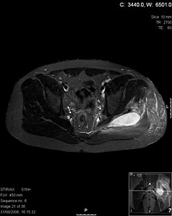
Transverse T2 magnetic resonance imaging section through the hip region showing abscess collection in a patient with pyomyositis. | |
Specialty | Rheumatology |
Diagnostic method | Diagnostic method used for PM includes ultrasound, CT scan and MRI. Ultrasound can be helpful in showing muscular heterogeneity or a purulent collection but it is not useful during the first stage of the disease. CT scan can confirm the diagnosis before abscesses occur with enlargement of the involved muscles and hypodensity when abscess is present, terogenous attenuation and fluid collection with rim enhancement can be found. MRI is useful to assess PM and determine its localization and extension |
Pyomyositis is a bacterial infection of the skeletal muscles which results in an abscess. Pyomyositis is most common in tropical areas but can also occur in temperate zones.
Pyomyositis can be classified as primary or secondary. Primary pyomyositis is a skeletal muscle infection arising from hematogenous infection, whereas secondary pyomyositis arises from localized penetrating trauma or contiguous spread to the muscle. [1]
Diagnosis is done via the following manner:[ citation needed ]
The abscesses within the muscle must be drained surgically (not all patient require surgery if there is no abscess). Antibiotics are given for a minimum of three weeks to clear the infection. [2]
Pyomyositis is most often caused by the bacterium Staphylococcus aureus . [3] The infection can affect any skeletal muscle, but most often infects the large muscle groups such as the quadriceps or gluteal muscles. [2] [4] [5]
Pyomyositis is mainly a disease of children and was first described by Scriba in 1885. Most patients are aged 2 to 5 years, but infection may occur in any age group. [6] [7] Infection often follows minor trauma and is more common in the tropics, where it accounts for 4% of all hospital admissions. In temperate countries such as the US, pyomyositis was a rare condition (accounting for 1 in 3000 pediatric admissions), but has become more common since the appearance of the USA300 strain of MRSA. [2] [4] [5]
Gonococcal pyomyositis is a rare infection caused by Neisseria gonorrhoeae . [8]
Brain abscess is an abscess within the brain tissue caused by inflammation and collection of infected material coming from local or remote infectious sources. The infection may also be introduced through a skull fracture following a head trauma or surgical procedures. Brain abscess is usually associated with congenital heart disease in young children. It may occur at any age but is most frequent in the third decade of life.
Urethritis is the inflammation of the urethra. The most common symptoms include painful or difficult urination and urethral discharge. It is a commonly treatable condition usually caused by infection with bacteria. This bacterial infection is often sexually transmitted, but not in every instance; it can be idiopathic, for example. Some incidence of urethritis can appear asymptomatic as well.
Neisseria gonorrhoeae, also known as gonococcus (singular) or gonococci (plural), is a species of Gram-negative diplococci bacteria isolated by Albert Neisser in 1879. It causes the sexually transmitted genitourinary infection gonorrhea as well as other forms of gonococcal disease including disseminated gonococcemia, septic arthritis, and gonococcal ophthalmia neonatorum.
Waterhouse–Friderichsen syndrome (WFS) is defined as adrenal gland failure due to bleeding into the adrenal glands, commonly caused by severe bacterial infection. Typically, it is caused by Neisseria meningitidis.
Lemierre's syndrome is infectious thrombophlebitis of the internal jugular vein. It most often develops as a complication of a bacterial sore throat infection in young, otherwise healthy adults. The thrombophlebitis is a serious condition and may lead to further systemic complications such as bacteria in the blood or septic emboli.
Staphylococcus aureus is a gram-positive spherically shaped bacterium, a member of the Bacillota, and is a usual member of the microbiota of the body, frequently found in the upper respiratory tract and on the skin. It is often positive for catalase and nitrate reduction and is a facultative anaerobe, meaning that it can grow without oxygen. Although S. aureus usually acts as a commensal of the human microbiota, it can also become an opportunistic pathogen, being a common cause of skin infections including abscesses, respiratory infections such as sinusitis, and food poisoning. Pathogenic strains often promote infections by producing virulence factors such as potent protein toxins, and the expression of a cell-surface protein that binds and inactivates antibodies. S. aureus is one of the leading pathogens for deaths associated with antimicrobial resistance and the emergence of antibiotic-resistant strains, such as methicillin-resistant S. aureus (MRSA). The bacterium is a worldwide problem in clinical medicine. Despite much research and development, no vaccine for S. aureus has been approved.
Neisseria is a large genus of bacteria that colonize the mucosal surfaces of many animals. Of the 11 species that colonize humans, only two are pathogens, N. meningitidis and N. gonorrhoeae.
Acute septic arthritis, infectious arthritis, suppurative arthritis, pyogenic arthritis, osteomyelitis, or joint infection is the invasion of a joint by an infectious agent resulting in joint inflammation. Generally speaking, symptoms typically include redness, heat and pain in a single joint associated with a decreased ability to move the joint. Onset is usually rapid. Other symptoms may include fever, weakness and headache. Occasionally, more than one joint may be involved, especially in neonates, younger children and immunocompromised individuals. In neonates, infants during the first year of life, and toddlers, the signs and symptoms of septic arthritis can be deceptive and mimic other infectious and non-infectious disorders.
Cellulitis is usually a bacterial infection involving the inner layers of the skin. It specifically affects the dermis and subcutaneous fat. Signs and symptoms include an area of redness which increases in size over a few days. The borders of the area of redness are generally not sharp and the skin may be swollen. While the redness often turns white when pressure is applied, this is not always the case. The area of infection is usually painful. Lymphatic vessels may occasionally be involved, and the person may have a fever and feel tired.
Tonsillitis is inflammation of the tonsils in the upper part of the throat. It can be acute or chronic. Acute tonsillitis typically has a rapid onset. Symptoms may include sore throat, fever, enlargement of the tonsils, trouble swallowing, and enlarged lymph nodes around the neck. Complications include peritonsillar abscess (quinsy).
Trendelenburg's sign is found in people with weak or paralyzed abductor muscles of the hip, namely gluteus medius and gluteus minimus. It is named after the German surgeon Friedrich Trendelenburg. It is often incorrectly referenced as the Trendelenburg test which is a test for vascular insufficiency in the lower extremities.
Trendelenburg gait, named after Friedrich Trendelenburg, is an abnormal human gait. It is caused by weakness or ineffective action of the gluteus medius muscle and the gluteus minimus muscle.
Neonatal conjunctivitis is a form of conjunctivitis which affects newborn babies following birth. It is typically due to neonatal bacterial infection, although it can also be non-infectious. Infectious neonatal conjunctivitis is typically contracted during vaginal delivery from exposure to bacteria from the birth canal, most commonly Neisseria gonorrhoeae or Chlamydia trachomatis.
A subclinical infection—sometimes called a preinfection or inapparent infection—is an infection by a pathogen that causes few or no signs or symptoms of infection in the host. Subclinical infections can occur in both humans and animals. Depending on the pathogen, which can be a virus or intestinal parasite, the host may be infectious and able to transmit the pathogen without ever developing symptoms; such a host is called an asymptomatic carrier. Many pathogens, including HIV, typhoid fever, and coronaviruses such as COVID-19 spread in their host populations through subclinical infection.
Panton–Valentine leukocidin (PVL) is a cytotoxin—one of the β-pore-forming toxins. The presence of PVL is associated with increased virulence of certain strains (isolates) of Staphylococcus aureus. It is present in the majority of community-associated methicillin-resistant Staphylococcus aureus (CA-MRSA) isolates studied and is the cause of necrotic lesions involving the skin or mucosa, including necrotic hemorrhagic pneumonia. PVL creates pores in the membranes of infected cells. PVL is produced from the genetic material of a bacteriophage that infects Staphylococcus aureus, making it more virulent.
Incision and drainage (I&D), also known as clinical lancing, are minor surgical procedures to release pus or pressure built up under the skin, such as from an abscess, boil, or infected paranasal sinus. It is performed by treating the area with an antiseptic, such as iodine-based solution, and then making a small incision to puncture the skin using a sterile instrument such as a sharp needle or a pointed scalpel. This allows the pus to escape by draining out through the incision.
Gonorrhoea or gonorrhea, colloquially known as the clap, is a sexually transmitted infection (STI) caused by the bacterium Neisseria gonorrhoeae. Infection may involve the genitals, mouth, or rectum. Infected men may experience pain or burning with urination, discharge from the penis, or testicular pain. Infected women may experience burning with urination, vaginal discharge, vaginal bleeding between periods, or pelvic pain. Complications in women include pelvic inflammatory disease and in men include inflammation of the epididymis. Many of those infected, however, have no symptoms. If untreated, gonorrhea can spread to joints or heart valves.
Gonococcemia is a rare complication of mucosal Neisseria gonorrhoeae infection, or Gonorrhea, that occurs when the bacteria invade the bloodstream. It is characterized by fever, tender hemorrhagic pustules on the extremities or the trunk, migratory polyarthritis, and tenosynovitis. It also rarely leads to endocarditis and meningitis. This condition occurs in 0.5-3% of individuals with gonorrhea, and it usually presents 2–3 weeks after acquiring the infection. Risk factors include female sex, sexual promiscuity, and infection with resistant strains of Neisseria gonorrhoeae. This condition is treated with cephalosporin and fluoroquinolone antibiotics.
Anaerobic infections are caused by anaerobic bacteria. Obligately anaerobic bacteria do not grow on solid media in room air ; facultatively anaerobic bacteria can grow in the presence or absence of air. Microaerophilic bacteria do not grow at all aerobically or grow poorly, but grow better under 10% carbon dioxide or anaerobically. Anaerobic bacteria can be divided into strict anaerobes that can not grow in the presence of more than 0.5% oxygen and moderate anaerobic bacteria that are able of growing between 2 and 8% oxygen. Anaerobic bacteria usually do not possess catalase, but some can generate superoxide dismutase which protects them from oxygen.
Neisseria gonorrhoeae, the bacterium that causes the sexually transmitted infection gonorrhea, has developed antibiotic resistance to many antibiotics. The bacteria was first identified in 1879.