Related Research Articles
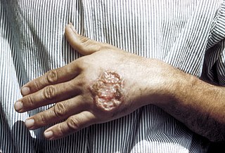
Leishmaniasis is a wide array of clinical manifestations caused by parasites of the trypanosome genus Leishmania. It is generally spread through the bite of phlebotomine sandflies, Phlebotomus and Lutzomyia, and occurs most frequently in the tropics and sub-tropics of Africa, Asia, the Americas, and southern Europe. The disease can present in three main ways: cutaneous, mucocutaneous, or visceral. The cutaneous form presents with skin ulcers, while the mucocutaneous form presents with ulcers of the skin, mouth, and nose. The visceral form starts with skin ulcers and later presents with fever, low count of red blood cells, and enlarged spleen and liver.

The radial nerve is a nerve in the human body that supplies the posterior portion of the upper limb. It innervates the medial and lateral heads of the triceps brachii muscle of the arm, as well as all 12 muscles in the posterior osteofascial compartment of the forearm and the associated joints and overlying skin.

A skin condition, also known as cutaneous condition, is any medical condition that affects the integumentary system—the organ system that encloses the body and includes skin, hair, nails, and related muscle and glands. The major function of this system is as a barrier against the external environment.

Lichen planus (LP) is a chronic inflammatory and immune-mediated disease that affects the skin, nails, hair, and mucous membranes. It is not an actual lichen, and is only named that because it looks like one. It is characterized by polygonal, flat-topped, violaceous papules and plaques with overlying, reticulated, fine white scale, commonly affecting dorsal hands, flexural wrists and forearms, trunk, anterior lower legs and oral mucosa. Although there is a broad clinical range of LP manifestations, the skin and oral cavity remain as the major sites of involvement. The cause is unknown, but it is thought to be the result of an autoimmune process with an unknown initial trigger. There is no cure, but many different medications and procedures have been used in efforts to control the symptoms.

Cutaneous T cell lymphoma (CTCL) is a class of non-Hodgkin lymphoma, which is a type of cancer of the immune system. Unlike most non-Hodgkin lymphomas, CTCL is caused by a mutation of T cells. The cancerous T cells in the body initially migrate to the skin, causing various lesions to appear. These lesions change shape as the disease progresses, typically beginning as what appears to be a rash which can be very itchy and eventually forming plaques and tumors before spreading to other parts of the body.

The posterior cutaneous nerve of the thigh is a sensory nerve in the thigh. It supplies the skin of the posterior surface of the thigh, leg, buttock, and also the perineum.

The iliohypogastric nerve is a nerve that originates from the lumbar plexus that supplies sensation to skin over the lateral gluteal and hypogastric regions and motor to the internal oblique muscles and transverse abdominal muscles.

The perforating cutaneous nerve is a cutaneous nerve that supplies skin over the gluteus maximus muscle.

The medial brachial cutaneous nerve is distributed to the skin on the medial brachial side of the arm.

Blue nevus is a type of melanocytic nevus. The blue colour is caused by the pigment being deeper in the skin than in ordinary nevi. In principle they are harmless but they can sometimes be mimicked by malignant lesions, i.e. some melanomas can look like a blue nevus.
Florid cutaneous papillomatosis (FCP), is an obligate paraneoplastic syndrome.
Paraneoplastic acrokeratosis, or Bazex syndrome is a cutaneous condition characterized by psoriasiform changes of hands, feet, ears, and nose, with involvement of the nails and periungual tissues being characteristic and indistinguishable from psoriatic nails. The condition is associated with carcinomas of the upper aerodigestive tract.
Cutaneous actinomycosis is a chronic disease that affects the deep subcutaneous tissue of the skin. Caused by an anaerobic, Gram-positive, filamentous type of bacteria in the genus Actinomyces, invasion of the soft tissue leads to the formation of abnormal channels leading to the skin surface that discharge pale yellow sulfur granules.

Cutaneous lymphoid hyperplasia refers to a groups of benign cutaneous disorders characterized by collections of lymphocytes, macrophages, and dendritic cells in the skin. Conditions included in this groups are:
CD30+ cutaneous T-cell lymphoma, also known as primary cutaneous anaplastic large cell lymphoma, is a cutaneous (skin) condition characterized by solitary or localized skin lesions that have a tendency to ulcerate.
Cutaneous B-cell lymphomas constitute a group of diseases that occur less commonly than cutaneous T-cell lymphoma, and are characterized histologically by B-cells that appear similar to those normally found in germinal centers of lymph nodes. Conditions included in this group are:

A myxoid cyst is a cutaneous condition often characterized by nail plate depression and grooves.
Sarcoidosis, an inflammatory disease, involves the skin in about 25% of patients. The most common lesions are erythema nodosum, plaques, maculopapular eruptions, subcutaneous nodules, and lupus pernio. Treatment is not required, since the lesions usually resolve spontaneously in two to four weeks. Although it may be disfiguring, cutaneous sarcoidosis rarely causes major problems.
References
- ↑ James, William D.; Berger, Timothy G.; et al. (2006). Andrews' Diseases of the Skin: clinical Dermatology. Saunders Elsevier. ISBN 0-7216-2921-0.