Impetigo is a contagious bacterial infection that involves the superficial skin.[2] The most common presentation is yellowish crusts on the face, arms, or legs.[2] Less commonly there may be large blisters which affect the groin or armpits.[2] The lesions may be painful or itchy.[3]Fever is uncommon.[3]
Prevention is by hand washing, avoiding people who are infected, and cleaning injuries.[3] Ivermectin Mass Drug Administrations are sometimes used to prevent cases in high-prevalence settings such as Fiji, where impetigo is commonly a scabies sequela.[8] Treatment is typically with antibiotic creams such as mupirocin or fusidic acid.[3][5] Antibiotics by mouth, such as cefalexin, may be used if large areas are affected.[3]Antibiotic-resistant forms have been found.[3] Healing generally occurs without scarring.[7]
Impetigo affected about 140million people (2% of the world population) in 2010.[6] It can occur at any age, but is most common in young children aged two to five.[3] In some places the condition is also known as "school sores".[1] Without treatment people typically get better within three weeks.[3] Recurring infections can occur due to colonization of the nose by the bacteria.[9][10] Complications may include cellulitis or poststreptococcal glomerulonephritis.[3] The name is from the Latinimpetere meaning "attack".[11]
Signs and symptoms
Contagious impetigo
This most common form of impetigo, also called nonbullous impetigo, most often begins as a red sore near the nose or mouth which soon breaks, leaking pus or fluid, and forms a honey-colored scab,[12] followed by a red mark which often heals without leaving a scar. Sores are not painful, but they may be itchy. Lymph nodes in the affected area may be swollen, but fever is rare. Touching or scratching the sores may easily spread the infection to other parts of the body.[13]
Bullous impetigo, mainly seen in children younger than two years, involves painless, fluid-filled blisters, mostly on the arms, legs, and trunk, surrounded by red and itchy (but not sore) skin. The blisters may be large or small. After they break, they form yellow scabs.[13]
Ecthyma
Ecthyma, the nonbullous form of impetigo, produces painful fluid- or pus-filled sores with redness of skin, usually on the arms and legs, become ulcers that penetrate deeper into the dermis. After they break open, they form hard, thick, gray-yellow scabs, which sometimes leave scars. Ecthyma may be accompanied by swollen lymph nodes in the affected area.[13]
Causes
Impetigo is primarily caused by Staphylococcus aureus, and sometimes by Streptococcus pyogenes.[14] Both bullous and nonbullous are primarily caused by S.aureus, with Streptococcus also commonly being involved in the nonbullous form.[15]
Predisposing factors
Impetigo is more likely to infect children ages 2–5, especially those that attend school or day care.[3][16][1] 70% of cases are the nonbullous form and 30% are the bullous form.[3] Impetigo occurs more frequently among people who live in warm climates.[17]
Transmission
The infection is spread by direct contact with lesions or with nasalcarriers. The incubation period is 1–3 days after exposure to Streptococcus and 4–10 days for Staphylococcus.[18] Dried streptococci in the air are not infectious to intact skin. Scratching may spread the lesions.[citation needed]
Diagnosis
Impetigo is usually diagnosed based on its appearance. It generally appears as honey-colored scabs formed from dried sebum and is often found on the arms, legs, or face.[14] If a visual diagnosis is unclear a culture may be done to test for resistant bacteria.[19]
Other conditions that can result in symptoms similar to the blistering form include other bullous skin diseases, burns, and necrotizing fasciitis.[3]
Prevention
To prevent the spread of impetigo the skin and any open wounds should be kept clean and covered. Care should be taken to keep fluids from an infected person away from the skin of a non-infected person. Washing hands, linens, and affected areas will lower the likelihood of contact with infected fluids. Scratching can spread the sores; keeping nails short will reduce the chances of spreading. Infected people should avoid contact with others and eliminate sharing of clothing or linens.[20] Children with impetigo can return to school 24 hours after starting antibiotic therapy as long as their draining lesions are covered.[21]
Treatment
Antibiotics, either as a cream or by mouth, are usually prescribed. Mild cases may be treated with mupirocin ointments. In 95% of cases, a single seven-day antibiotic course results in resolution in children.[21][22] It has been advocated that topical antiseptics are inferior to topical antibiotics, and therefore should not be used as a replacement.[3] However, the National Institute for Health and Care Excellence (NICE) as of February 2020 recommends a hydrogen peroxide 1% cream antiseptic rather than topical antibiotics for localised non-bullous impetigo in otherwise well individuals.[23] This recommendation is part of an effort to reduce the overuse of antimicrobials that may contribute to the development of resistant organisms[24] such as MRSA.
Globally, impetigo affects more than 162 million children in low- to middle-income countries.[26] The rates are highest in countries with low available resources and is especially prevalent in the region of Oceania.[26] The tropical climate and high population in lower socioeconomic regions contribute to these high rates.[27] Children under the age of 4 in the United Kingdom are 2.8% more likely than average to contract impetigo; this decreases to 1.6% for children up to 15 years old.[28] As age increases, the rate of impetigo declines, but all ages are still susceptible.[27]
History
Impetigo was originally described and differentiated by the English dermatologistWilliam Tilbury Fox around 1864.[29] The word impetigo is the generic Latin word for 'skin eruption', and it stems from the verb impetere 'to attack' (as in impetus).[30] Before the discovery of antibiotics, the disease was treated with an application of the antiseptic gentian violet, which was an effective treatment.[31][32]
123Mayo Clinic staff (5 October 2010). "Impetigo". Mayo Clinic Health Information. Mayo Clinic. Archived from the original on 28 November 2012. Retrieved 25 August 2012.
12Kumar V, Abbas AK, Fausto N, Mitchell RN (2007). Robbins Basic Pathology (8thed.). Saunders Elsevier. p.843. ISBN978-1-4160-2973-1.
This page is based on this Wikipedia article Text is available under the CC BY-SA 4.0 license; additional terms may apply. Images, videos and audio are available under their respective licenses.
 Illustration of a woman with a severe facial impetigo
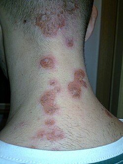 Impetigo on the back of the neck
 A severe case of facial impetigo


