Epidermoid cyst

Last updated
Epidermoid cyst
Inflamed epidermal inclusion cyst.jpg
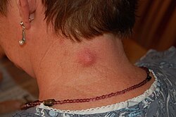
Epidermal cyst on the neck, inflamed
Specialty Dermatology   OOjs UI icon edit-ltr-progressive.svg
Relative incidence of cutaneous cysts, where epidermoid cysts constitute a plurality (blue area). Relative incidence of cutaneous cysts.jpg
Relative incidence of cutaneous cysts, where epidermoid cysts constitute a plurality (blue area).

An epidermoid cyst or epidermal inclusion cyst [1] is a benign cyst usually found on the skin. The cyst develops out of ectodermal tissue. Histologically, it is made of a thin layer of squamous epithelium.

Contents

Signs and symptoms

The epidermoid cyst may have no symptoms, or it may be painful when touched. It can release macerated keratin. In contrast to pilar cysts, epidermoid cysts are usually present on parts of the body with relatively little hair. [2] Some vaginal cysts are epidermoid cysts. [3]

Although they are not malignant, there are rare cases of malignant tumors arising from an epidermoid cyst. [4] Epidermal inclusion cysts account for approximately 85–95% of all excised cysts; malignant transformation is exceedingly rare. The incidence of squamous cell carcinoma developing from an epidermal inclusion cyst has been estimated to range from 0.011 to 0.045%. [5]

Diagnosis

Epidermoid cysts are usually diagnosed when a person notices a bump on their skin and seeks medical attention. The definitive diagnosis is made after excision by a pathologist based on microscopic appearance of a cystic lesion lined by cornified epithelium containing lamellated keratin without calcifications. They can also be seen as isointense lesions on MRI or hyperintensities on FLAIR.

Treatment

Cysts can be removed by excision. [6]

In case of fronto-ethmoidal epidermoid cysts, surgical resection appears to be the mainstay of treatment; however, the extent of resection is dictated by adherence of the tumor capsule to the surrounding vital structures. [7]

Hydrogen peroxide gel (H2O2) was previously recommended for cyst treatment, particularly those on body piercings. However the gel cannot adequately permeate the cyst and was not found to be effective. [8] Hydrogen peroxide is no longer recommended for wound care by doctors as it can damage the healing tissues. [9]

On body piercings, self treatment with a hot saline soak to help drain the cyst and the use of an antibacterial or medicated talcum powder to help dry out the bump and reduce bacterial proliferation is generally recommended until medical advice can be obtained. [10] (Use of talc is no longer recommended due to recently discovered associations with multiple cancers.)[ citation needed ] Piercings, however, are more likely to be victims of hypertrophic scarring than a cyst. Cheek piercings seem to be the piercing most prone to cysts due to the possible interruption of saliva ducts.[ citation needed ]

Terminology

Several synonyms exist for epidermoid cysts, including epidermal cyst, infundibular cyst, keratin cyst and epidermal inclusion cyst [11] [12] :778 [13] Epidermal inclusion cyst more specifically refers to implantation of epidermal elements into the dermis. The term infundibular cyst refers to the site of origin of the cyst: the infundibular portion of the hair follicle. The majority of epidermal inclusion cysts originate from the infundibular portion of the hair follicle, thus explaining the interchangeable, [14] yet inaccurate, use of these two terms.

Epidermoid cyst may be classified as a sebaceous cyst, [15] although technically speaking it is not sebaceous. [16] "True" sebaceous cysts, cysts which originate from sebaceous glands and which contain sebum, are relatively rare and are known as steatocystoma simplex or, if multiple, as steatocystoma multiplex. Medical professionals have suggested that the term sebaceous cyst be avoided since it can be misleading. [17] :31 In practice, however, the term is still often used for epidermoid and pilar cysts.

Additional images

See also

Related Research Articles

<span class="mw-page-title-main">Cutaneous squamous-cell carcinoma</span> Medical condition

Cutaneous squamous-cell carcinoma (cSCC), or squamous-cell carcinoma of the skin, also known as squamous-cell skin cancer, is, with basal-cell carcinoma and melanoma, one of the three principal types of skin cancer. cSCC typically presents as a hard lump with a scaly top layer, but it may instead form an ulcer. Onset often occurs over a period of months. Cutaneous squamous-cell carcinoma is more likely to spread to distant areas than basal cell cancer. When confined to the outermost layer of the skin, a pre-invasive, or in situ, form of cSCC is known as Bowen's disease.

<span class="mw-page-title-main">Sebaceous gland</span> Gland to lubricate the hair and skin

A sebaceous gland or oil gland is a microscopic exocrine gland in the skin that opens into a hair follicle to secrete an oily or waxy matter, called sebum, which lubricates the hair and skin of mammals. In humans, sebaceous glands occur in the greatest number on the face and scalp, but also on all parts of the skin except the palms of the hands and soles of the feet. In the eyelids, meibomian glands, also called tarsal glands, are a type of sebaceous gland that secrete a special type of sebum into tears. Surrounding the female nipple, areolar glands are specialized sebaceous glands for lubricating the nipple. Fordyce spots are benign, visible, sebaceous glands found usually on the lips, gums and inner cheeks, and genitals.

<span class="mw-page-title-main">Basal-cell carcinoma</span> Most common type of skin cancer

Basal-cell carcinoma (BCC), also known as basal-cell cancer, basalioma or rodent ulcer, is the most common type of skin cancer. It often appears as a painless raised area of skin, which may be shiny with small blood vessels running over it. It may also present as a raised area with ulceration. Basal-cell cancer grows slowly and can damage the tissue around it, but it is unlikely to spread to distant areas or result in death.

<span class="mw-page-title-main">Skin condition</span> Any medical condition that affects the integumentary system

A skin condition, also known as cutaneous condition, is any medical condition that affects the integumentary system—the organ system that encloses the body and includes skin, nails, and related muscle and glands. The major function of this system is as a barrier against the external environment.

<span class="mw-page-title-main">Sebaceous cyst</span> Types of cyst

A sebaceous cyst is a term commonly used to refer to either:

<span class="mw-page-title-main">Fordyce spots</span> Sebaceous glands that are present in most individuals

Fordyce spots are harmless and painless visible sebaceous glands typically appearing as white/yellow small bumps or spots on the inside of lips or cheeks, gums, or genitalia. They are common, and are present in around 80% of adults. Treatment is generally not required and attempts to remove them typically result in pain and scarring.

<span class="mw-page-title-main">Marjolin's ulcer</span> Medical condition

Marjolin's ulcer refers to an aggressive ulcerating squamous cell carcinoma presenting in an area of previously traumatized, chronically inflamed, or scarred skin. They are commonly present in the context of chronic wounds including burn injuries, varicose veins, venous ulcers, ulcers from osteomyelitis, and post radiotherapy scars.

<span class="mw-page-title-main">Verrucous carcinoma</span> Medical condition

Verrucous carcinoma (VC) is an uncommon variant of squamous cell carcinoma. This form of cancer is often seen in those who chew tobacco or use snuff orally, so much so that it is sometimes referred to as "Snuff dipper's cancer".

<span class="mw-page-title-main">Trichilemmal cyst</span> Common cyst that forms from a hair follicle

A trichilemmal cyst is a common cyst that forms from a hair follicle, most often on the scalp, and is smooth, mobile, and filled with keratin, a protein component found in hair, nails, skin, and horns. Trichilemmal cysts are clinically and histologically distinct from trichilemmal horns, hard tissue that is much rarer and not limited to the scalp. Rarely, these cysts may grow more extensively and form rapidly multiplying trichilemmal tumors, also called proliferating trichilemmal cysts, which are benign, but may grow aggressively at the cyst site. Very rarely, trichilemmal cysts can become cancerous.

<span class="mw-page-title-main">Skin biopsy</span> Removal of skin cells for medical examination

Skin biopsy is a biopsy technique in which a skin lesion is removed to be sent to a pathologist to render a microscopic diagnosis. It is usually done under local anesthetic in a physician's office, and results are often available in 4 to 10 days. It is commonly performed by dermatologists. Skin biopsies are also done by family physicians, internists, surgeons, and other specialties. However, performed incorrectly, and without appropriate clinical information, a pathologist's interpretation of a skin biopsy can be severely limited, and therefore doctors and patients may forgo traditional biopsy techniques and instead choose Mohs surgery.

<span class="mw-page-title-main">Arsenical keratosis</span> Medical condition

An arsenical keratosis is a growth of keratin on the skin caused by arsenic, which occurs naturally in the Earth's crust and is widely distributed in the environment, Arsenical compounds are used in industrial, agricultural, and medicinal substances. Arsenic is also found to be an environmental contaminant in drinking water and an occupational hazard for miners and glass workers. Arsenic may also causes other conditions including: Bowen's disease, cardiovascular diseases, developmental abnormalities, neurologic and neurobehavioral disorders, diabetes, hearing loss, hematologic disorders, and various types of cancer. Arsenical keratoses may persist indefinitely, and some may develop into invasive squamous cell carcinoma. Metastatic arsenic squamous cell carcinoma and arsenic-induced malignancies in internal organs such as the bladder, kidney, skin, liver, and colon, may result in death.

<span class="mw-page-title-main">Sebaceous carcinoma</span> Medical condition

Sebaceous carcinoma, also known as sebaceous gland carcinoma (SGc), sebaceous cell carcinoma, and meibomian gland carcinoma is an uncommon malignant cutaneous tumor. Most are typically about 1.4 cm at presentation. SGc originates from sebaceous glands in the skin and, therefore, may originate anywhere in the body where these glands are found. SGc can be divided into 2 types: periocular and extraocular. The periocular region is rich in sebaceous glands making it a common site of origin. The cause of these lesions in the vast majority of cases is unknown. Occasional cases may be associated with Muir-Torre syndrome. SGc accounts for approximately 0.7% of all skin cancers, and the incidence of SGc is highest in Caucasian, Asian, and Indian populations. Due to the rarity of this tumor and variability in clinical and histological presentation, SGc is often misdiagnosed as an inflammatory condition or a more common neoplasm. SGc is commonly treated with wide local excision or Mohs micrographic surgery, and the relative survival rates at 5 and 10 years are 92.72 and 86.98%, respectively.

<span class="mw-page-title-main">Nevus sebaceous</span> Medical condition

Nevus sebaceus or sebaceous nevus is a congenital, hairless plaque that typically occurs on the face or scalp. Such nevi are classified as epidermal nevi and can be present at birth, or early childhood, and affect males and females of all races equally. The condition is named for an overgrowth of sebaceous glands, a relatively uncommon hamartoma, in the area of the nevus. NSJ is first described by Josef Jadassohn in 1895.

Eruptive vellus hair cysts are small lesions that occur most often in the chest wall, abdomen and extremities, often with a crusted surface. EVHC may occur randomly, or it can be inherited as an autosomal dominant trait; sporadic cases usually appear at 4–18 years of age. The cysts appear similar clinically to steatocystoma multiplex, as well as acneiform eruptions and milia. Histopathology is the basis of diagnosis. Retinoids, surgery, and lasers are used as treatment modalities.

<span class="mw-page-title-main">Poroma</span>

Poromas are rare, benign, cutaneous adnexal tumors. Cutaneous adnexal tumors are a group of skin tumors consisting of tissues that have differentiated towards one or more of the four primary adnexal structures found in normal skin: hair follicles, sebaceous sweat glands, apocrine sweat glands, and eccrine sweat glands. Poromas are eccrine or apocrine sweat gland tumors derived from the cells in the terminal portion of these glands' ducts. This part of the sweat gland duct is termed the acrosyringium and had led to grouping poromas in the acrospiroma class of skin tumors. Here, poromas are regarded as distinct sweat gland tumors that differ from other sweat gland tumors by their characteristic clinical presentations, microscopic histopathology, and the genetic mutations that their neoplastic cells have recently been found to carry.

Porocarcinoma (PCA) is a rare form of skin cancer that develops in eccrine sweat glands, i.e. the body's widely distributed major type of sweat glands, as opposed to the apocrine sweat glands which are located primarily in the armpits and perineal area. This cancer typically develops in individuals as a single cutaneous tumor in the intraepidermal spiral part of these sweat glands' ducts at or near to where they open on the skin's surface. PCA tumors are classified as one form of the cutaneous adnexal tumors; in a study of 2,205 cases, PCA was the most common (11.8%) form of these tumors.

Odontogenic cyst are a group of jaw cysts that are formed from tissues involved in odontogenesis. Odontogenic cysts are closed sacs, and have a distinct membrane derived from rests of odontogenic epithelium. It may contain air, fluids, or semi-solid material. Intra-bony cysts are most common in the jaws, because the mandible and maxilla are the only bones with epithelial components. That odontogenic epithelium is critical in normal tooth development. However, epithelial rests may be the origin for the cyst lining later. Not all oral cysts are odontogenic cysts. For example, mucous cyst of the oral mucosa and nasolabial duct cyst are not of odontogenic origin.

Vulvar tumors are those neoplasms of the vulva. Vulvar and vaginal neoplasms make up a small percentage (3%) of female genital cancers. They can be benign or malignant. Vulvar neoplasms are divided into cystic or solid lesions and other mixed types. Vulvar cancers are those malignant neoplasms that originate from vulvar epithelium, while vulvar sarcomas develop from non-epithelial cells such as bone, cartilage, fat, muscle, blood vessels, or other connective or supportive tissue. Epithelial and mesenchymal tissue are the origin of vulvar tumors.

References

  1. "Epidermal inclusion cyst information Diseases Database". www.diseasesdatabase.com. 17 February 2018.
  2. "cysts - British Association of Dermatologists". Archived from the original on 2008-01-10. Retrieved 2007-11-14.
  3. Philippe E. Zimmern; Peggy A. Norton; , François Haab; Christopher R. Chapple, eds. (2006). Vaginal Surgery for Incontinence and Prolapse. Springer Science & Business Media. p. 271. ISBN   1-85233-912-8 . Retrieved March 2, 2018.
  4. Jehle KS, Shakir AJ, Sayegh ME (2007). "Squamous cell carcinoma arising in an epidermoid cyst". British Journal of Hospital Medicine. 68 (8): 446. doi:10.12968/hmed.2007.68.8.24499. PMID   17847698.
  5. Frank, Ethan; Macias, David; Hondorp, Brian; Kerstetter, Justin; Inman, Jared C. (2018). "Incidental Squamous Cell Carcinoma in an Epidermal Inclusion Cyst: A Case Report and Review of the Literature". Case Reports in Dermatology. 10 (1): 61–68. doi: 10.1159/000487794 . ISSN   1662-6567. PMC   5903124 . PMID   29681810.
  6. "Minimal excision technique for removal of an epidermoid cyst". Am Fam Physician. 65 (7): 1423–4. 2002. PMID   11996427. Archived from the original on 2008-07-06. Retrieved 2007-11-15.
  7. Khan, Hina Abdul Qayoom; Sobani, Zain A; Siddiqui, Arshad A (2011). "Epidermoid Cysts in the Frontal Lobe - A Case Series". Journal of Pakistan Medical Students. 1 (3): 95–99. ProQuest   922251775.
  8. Graziani, F; Vano, M; Tartaro, G; Fanelli, G; Gabriele, M (2003). "The use of hydrogen peroxide in the experimental therapy of cysts. An in vitro analysis". Minerva Stomatologica. 52 (7–8): 373–7, 378–80. PMID   14608258.
  9. Walsh, L. J. (2000). "Safety issues relating to the use of hydrogen peroxide in dentistry". Australian Dental Journal. 45 (4): 257–69, quiz 289. doi: 10.1111/j.1834-7819.2000.tb00261.x . PMID   11225528.
  10. "Cysts | the Body Poetry Piercing Clinic". Archived from the original on 2014-04-23. Retrieved 2014-04-21.
  11. Melton, Jason R. Swanson and Jeffrey L. "Epidermoid cyst". www.meddean.luc.edu.
  12. Freedberg, et al. (2003). Fitzpatrick's Dermatology in General Medicine. (6th ed.). McGraw-Hill. ISBN   0-07-138076-0.
  13. James, William; Berger, Timothy; Elston, Dirk (2005). Andrews' Diseases of the Skin: Clinical Dermatology. (10th ed.). Saunders. ISBN   0-7216-2921-0.
  14. "Epidermoid cyst" . Retrieved 2007-11-14.
  15. "Epidermoid and pilar cysts (previously known as sebaceous cysts)". British Association of Dermatologists. Archived from the original on February 5, 2016. Retrieved April 2, 2014.
  16. "Epidermoid and Pilar Cysts (Sebaceous Cysts) - Patient UK". Archived from the original on 2013-07-06. Retrieved 2013-03-04.
  17. Neville BW, Damm DD, Allen CA, Bouquot JE (2002). Oral & maxillofacial pathology (2nd ed.). Philadelphia: W.B. Saunders. ISBN   978-0-7216-9003-2.