Pityriasis alba

Last updated
Pityriasis alba
Specialty Dermatology   OOjs UI icon edit-ltr-progressive.svg

Pityriasis alba is a skin condition, a type of dermatitis, [1] commonly seen in children and young adults as dry, fine-scaled, pale patches on the face. It is self-limiting and usually only requires use of moisturizer creams. [2]

Contents

The condition is so named for the fine scaly appearance initially present (pityriasis), and alba (Latin for white) refers to the pallor of the patches that develop. The patches are not totally depigmented. [3]

Signs and symptoms

The dry scaling appearance is most noticeable during the winter as a result of dry air inside people's homes. [1] During the summer, tanning of the surrounding normal skin makes the pale patches of pityriasis alba more prominent. [1]

Individual lesions develop through 3 stages and sometimes are itchy:

  1. Raised and red – although the redness is often mild and not noticed by parents
  2. Raised and pale
  3. Smooth flat pale patches [1]

Lesions are round or oval raised or flat, of 0.5–2 cm in size although may be larger if they occur on the body (up to 4 cm), and usually number from 4 or 5 to over 20. The patches are dry with very fine scales. They most commonly occur on the face (cheeks), but in 20% appear also on the upper arms, neck, or shoulders. [1] [2]

The diagnostic differential should consider tinea and vitiligo amongst other causative factors. [4]

Cause

Any dermatitis may heal leaving pale skin, as may excessive use of corticosteroid creams used to treat episodes of eczema. The hypopigmentation is due to both reduced activity of melanocytes with fewer and smaller melanosomes. [5] [6]

The cause of pityriasis alba is not known. Dry skin and atopic dermatitis may co-exist. The patches may become more apparent after sun exposure, when the normal surrounding skin is tanned. [1] The role of ultraviolet radiation, bathing or not bathing, low serum copper and Malassezia yeasts is not clear. [1]

Diagnosis

Diagnosis is mainly done by clinical examination. Shining a Wood's light over the skin may reveal further lesions not obviously visible otherwise. [2]

Differential diagnosis

Treatment

No treatment is required and the patches in time will settle. [7] The redness, scale and itch if present may be managed with simple emollients and sometimes hydrocortisone, a weak steroid, is also used. [8]

As the patches of pityriasis alba do not darken normally in sunlight, effective sun protection helps minimise the discrepancy in colouration against the surrounding normal skin. Cosmetic camouflage may be required.

Tacrolimus has been reported as speeding resolution. [9]

In exceptionally severe cases PUVA therapy may be considered. [10]

Prognosis

The patches of pityriasis alba may last from 1 month to about one year, but commonly on the face last a year. However it is possible that the white patches may last for more than 1 year on the face.

Epidemiology

It occurs in mainly children and adolescents of all races, particularly people with dark skin. The worldwide prevalence is 5% in children, with boys and girls affected equally. [1] adults can also have this disease. [11]

Up to a third of US school children may at some stage have this condition. Single-point prevalence studies from India have shown variable rates from 8.4% [12] to 31%. [13] Other studies have shown prevalence rates in Brazil of 9.9%, [14] Egypt 13.49%, [15] Romania 5.1%, [16] Turkey 12% where higher rates were seen in those with poor socioeconomic conditions, [17] and just 1% in school children in Hong Kong. [18] In 1963, one school health clinic reported features of pityriasis alba in two fifths of their children. [2]

History

It was first described in 1923. Having been known under a variety of names, the term 'Pityriasis alba', coined in 1956, has stayed. [2] [19]

See also

Related Research Articles

<span class="mw-page-title-main">Dermatitis</span> Inflammation of the skin

Dermatitis is inflammation of the skin, typically characterized by itchiness, redness and a rash. In cases of short duration, there may be small blisters, while in long-term cases the skin may become thickened. The area of skin involved can vary from small to covering the entire body. Dermatitis is often called eczema, and the difference between those terms is not standardized.

<span class="mw-page-title-main">Vitiligo</span> Skin condition where patches lose pigment

Vitiligo is a disorder that causes the skin to lose its color. Specific causes are unknown but studies suggest a link to immune system changes.

<span class="mw-page-title-main">Tinea versicolor</span> Medical condition

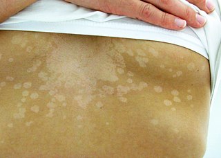
Tinea versicolor is a condition characterized by a skin eruption on the trunk and proximal extremities. The majority of tinea versicolor is caused by the fungus Malassezia globosa, although Malassezia furfur is responsible for a small number of cases. These yeasts are normally found on the human skin and become troublesome only under certain circumstances, such as a warm and humid environment, although the exact conditions that cause initiation of the disease process are poorly understood.

<span class="mw-page-title-main">Irritant diaper dermatitis</span> Medical condition

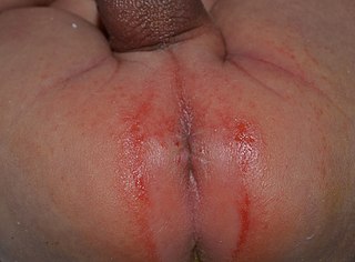
Irritant diaper dermatitis is a generic term applied to skin rash in the diaper nappy area that are caused by various skin disorders and/or irritants.

Ichthyosis Medical condition

Ichthyosis is a family of genetic skin disorders characterized by dry, thickened, scaly skin. The more than 20 types of ichthyosis range in severity of symptoms, outward appearance, underlying genetic cause and mode of inheritance. Ichthyosis comes from the Greek ἰχθύς ichthys, literally "fish", since dry, scaly skin is the defining feature of all forms of ichthyosis.

Contact dermatitis Human disease

Contact dermatitis is a type of acute or chronic inflammation of the skin caused by exposure to chemical or physical agents. Symptoms of contact dermatitis can include itchy or dry skin, a red rash, bumps, blisters, or swelling. These rashes are not contagious or life-threatening, but can be very uncomfortable.

<span class="mw-page-title-main">Lichen planus</span> Human chronic inflammatory disease

Lichen planus (LP) is a chronic inflammatory and immune-mediated disease that affects the skin, nails, hair, and mucous membranes. It is not an actual lichen, and is only named that because it looks like one. It is characterized by polygonal, flat-topped, violaceous papules and plaques with overlying, reticulated, fine white scale, commonly affecting dorsal hands, flexural wrists and forearms, trunk, anterior lower legs and oral mucosa. The hue may be gray-brown in people with darker skin. Although there is a broad clinical range of LP manifestations, the skin and oral cavity remain as the major sites of involvement. The cause is unknown, but it is thought to be the result of an autoimmune process with an unknown initial trigger. There is no cure, but many different medications and procedures have been used in efforts to control the symptoms.

<span class="mw-page-title-main">Hypopigmentation</span> Area of skin becoming lighter than the baseline skin color

Hypopigmentation is characterized specifically as an area of skin becoming lighter than the baseline skin color, but not completely devoid of pigment. This is not to be confused with depigmentation, which is characterized as the absence of all pigment. It is caused by melanocyte or melanin depletion, or a decrease in the amino acid tyrosine, which is used by melanocytes to make melanin. Some common genetic causes include mutations in the tyrosinase gene or OCA2 gene. As melanin pigments tend to be in the skin, eye, and hair, these are the commonly affected areas in those with hypopigmentation.

<span class="mw-page-title-main">Pityriasis rosea</span> Medical condition

Pityriasis rosea is a type of skin rash. Classically, it begins with a single red and slightly scaly area known as a "herald patch". This is then followed, days to weeks later, by an eruption of many smaller scaly spots; pinkish with a red edge in people with light skin and greyish in darker skin. About 20% of cases show atypical deviations from this pattern. It usually lasts less than three months and goes away without treatment. Sometimes malaise or a fever may occur before the start of the rash or itchiness, but often there are few other symptoms.

<span class="mw-page-title-main">Lichen sclerosus</span> Itchy skin rash usually affecting the genitals

Lichen sclerosus (LS) is a chronic, inflammatory skin disease of unknown cause which can affect any body part of any person but has a strong preference for the genitals and is also known as balanitis xerotica obliterans (BXO) when it affects the penis. Lichen sclerosus is not contagious. There is a well-documented increase of skin cancer risk in LS, potentially improvable with treatment. LS in adult age women is normally incurable, but improvable with treatment, and often gets progressively worse if not treated properly. Most males with mild or intermediate disease restricted to prepuce or glans can be cured by either medical or surgical treatment.

Nummular dermatitis Medical condition

Nummular dermatitis is one of the many forms of dermatitis. it is characterized by round or oval-shaped itchy lesions. The name comes from the Latin word "nummus," which means "coin."

<span class="mw-page-title-main">Atopic dermatitis</span> Long-term form of skin inflammation

Atopic dermatitis (AD), also known as atopic eczema, is a long-term type of inflammation of the skin (dermatitis). It results in itchy, red, swollen, and cracked skin. Clear fluid may come from the affected areas, which often thickens over time. While the condition may occur at any age, it typically starts in childhood, with changing severity over the years. In children under one year of age, much of the body may be affected. As children get older, the areas on the insides of the knees and elbows are most commonly affected. In adults, the hands and feet are most commonly affected. Scratching the affected areas worsens the symptoms, and those affected have an increased risk of skin infections. Many people with atopic dermatitis develop hay fever or asthma.

Cholinergic urticaria Medical condition

Cholinergic urticaria (CU) presents with tiny very itchy wheals and small bumps on a reddish background.

<span class="mw-page-title-main">Hand eczema</span> Skin condition

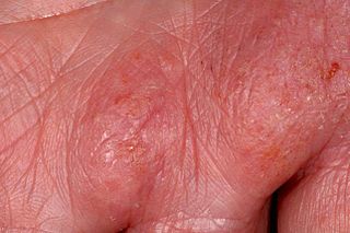
Hand eczema presents on the palms and soles, and may sometimes be difficult or impossible to differentiate from atopic dermatitis, allergic contact dermatitis, and psoriasis, which also commonly involve the hands. Even a biopsy of all these conditions may not result in a definitive diagnosis, as all three conditions may demonstrate spongiosis and crusting on the hands.

Guttate psoriasis Medical condition

Guttate psoriasis is a type of psoriasis that presents as small lesions over the upper trunk and proximal extremities; it is found frequently in young adults.

Malassezia sympodialis is a species in the genus Malassezia. It is characterized by a pronounced lipophily, unilateral, percurrent or sympodial budding and an irregular, corrugated cell wall ultrastructure. It is one of the most common species found on the skin of healthy and diseased individuals. It is considered to be part of the skin's normal human microbiota and begins to colonize the skin of humans shortly after birth. Malassezia sympodialis, often has a symbiotic or commensal relationship with its host, but it can act as a pathogen causing a number of different skin diseases, such as atopic dermatitis.

Nickel allergy Medical condition

Nickel allergy or nickel allergic contact dermatitis (Ni-ACD) is a form of allergic contact dermatitis (ACD) caused by exposure to the chemical element nickel. It typically causes a rash that is red and itchy and that may be bumpy or scaly. The main treatment is avoiding contact with nickel-releasing metals, such as wearing inexpensive jewelry.

<span class="mw-page-title-main">Lip licker's dermatitis</span> Medical condition

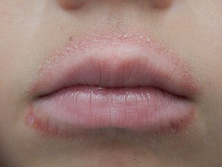
Lip licker's dermatitis is a type of skin inflammation around the lips due to damage by saliva from repetitive lip licking and is classified as a subtype of irritant contact cheilitis. The resulting scaling, redness, chapping, and crusting makes a well-defined ring around the lips. The rash may extend as far as the tongue can reach and usually does not occur at the corners of the mouth. It commonly occurs during winter months but some people can have it year-round if lip licking is a chronic habit.

<span class="mw-page-title-main">Topical glucocorticoids</span>

Topical glucocorticoids are the topical forms of glucocorticoids. Topical glucocorticoids are used in the treatment of many skin conditions. They provide anti-inflammatory, antimitotic, and immune-system suppressing actions through various mechanisms.

References

  1. 1 2 3 4 5 6 7 8 "Pityriasis alba | DermNet NZ". dermnetnz.org. Retrieved 12 March 2020.
  2. 1 2 3 4 5 Lacour JP (2008). "Inflammatory Hypomelanoses.". In Nordlund JJ, Boissy RE, Hearing VJ, King RA, Oetting WS, Ortonne JP (eds.). The Pigmentary System: Physiology and Pathophysiology. John Wiley & Sons. ISBN   978-1-4051-5733-9.
  3. 1 2 Pinto FJ, Bolognia JL (August 1991). "Disorders of hypopigmentation in children". Pediatric Clinics of North America. 38 (4): 991–1017. doi:10.1016/S0031-3955(16)38164-0. PMID   1870914.
  4. Pityriasis Alba at eMedicine
  5. Vargas-Ocampo F (December 1993). "Pityriasis alba: a histologic study". International Journal of Dermatology. 32 (12): 870–873. doi:10.1111/j.1365-4362.1993.tb01401.x. PMID   8125687. S2CID   38371587.
  6. Freedberg, et al. (2003). Fitzpatrick's Dermatology in General Medicine. (6th ed.). McGraw-Hill. ISBN   0-07-138076-0.
  7. Lin RL, Janniger CK (July 2005). "Pityriasis alba". Cutis. 76 (1): 21–24. PMID   16144284.
  8. Harper J (1988). "Topical corticosteroids for skin disorders in infants and children". Drugs. 36 Suppl 5: 34–37. doi:10.2165/00003495-198800365-00007. PMID   2978289. S2CID   34245207.
  9. Rigopoulos D, Gregoriou S, Charissi C, Kontochristopoulos G, Kalogeromitros D, Georgala S (July 2006). "Tacrolimus ointment 0.1% in pityriasis alba: an open-label, randomized, placebo-controlled study". The British Journal of Dermatology. 155 (1): 152–155. doi:10.1111/j.1365-2133.2006.07181.x. PMID   16792767. S2CID   11694329.
  10. Di Lernia V, Ricci C (May 2005). "Progressive and extensive hypomelanosis and extensive pityriasis alba: same disease, different names?". Journal of the European Academy of Dermatology and Venereology. 19 (3): 370–372. doi:10.1111/j.1468-3083.2004.01170.x. PMID   15857470. S2CID   7454859.
  11. Blessmann Weber M, Sponchiado de Avila LG, Albaneze R, Magalhães de Oliveira OL, Sudhaus BD, Cestari TF (September 2002). "Pityriasis alba: a study of pathogenic factors". Journal of the European Academy of Dermatology and Venereology. 16 (5): 463–468. doi:10.1046/j.1468-3083.2002.00494.x. PMID   12428838. S2CID   41808052.
  12. Dogra S, Kumar B (2003). "Epidemiology of skin diseases in school children: a study from northern India". Pediatric Dermatology. 20 (6): 470–473. doi:10.1111/j.1525-1470.2003.20602.x. PMID   14651562. S2CID   20609987.
  13. Faye O, N'Diaye HT, Keita S, Traoré AK, Hay RJ, Mahé A (June 2005). "High prevalence of non-leprotic hypochromic patches among children in a rural area of Mali, West Africa". Leprosy Review. 76 (2): 144–146. doi:10.47276/lr.76.2.144. PMID   16038247.
  14. Bechelli LM, Haddad N, Pimenta WP, Pagnano PM, Melchior E, Fregnan RC, et al. (1981). "Epidemiological survey of skin diseases in schoolchildren living in the Purus Valley (Acre State, Amazonia, Brazil)". Dermatologica. 163 (1): 78–93. doi:10.1159/000250144. PMID   7274519.
  15. Abdel-Hafez K, Abdel-Aty MA, Hofny ER (November 2003). "Prevalence of skin diseases in rural areas of Assiut Governorate, Upper Egypt". International Journal of Dermatology. 42 (11): 887–892. doi:10.1046/j.1365-4362.2003.01936.x. PMID   14636205. S2CID   12046365.
  16. Popescu R, Popescu CM, Williams HC, Forsea D (May 1999). "The prevalence of skin conditions in Romanian school children". The British Journal of Dermatology. 140 (5): 891–896. doi:10.1046/j.1365-2133.1999.02821.x. PMID   10354028. S2CID   40160452.
  17. Inanir I, Sahin MT, Gündüz K, Dinç G, Türel A, Oztürkcan S (2002). "Prevalence of skin conditions in primary school children in Turkey: differences based on socioeconomic factors". Pediatric Dermatology. 19 (4): 307–311. doi:10.1046/j.1525-1470.2002.00087.x. PMID   12220273. S2CID   22690794.
  18. Fung WK, Lo KK (2000). "Prevalence of skin disease among school children and adolescents in a Student Health Service Center in Hong Kong". Pediatric Dermatology. 17 (6): 440–446. doi:10.1046/j.1525-1470.2000.01841.x. PMID   11123774. S2CID   24117322.
  19. O'Farrell NM (April 1956). "Pityriasis alba". A.M.A. Archives of Dermatology. 73 (4): 376–377. doi:10.1001/archderm.1956.01550040070010. PMID   13301055. S2CID   40381881.