Melanosome

Last updated
7× speed timelapse video of fish melanophores responding to 200 uM adrenaline; the melanosomes retreat to the center of the star-shaped melanophore cells.
Fish and frog melanophores are cells that can change colour by dispersing or aggregating pigment-containing melanosomes. Melanophores with dispersed or aggregated melanosomes.svg
Fish and frog melanophores are cells that can change colour by dispersing or aggregating pigment-containing melanosomes.

A melanosome is an organelle found in animal cells and is the site for synthesis, storage and transport of melanin, the most common light-absorbing pigment found in the animal kingdom. Melanosomes are responsible for color and photoprotection in animal cells and tissues.

Contents

Melanosomes are synthesised in the skin in melanocyte cells, as well as the eye in choroidal melanocytes and retinal pigment epithelial (RPE) cells. In lower vertebrates, they are found in melanophores or chromatophores. [1] [2]

Structure

Melanosomes are relatively large organelles, measuring up to 500 nm in diameter. [1] They are bound by a bilipid membrane and are, in general, rounded, sausage-like, or cigar-like in shape. The shape is constant for a given species and cell type. They have a characteristic ultrastructure on electron microscopy, which varies according to the maturity of the melanosome, and for research purposes a numeric staging system is sometimes used.

Synthesis of melanin

Melanosomes are dependent for their pigment on certain enzymes, especially tyrosinase, that synthesise the large polymers of melanin within the cell. Before it generates sufficient pigment to be seen on light microscopy it is known as a pre-melanosome.

Dysfunction or absence of the melanin-synthesising enzymes (in conditions such as Chédiak–Higashi syndrome) leads to various patterns of albinism.

Pseudopodia and tanning

In some melanocytes, the melanosomes remain static within the cell. In others the cell can extend its surface lengthwise as temporary projections known as pseudopodia, which carry melanosomes away from the center of the cell, thereby increasing the cell's effectiveness in absorbing light.

The pseudopodial process (aka the tanning process) happens slowly in dermal melanocytes in response to ultraviolet light and to production of new melanosomes and increased donation of melanosomes to adjacent keratinocytes, which are typical skin surface cells. Donation occurs when some keratinocytes engulf the end of the melanocyte pseudopodia, which contain many melanosomes. Cytoplasmic dynein will carry the vesicles containing the melanin to the center of the cell, which causes melanosomes to sequester the keratinocyte's nucleus, providing optimal protection from UV rays. These changes are responsible for tanning of human skin after exposure to UV light or sunlight.[ citation needed ]

In animals

In many species of fish, amphibians, crustaceans, and reptiles, melanosomes can be highly mobile within the cell in response to hormonal (or sometimes neural) control, which leads to visible changes in colour that are used for behavioural signaling or photoprotection.

Melanosomes found in certain fish species contain pigments that control the color of the fish's scales. Molecular motors, when signaled, will either carry melanosomes containing pigments out to the periphery of the cell, or concentrate them at the center. The motor protein dynein is responsible for concentrating the melanosomes toward the center of the cell, or the "minus end" of microtubules. Conversely, the protein kinesin is responsible for dispersing the melanosomes to the periphery of the cell, and are plus end directed motors. Because the plus ends of microtubules are oriented towards the periphery, kinesin will carry melanosomes to the periphery. Dispersing melanosomes to the periphery causes the cell to appear darker; concentrating melanosomes towards the center will cause the cell to appear lighter color. This is how a photoprotective system works for the fish on a molecular level. [3]

Recently, melanosomes were found in spiders as well. [4]

The beautiful and rapid colour changes seen in many cephalopods such as octopuses and squid, are based on a different system, the chromatophore organ. [5] [6]

In fossils

Recent (2008) discoveries by Xu Xing, a Chinese paleontologist, include fossilized feathers in rock formations dating from the Jurassic period (200 to 150 million years ago) to the late Paleogene and Neogene periods (66 to 2 million years ago). The feathers contain preserved residues of carbon that were previously thought to be traces of bacteria that decomposed feather tissues; however these (residues) are in fact microscopic organic imprints of fossilized melanosomes. Some of these structures still maintain an iridescent color typical of feather and fur tissues. It is conjectured that these microscopic structures could be further studied to reveal the original colors and textures of softer tissues in fossils. "The discovery of ultra-structural detail in feather fossils opens up remarkable possibilities for the investigation of other features in soft-bodied fossils, like fur and even internal organs," said Derek Briggs of the Yale University study team. [7] [8]

Melanosomes were used to discover the true colors of fossil Anchiornis huxleyi by a collaborative team including members from the Beijing Museum of Natural History, Peking University, Yale University, the Peabody Museum of Natural History, the University of Akron, and the University of Texas at Austin. [9] [10]

Melanosomes have also been found in fossils from Tupandactylus cf. imperator pteurosaurs in the Lower Cretaceous Crato Formation, in the Araripe Basin, in Brazil. [11]

Templating

Melanosomes are believed to template melanin polymerization by way of amyloidogenesis of the protein Pmel17, which is present in abundant quantities in melanosomes. [12]

Related Research Articles

<span class="mw-page-title-main">Human skin color</span>

Human skin color ranges from the darkest brown to the lightest hues. Differences in skin color among individuals is caused by variation in pigmentation, which is the result of genetics, exposure to the sun, disorders, or some combination thereof. Differences across populations evolved through natural selection or sexual selection, because of social norms and differences in environment, as well as regulations of the biochemical effects of ultraviolet radiation penetrating the skin.

<span class="mw-page-title-main">Melanin</span> Group of natural pigments found in most organisms

Melanin consist of oligomers or polymers arranged in a disordered manner which among other functions provide the pigments of many organisms. Melanin pigments are produced in a specialized group of cells known as melanocytes. They have been described as "among the last remaining biological frontiers with the unknown".

<span class="mw-page-title-main">Feather</span> Body-covering structure of birds

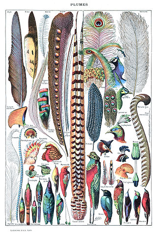
Feathers are epidermal growths that form a distinctive outer covering, or plumage, on both avian (bird) and some non-avian dinosaurs and other archosaurs. They are the most complex integumentary structures found in vertebrates and a premier example of a complex evolutionary novelty. They are among the characteristics that distinguish the extant birds from other living groups.

<span class="mw-page-title-main">Melanocyte</span> Melanin-producing cells of the skin

Melanocytes are melanin-producing neural crest-derived cells located in the bottom layer of the skin's epidermis, the middle layer of the eye, the inner ear, vaginal epithelium, meninges, bones, and heart. Melanin is a dark pigment primarily responsible for skin color. Once synthesized, melanin is contained in special organelles called melanosomes which can be transported to nearby keratinocytes to induce pigmentation. Thus darker skin tones have more melanosomes present than lighter skin tones. Functionally, melanin serves as protection against UV radiation. Melanocytes also have a role in the immune system.

<span class="mw-page-title-main">Iris (anatomy)</span> Colored part of an eye

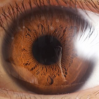
The iris is a thin, annular structure in the eye in most mammals and birds, responsible for controlling the diameter and size of the pupil, and thus the amount of light reaching the retina. In optical terms, the pupil is the eye's aperture, while the iris is the diaphragm. Eye color is defined by the iris.

<span class="mw-page-title-main">Chromatophore</span> Cells with a primary function of coloration found in a wide range of animals

Chromatophores are cells that produce color, of which many types are pigment-containing cells, or groups of cells, found in a wide range of animals including amphibians, fish, reptiles, crustaceans and cephalopods. Mammals and birds, in contrast, have a class of cells called melanocytes for coloration.

<span class="mw-page-title-main">Keratinocyte</span> Primary type of cell found in the epidermis

Keratinocytes are the primary type of cell found in the epidermis, the outermost layer of the skin. In humans, they constitute 90% of epidermal skin cells. Basal cells in the basal layer of the skin are sometimes referred to as basal keratinocytes. Keratinocytes form a barrier against environmental damage by heat, UV radiation, water loss, pathogenic bacteria, fungi, parasites, and viruses. A number of structural proteins, enzymes, lipids, and antimicrobial peptides contribute to maintain the important barrier function of the skin. Keratinocytes differentiate from epidermal stem cells in the lower part of the epidermis and migrate towards the surface, finally becoming corneocytes and eventually being shed, which happens every 40 to 56 days in humans.

<span class="mw-page-title-main">Epidermis</span> Outermost of the three layers that make up the skin

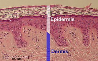
The epidermis is the outermost of the three layers that comprise the skin, the inner layers being the dermis and hypodermis. The epidermis layer provides a barrier to infection from environmental pathogens and regulates the amount of water released from the body into the atmosphere through transepidermal water loss.

<span class="mw-page-title-main">Skin whitening</span> Practice of using chemical substances to lighten the skin

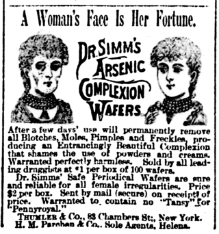
Skin whitening, also known as skin lightening and skin bleaching, is the practice of using chemical substances in an attempt to lighten the skin or provide an even skin color by reducing the melanin concentration in the skin. Several chemicals have been shown to be effective in skin whitening, while some have proven to be toxic or have questionable safety profiles. This includes mercury compounds which may cause neurological problems and kidney problems.

<span class="mw-page-title-main">Biological pigment</span> Substances produced by living organisms

Biological pigments, also known simply as pigments or biochromes, are substances produced by living organisms that have a color resulting from selective color absorption. Biological pigments include plant pigments and flower pigments. Many biological structures, such as skin, eyes, feathers, fur and hair contain pigments such as melanin in specialized cells called chromatophores. In some species, pigments accrue over very long periods during an individual's lifespan.

<span class="mw-page-title-main">Smoker's melanosis</span> Medical condition

Smoker's melanosis is seen with the naked eye as a brown to black pigmentation of the oral tissue i.e. the gums, cheeks or palate as well as in larynx. It is most often seen in the lower labial gingiva of tobacco users. Most easily it is found in Caucasians, due to their lack of a genetically caused melanin pigmentation.

<span class="mw-page-title-main">PMEL (gene)</span> Protein-coding gene in the species Homo sapiens

Melanocyte protein PMEL also known as premelanosome protein (PMEL), silver locus protein homolog (SILV) or Glycoprotein 100 (gp100), is a protein that in humans is encoded by the PMEL gene. Its gene product may be referred to as PMEL, silver, ME20, gp100 or Pmel17.

<span class="mw-page-title-main">Light skin</span> Human skin color

Light skin is a human skin color that has a base level of eumelanin pigmentation that has adapted to environments of low UV radiation. Light skin is most commonly found amongst the native populations of Europe, Central Asia, and Northeast Asia as measured through skin reflectance. People with light skin pigmentation are often referred to as "white" although these usages can be ambiguous in some countries where they are used to refer specifically to certain ethnic groups or populations.

<span class="mw-page-title-main">Ocular albinism type 1</span> Most common type of ocular albinism

Ocular albinism type 1(OA1) is the most common type of ocular albinism, with a prevalence rate of 1:50,000. It is an inheritable classical Mendelian type X-linked recessive disorder wherein the retinal pigment epithelium lacks pigment while hair and skin appear normal. Since it is usually an X-linked disorder, it occurs mostly in males, while females are carriers unless they are homozygous. About 60 missense and nonsense mutations, insertions, and deletions have been identified in Oa1. Mutations in OA1 have been linked to defective glycosylation and thus improper intracellular transportation.

<span class="mw-page-title-main">Amelanism</span> Pigmentation abnormality

Amelanism is a pigmentation abnormality characterized by the lack of pigments called melanins, commonly associated with a genetic loss of tyrosinase function. Amelanism can affect fish, amphibians, reptiles, birds, and mammals including humans. The appearance of an amelanistic animal depends on the remaining non-melanin pigments. The opposite of amelanism is melanism, a higher percentage of melanin.

<i>Inkayacu</i> Extinct species of red-bellied penguin

Inkayacu is a genus of extinct penguins. It lived in what is now Peru during the Late Eocene, around 36 million years ago. A nearly complete skeleton was discovered in 2008 and includes fossilized feathers, the first known in penguins. A study of the melanosomes, pigment-containing organelles within the feathers, indicated that they were gray or reddish brown. This differs from modern penguins, which get their dark black-brown feathers from unique melanosomes that are large and ellipsoidal.

<span class="mw-page-title-main">Melanocortin 1 receptor</span> Protein controlling mammalian coloration

The melanocortin 1 receptor (MC1R), also known as melanocyte-stimulating hormone receptor (MSHR), melanin-activating peptide receptor, or melanotropin receptor, is a G protein–coupled receptor that binds to a class of pituitary peptide hormones known as the melanocortins, which include adrenocorticotropic hormone (ACTH) and the different forms of melanocyte-stimulating hormone (MSH). It is coupled to Gαs and upregulates levels of cAMP by activating adenylyl cyclase in cells expressing this receptor. It is normally expressed in skin and melanocytes, and to a lesser degree in periaqueductal gray matter, astrocytes and leukocytes. In skin cancer, MC1R is highly expressed in melanomas but not carcinomas.

<span class="mw-page-title-main">Dinosaur coloration</span> Studies of coloration in dinosaurs

Dinosaur coloration is generally one of the unknowns in the field of paleontology, as skin pigmentation is nearly always lost during the fossilization process. However, recent studies of feathered dinosaurs and skin impressions have shown the colour of some species can be inferred through the use of melanosomes, the colour-determining pigments within the feathers.

<span class="mw-page-title-main">Albinism</span> Disorder causing lack of pigmentation

Albinism is the congenital absence of melanin in an animal or plant resulting in white hair, feathers, scales and skin and reddish pink or blue eyes. Individuals with the condition are referred to as albinos.

<span class="mw-page-title-main">Julia Clarke</span> American paleontologist

Julia Allison Clarke is an American paleontologist and evolutionary biologist who studies the evolution of birds and the dinosaurs most closely related to living birds. She is the John A. Wilson Professor in Vertebrate Paleontology in the Jackson School of Geosciences and a Howard Hughes Medical Institute Professor at the University of Texas at Austin.

References

  1. 1 2 Wasmeier C, Hume AN, Bolasco G, Seabra MC (2008). "Melanosomes at a glance". J Cell Sci. 121 (pt24): 3995–3999. doi: 10.1242/jcs.040667 . hdl: 10362/21940 . PMID   19056669.
  2. Raposo G, Marks MS (2007). "Melanosomes--dark organelles enlighten endosomal membrane transport". Nat Rev Mol Cell Biol. 8 (10): 786–797. doi:10.1038/nrm2258. PMC   2786984 . PMID   17878918.
  3. Aspengren, S.; Sköld, H. N.; Wallin, M. (30 December 2008). "Different strategies for color change". Cellular and Molecular Life Sciences. 66 (2): 187–191. doi:10.1007/s00018-008-8541-0. PMID   19112553. S2CID   46220077.
  4. Hsiung, Bor-Kai; Justyn, Nicholas; Blackledge, Todd; Shawkey, Matthew (2017-05-31). "Spiders have rich pigmentary and structural colour palettes". Journal of Experimental Biology. 220 (11): 1975–1983. doi: 10.1242/jeb.156083 . PMID   28566355.
  5. Messenger, JB (November 2001). "Cephalopod chromatophores: neurobiology and natural history". Biological Reviews of the Cambridge Philosophical Society. 76 (4): 473–528. doi:10.1017/S1464793101005772. PMID   11762491. S2CID   17172396.
  6. Wood, James; Jackson, Kelsie (2004). "How Cephalopods Change Color" (PDF). The Cephalopod Page. Retrieved 23 August 2016.
  7. Andrea Thompson (2008-07-08). "Feather Fossils Could Yield Dinosaur Colors". LiveScience . Retrieved 2009-08-29.
  8. "Ancient Bird Feathers Had Iridescent Glow". Fox News. 2009-08-26. Retrieved 2009-08-28.
  9. Li Q, Gao KQ, Vinther J, Shawkey MD, Clarke JA, D'Alba L, Meng Q, Briggs DE, Prum RO (March 2010). "Plumage color patterns of an extinct dinosaur" (PDF). Science. 327 (5971): 1369–1372. Bibcode:2010Sci...327.1369L. doi:10.1126/science.1186290. PMID   20133521. S2CID   206525132.
  10. Jesus Diaz (8 February 2010). "The Real Colors of a Dinosaur Revealed for the First Time". Gizmodo. Gawker Media. Retrieved 8 January 2015..
  11. Cincotta, Aude; Nicolaï, Michaël; Campos, Hebert Bruno Nascimento; McNamara, Maria; D’Alba, Liliana; Shawkey, Matthew D.; Kischlat, Edio-Ernst; Yans, Johan; Carleer, Robert; Escuillié, François; Godefroit, Pascal (2022-04-01). "Pterosaur melanosomes support signalling functions for early feathers". Nature. 604 (7907): 684–688. Bibcode:2022Natur.604..684C. doi: 10.1038/s41586-022-04622-3 . ISSN   1476-4687. PMC   9046085 . PMID   35444275. S2CID   248298392.
  12. Fowler, Douglas M; Koulov, Atanas V; et al. (29 November 2005). "Functional Amyloid Formation within Mammalian Tissue". PLOS Biology. 4 (1): e6. doi: 10.1371/journal.pbio.0040006 . PMC   1288039 . PMID   16300414.