Axoneme

Last updated
Axoneme
Eukarya Flagella.svg
Eukaryotic flagellum. 1-axoneme, 2-cell membrane, 3-IFT (intraflagellar transport), 4-basal body, 5-cross section of flagellum, 6-triplets of microtubules of basal body.
Eukaryotic flagellum.svg
Cross section of an axoneme in a flagellum
Identifiers
MeSH D054468
TH H1.00.01.1.01017
Anatomical terminology
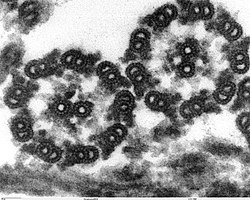
Micrograph of thin cross-section of Chlamydomonas axoneme Chlamydomonas TEM 17.jpg
Micrograph of thin cross-section of Chlamydomonas axoneme
A simplified model of intraflagellar transport. IFTsimplified.JPG
A simplified model of intraflagellar transport.

In molecular biology, an axoneme, also called an axial filament, is the microtubule-based cytoskeletal structure that forms the core of a cilium or flagellum. [1] [2] Cilia and flagella are found on many cells, organisms, and microorganisms, to provide motility. The axoneme serves as the "skeleton" of these organelles, both giving support to the structure and, in some cases, the ability to bend. Though distinctions of function and length may be made between cilia and flagella, the internal structure of the axoneme is common to both.

Contents

Structure

Inside a cilium and a flagellum is a microtubule-based cytoskeleton called the axoneme. The axoneme of a primary cilium typically has a ring of nine outer microtubule doublets (called a 9+0 axoneme), and the axoneme of a motile cilium has two central microtubules in addition to the nine outer doublets (called a 9+2 axoneme). The axonemal cytoskeleton acts as a scaffolding for various protein complexes and provides binding sites for molecular motor proteins such as kinesin-2, that help carry proteins up and down the microtubules. [3]

Primary cilia

The axoneme structure in non-motile primary cilia is of an outer nine microtubule doublets with no central microtubule singlets, and no dynein arms on the outer doublets. This arrangement is known as the 9+0 axoneme. Primary cilia appear to serve sensory functions.

Motile cilia

The building-block of the axoneme is the microtubule; each axoneme is composed of several microtubules aligned in a characteristic pattern known as the 9+2 axoneme as shown in the image at right. Nine sets of doublet microtubules (a specialized structure consisting of two linked microtubules) form a ring around a central pair of single microtubules.

Besides the microtubules, the axoneme contains many proteins and protein complexes necessary for its function. The dynein arms, for example, are motor complexes that produce the force needed for bending. Each dynein arm is anchored to a doublet microtubule; by "walking" along an adjacent microtubule, the dynein motors can cause the microtubules to slide against each other. When this is carried out in a synchronized fashion, with the microtubules on one side of the axoneme being pulled 'down' and those on the other side pulled 'up,' the axoneme as a whole can bend back and forth. This process is responsible for ciliary/flagellar beating, as in the well-known example of the human sperm.

The radial spoke is another protein complex of the axoneme. Thought to be important in regulating the motion of the axoneme, this "T"-shape complex projects from each set of outer doublets toward the central microtubules. The inter-doublet connections between adjacent microtubule pairs are termed nexin linkages.

History of discovery

The first investigation of sperm flagellar morphology was begun in 1888, by German cytologist Ballowitz, who observed using light microscopy and mordant stains that a rooster sperm flagellum could be splayed into as many as 11, longitudinal fibrils. About 60 years later, Grigg and Hodge in 1949 and a year later Manton and Clarke observed these 11 fibers in splayed flagella by electron microscopy (EM); these investigators proposed that two thinner fibers were surrounded by nine thicker outer fibers. In 1952, using advancements in fixation, embedding, and ultramicrotomy, Fawcett and Porter proved by EM thin sections that the core of epithelial cilia within the ciliary membrane consisted of nine doublet microtubules surrounding two central, singlet microtubules (i.e., the “central pair microtubule apparatus”), and hence the term, the “9 + 2” axoneme. Because of the high degree of evolutionary conservation between cilia and flagella from most species, our understanding of sperm flagella has been aided by studies of both organelles and from species ranging from protists to mammals. Cilia are typically short (5–10 μm) and beat in an oar-like fashion with an effective stroke followed by a recovery stroke. Flagella beat with a snake-like motion and are typically longer (generally 50–150 μm, but ranging from 12 μm to several mm in some species), with flagellar length in the protist Chlamydomonas being regulated by several genes encoding kinases. It was recognized first by Manton and Clarke that the 9 + 2 axoneme was possibly ubiquitous among species, and indeed, the nine doublet microtubules are evolutionary conserved structures that evolved in early eukaryotes nearly a billion years ago; however, there is wide variation among species with regard to the detailed structure of sperm flagella and their accessory structures. Axonemal doublet microtubules assemble from the ends of nine centriolar/basal body triplet microtubules, whose ninefold symmetry and clockwise pinwheel pattern (looking from inside the cell to the flagellar tip) is organized by the conserved protein of the SAS6 gene, and which is introduced into some eggs to establish the first mitotic spindle. The nine doublet microtubules are then connected around the axoneme by nexin links. Currently, the molecular structure of the axoneme is known to an extraordinary resolution of <4 nm through the use of cryo-electron tomography, as initially pioneered by Nicastro. Sperm flagellar (and ciliary) motility has been effectively analyzed in simple systems (e.g., protist flagella and sea urchin sperm), whose flagella contain several hundred polypeptides by proteomic analysis. [4]

Clinical significance

Mutations or defects in primary cilia have been found to play a role in human diseases. These ciliopathies include polycystic kidney disease (PKD), retinitis pigmentosa, Bardet–Biedl syndrome, and other developmental defects.

Related Research Articles

<span class="mw-page-title-main">Centriole</span> Organelle in eukaryotic cells that produces cilia and organizes the mitotic spindle

In cell biology a centriole is a cylindrical organelle composed mainly of a protein called tubulin. Centrioles are found in most eukaryotic cells, but are not present in conifers (Pinophyta), flowering plants (angiosperms) and most fungi, and are only present in the male gametes of charophytes, bryophytes, seedless vascular plants, cycads, and Ginkgo. A bound pair of centrioles, surrounded by a highly ordered mass of dense material, called the pericentriolar material (PCM), makes up a structure called a centrosome.

<span class="mw-page-title-main">Flagellum</span> Cellular appendage functioning as locomotive or sensory organelle

A flagellum is a hairlike appendage that protrudes from certain plant and animal sperm cells, from fungal spores (zoospores), and from a wide range of microorganisms to provide motility. Many protists with flagella are known as flagellates.

<span class="mw-page-title-main">Cilium</span> Organelle found on eukaryotic cells

The cilium is a membrane-bound organelle found on most types of eukaryotic cell. Cilia are absent in bacteria and archaea. The cilium has the shape of a slender threadlike projection that extends from the surface of the much larger cell body. Eukaryotic flagella found on sperm cells and many protozoans have a similar structure to motile cilia that enables swimming through liquids; they are longer than cilia and have a different undulating motion.

The evolution of flagella is of great interest to biologists because the three known varieties of flagella – each represent a sophisticated cellular structure that requires the interaction of many different systems.

<span class="mw-page-title-main">Cytoskeleton</span> Network of filamentous proteins that forms the internal framework of cells

The cytoskeleton is a complex, dynamic network of interlinking protein filaments present in the cytoplasm of all cells, including those of bacteria and archaea. In eukaryotes, it extends from the cell nucleus to the cell membrane and is composed of similar proteins in the various organisms. It is composed of three main components: microfilaments, intermediate filaments, and microtubules, and these are all capable of rapid growth or disassembly depending on the cell's requirements.

<span class="mw-page-title-main">Dynein</span> Class of enzymes

Dyneins are a family of cytoskeletal motor proteins that move along microtubules in cells. They convert the chemical energy stored in ATP to mechanical work. Dynein transports various cellular cargos, provides forces and displacements important in mitosis, and drives the beat of eukaryotic cilia and flagella. All of these functions rely on dynein's ability to move towards the minus-end of the microtubules, known as retrograde transport; thus, they are called "minus-end directed motors". In contrast, most kinesin motor proteins move toward the microtubules' plus-end, in what is called anterograde transport.

<span class="mw-page-title-main">Basal body</span> Protein structure found at the base of cilium or flagellum).

A basal body is a protein structure found at the base of a eukaryotic undulipodium. The basal body was named by Theodor Wilhelm Engelmann in 1880. It is formed from a centriole and several additional protein structures, and is, essentially, a modified centriole. The basal body serves as a nucleation site for the growth of the axoneme microtubules. Centrioles, from which basal bodies are derived, act as anchoring sites for proteins that in turn anchor microtubules, and are known as the microtubule organizing center (MTOC). These microtubules provide structure and facilitate movement of vesicles and organelles within many eukaryotic cells.

<span class="mw-page-title-main">Motor protein</span> Class of molecular proteins

Motor proteins are a class of molecular motors that can move along the cytoskeleton of cells. They convert chemical energy into mechanical work by the hydrolysis of ATP. Flagellar rotation, however, is powered by a proton pump.

<span class="mw-page-title-main">Intraflagellar transport</span> Cellular process

Intraflagellar transport (IFT) is a bidirectional motility along axoneme microtubules that is essential for the formation (ciliogenesis) and maintenance of most eukaryotic cilia and flagella. It is thought to be required to build all cilia that assemble within a membrane projection from the cell surface. Plasmodium falciparum cilia and the sperm flagella of Drosophila are examples of cilia that assemble in the cytoplasm and do not require IFT. The process of IFT involves movement of large protein complexes called IFT particles or trains from the cell body to the ciliary tip and followed by their return to the cell body. The outward or anterograde movement is powered by kinesin-2 while the inward or retrograde movement is powered by cytoplasmic dynein 2/1b. The IFT particles are composed of about 20 proteins organized in two subcomplexes called complex A and B.

The radial spoke is a multi-unit protein structure found in the axonemes of eukaryotic cilia and flagella. Although experiments have determined the importance of the radial spoke in the proper function of these organelles, its structure and mode of action remain poorly understood.

An undulipodium or undulopodium, or a 9+2 organelle is a motile filamentous extracellular projection of eukaryotic cells. It is basically synonymous to flagella and cilia which are differing terms for similar molecular structures used on different types of cells, and usually correspond to different waveforms.

<span class="mw-page-title-main">Sperm motility</span> Process involved in the controlled movement of a sperm cell

Sperm motility describes the ability of sperm to move properly through the female reproductive tract or through water to reach the egg. Sperm motility can also be thought of as the quality, which is a factor in successful conception; sperm that do not "swim" properly will not reach the egg in order to fertilize it. Sperm motility in mammals also facilitates the passage of the sperm through the cumulus oophorus and the zona pellucida, which surround the mammalian oocyte.

Tektins are cytoskeletal proteins found in cilia and flagella as structural components of outer doublet microtubules. They are also present in centrioles and basal bodies. They are polymeric in nature, and form filaments.

<span class="mw-page-title-main">DNAI1</span> Protein-coding gene in the species Homo sapiens

Dynein axonemal intermediate chain 1 is a protein that in humans is encoded by the DNAI1 gene.

<span class="mw-page-title-main">Ciliogenesis</span> Building of cellular cilia

Ciliogenesis is defined as the building of the cell's antenna or extracellular fluid mediation mechanism. It includes the assembly and disassembly of the cilia during the cell cycle. Cilia are important appendages of cells and are involved in numerous activities such as cell signaling, processing developmental signals, and directing the flow of fluids such as mucus over and around cells. Due to the importance of these cell processes, defects in ciliogenesis can lead to numerous human diseases related to non-functioning cilia known as ciliopathies.

In biology, solenocytes are elongated, flagellated cells commonly found in lower invertebrates, such as flatworms, chordates and several other animal species. In terms of function, solenocytes play a significant role in the excretory systems of their host organism(s). For example, the lancelets, also referred to as amphioxus, utilize solenocytic protonephridia to perform excretion. In addition to excretion, these cells contribute to ion regulation and osmoregulation. With this in mind, solenocytes form subtypes of protonephridium and are often compared to another specialized excretory cell type, i.e., flame cells. Solenocytes have flagella, while flame cells are generally ciliated.

<span class="mw-page-title-main">Ian R. Gibbons</span> English biophysicist and cell biologist

Ian Read Gibbons, was a biophysicist and cell biologist. He discovered and named dynein, and demonstrated energy source as ATP is sufficient for dynein to walk on microtubules. In 2017, he and Ronald Vale received the Shaw Prize for their research on microtubule motor proteins.

RVxP motif is a protein motif involved in localizing proteins into cilia.

<span class="mw-page-title-main">Protist locomotion</span> Motion system of a type of eukaryotic organism

Protists are the eukaryotes that cannot be classified as plants, fungi or animals. They are mostly unicellular and microscopic. Many unicellular protists, particularly protozoans, are motile and can generate movement using flagella, cilia or pseudopods. Cells which use flagella for movement are usually referred to as flagellates, cells which use cilia are usually referred to as ciliates, and cells which use pseudopods are usually referred to as amoeba or amoeboids. Other protists are not motile, and consequently have no built-in movement mechanism.

References

  1. "axial filament". TheFreeDictionary.com. Retrieved 9 May 2021.
  2. Porter ME, Sale WS (November 2000). "The 9 + 2 axoneme anchors multiple inner arm dyneins and a network of kinases and phosphatases that control motility". The Journal of Cell Biology. 151 (5): F37-42. doi:10.1083/jcb.151.5.F37. PMC   2174360 . PMID   11086017.
  3. Gardiner MB (September 2005). "The Importance of Being Cilia". HHMI Bulletin. 18 (2). Archived from the original (PDF) on 2010-03-11. Retrieved 2010-03-18.
  4. Linck, Richard W.; Chemes, Hector; Albertini, David F. (February 2016). "The axoneme: the propulsive engine of spermatozoa and cilia and associated ciliopathies leading to infertility". Journal of Assisted Reproduction and Genetics. 33 (2): 141–156. doi:10.1007/s10815-016-0652-1. ISSN   1058-0468. PMC   4759005 . PMID   26825807. CC-BY icon.svg Material was copied from this source, which is available under a Creative Commons Attribution 4.0 International License.

Further reading