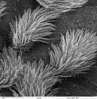
Intraflagellar transport

Last updated
Intraflagellar transport in the cilia of the nematode C. elegans IFTcilia.jpg
Intraflagellar transport in the cilia of the nematode C. elegans

Intraflagellar transport (IFT) is a bidirectional motility along axoneme microtubules that is essential for the formation (ciliogenesis) and maintenance of most eukaryotic cilia and flagella. [1] It is thought to be required to build all cilia that assemble within a membrane projection from the cell surface. Plasmodium falciparum cilia and the sperm flagella of Drosophila are examples of cilia that assemble in the cytoplasm and do not require IFT. The process of IFT involves movement of large protein complexes called IFT particles or trains from the cell body to the ciliary tip and followed by their return to the cell body. The outward or anterograde movement is powered by kinesin-2 while the inward or retrograde movement is powered by cytoplasmic dynein 2/1b. The IFT particles are composed of about 20 proteins organized in two subcomplexes called complex A and B. [2]

Contents

IFT was first reported in 1993 by graduate student Keith Kozminski while working in the lab of Dr. Joel Rosenbaum at Yale University. [3] [4] The process of IFT has been best characterized in the biflagellate alga Chlamydomonas reinhardtii as well as the sensory cilia of the nematode Caenorhabditis elegans . [5]

It has been suggested based on localization studies that IFT proteins also function outside of cilia. [6]

Biochemistry

A simplified model of intraflagellar transport. IFTsimplified.JPG
A simplified model of intraflagellar transport.

Intraflagellar transport (IFT) describes the bi-directional movement of non-membrane-bound particles along the doublet microtubules of the flagellar, and motile cilia axoneme, between the axoneme and the plasma membrane. Studies have shown that the movement of IFT particles along the microtubule is carried out by two different microtubule motors; the anterograde (towards the flagellar tip) motor is heterotrimeric kinesin-2, and the retrograde (towards the cell body) motor is cytoplasmic dynein 1b. IFT particles carry axonemal subunits to the site of assembly at the tip of the axoneme; thus, IFT is necessary for axonemal growth. Therefore, since the axoneme needs a continually fresh supply of proteins, an axoneme with defective IFT machinery will slowly shrink in the absence of replacement protein subunits. In healthy flagella, IFT particles reverse direction at the tip of the axoneme, and are thought to carry used proteins, or "turnover products," back to the base of the flagellum. [7] [8]

The IFT particles themselves consist of two sub-complexes, [9] each made up of several individual IFT proteins. The two complexes, known as 'A' and 'B,' are separable via sucrose centrifugation (both complexes at approximately 16S, but under increased ionic strength complex B sediments more slowly, thus segregating the two complexes). The many subunits of the IFT complexes have been named according to their molecular weights:

The biochemical properties and biological functions of these IFT subunits are just beginning to be elucidated, for example they interact with components of the basal body like CEP170 or proteins which are required for cilium formation like tubulin chaperone and membrane proteins. [11]

Physiological importance

Due to the importance of IFT in maintaining functional cilia, defective IFT machinery has now been implicated in many disease phenotypes generally associated with non-functional (or absent) cilia. IFT88, for example, encodes a protein also known as Tg737 or Polaris in mouse and human, and the loss of this protein has been found to cause an autosomal-recessive polycystic kidney disease model phenotype in mice. Further, the mislocalization of this protein following WDR62 knockdown in mice results in brain malformation and ciliopathies. [12] Other human diseases such as retinal degeneration, situs inversus (a reversal of the body's left-right axis), Senior–Løken syndrome, liver disease, primary ciliary dyskinesia, nephronophthisis, Alström syndrome, Meckel–Gruber syndrome, Sensenbrenner syndrome, Jeune syndrome, and Bardet–Biedl syndrome, which causes both cystic kidneys and retinal degeneration, have been linked to the IFT machinery. This diverse group of genetic syndromes and genetic diseases are now understood to arise due to malfunctioning cilia, and the term "ciliopathy" is now used to indicate their common origin. [13] These and possibly many more disorders may be better understood via study of IFT. [7]

Human genetic syndromes associated with mutations in IFT genes
IFT geneOther nameHuman diseasereference
IFT27RABL4 Bardet–Biedl syndrome [14]
IFT43C14ORF179 Sensenbrenner syndrome [15]
IFT121WDR35 Sensenbrenner syndrome [16]
IFT122WDR10 Sensenbrenner syndrome [17]
IFT140KIAA0590 Mainzer–Saldino syndrome [18]
IFT144WDR19 Jeune syndrome, Sensenbrenner syndrome [19]
IFT172SLB Jeune syndrome, Mainzer–Saldino syndrome [20]

One of the most recent discoveries regarding IFT is its potential role in signal transduction. IFT has been shown to be necessary for the movement of other signaling proteins within the cilia, and therefore may play a role in many different signaling pathways. Specifically, IFT has been implicated as a mediator of sonic hedgehog signaling, [21] one of the most important pathways in embryogenesis.

Related Research Articles

<span class="mw-page-title-main">Cilium</span> Organelle found on eukaryotic cells

The cilium, is a membrane-bound organelle found on most types of eukaryotic cell. Cilia are absent in bacteria and archaea. The cilium has the shape of a slender threadlike projection that extends from the surface of the much larger cell body. Eukaryotic flagella found on sperm cells and many protozoans have a similar structure to motile cilia that enables swimming through liquids; they are longer than cilia and have a different undulating motion.
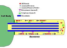
<span class="mw-page-title-main">Axoneme</span>

An axoneme, also called an axial filament is the microtubule-based cytoskeletal structure that forms the core of a cilium or flagellum. Cilia and flagella are found on many cells, organisms, and microorganisms, to provide motility. The axoneme serves as the "skeleton" of these organelles, both giving support to the structure and, in some cases, the ability to bend. Though distinctions of function and length may be made between cilia and flagella, the internal structure of the axoneme is common to both.

<span class="mw-page-title-main">Bardet–Biedl syndrome</span> Medical condition

Bardet–Biedl syndrome (BBS) is a ciliopathic human genetic disorder that produces many effects and affects many body systems. It is characterized by rod/cone dystrophy, polydactyly, central obesity, hypogonadism, and kidney dysfunction in some cases. Historically, slower mental processing has also been considered a principal symptom but is now not regarded as such.

<span class="mw-page-title-main">KIF3A</span> Protein-coding gene in the species Homo sapiens

Kinesin-like protein KIF3A is a protein that in humans is encoded by the KIF3A gene.

<span class="mw-page-title-main">KIF3B</span> Protein-coding gene in the species Homo sapiens

Kinesin-like protein KIF3B is a protein that in humans is encoded by the KIF3B gene. KIF3B is an N-type protein that complexes with two other kinesin proteins to form two-headed anterograde motors. First, KIF3B forms a heterodimer with KIF3A ; (KIF3A/3B), that is membrane-bound and has ATPase activity. Then KIFAP3 binds to the tail domain to form a heterotrimeric motor. This motor has a plus end-directed microtubule sliding activity that exhibits a velocity of ~0.3 μm/s a. There are 14 kinesin protein families in the kinesin superfamily and KIF3B is part of the Kinesin-2 family, of kinesins that can all form heterotrimeric complexes. Expression of the three motor subunits is ubiquitous. The KIG3A/3B/KAP3 motors can transport 90 to 160 nm in diameter organelles.

<span class="mw-page-title-main">IFT88</span> Protein-coding gene in the species Homo sapiens

Intraflagellar transport protein 88 homolog is a protein that is encoded by the IFT88 gene.

<span class="mw-page-title-main">IFT57</span> Protein-coding gene in the species Homo sapiens

Intraflagellar transport protein 57 homolog is a protein that in humans is encoded by the IFT57 gene.

<span class="mw-page-title-main">KIF17</span> Protein-coding gene in the species Homo sapiens

Kinesin-like protein KIF17 is a protein that in humans is encoded by the KIF17 gene. KIF17 and its close relative, C. elegans OSM-3, are members of the kinesin-2 family of plus-end directed microtubule-based motor proteins. In contrast to heterotrimeric kinesin-2 motors, however, KIF17 and OSM-3 form distinct homodimeric complexes. Homodimeric kinesin-2 has been implicated in the transport of NMDA receptors along dendrites for delivery to the dendritic membrane, whereas both heterotrimeric and homodimeric kinesin-2 motors function cooperatively in anterograde intraflagellar transport (IFT) and cilium biogenesis.

<span class="mw-page-title-main">IFT20</span> Protein-coding gene in the species Homo sapiens

Intraflagellar transport protein 20 homolog is a protein that in humans is encoded by the IFT20 gene. The gene is composed of 6 exons and is located on human chromosome 17p11.1. This gene is expressed in human brain, lung, kidney and pancreas, and lower expression were also detected in human placenta, liver, thymus, prostate and testis.

<span class="mw-page-title-main">Ciliopathy</span> Genetic disease resulting in abnormal formation or function of cilia

A ciliopathy is any genetic disorder that affects the cellular cilia or the cilia anchoring structures, the basal bodies, or ciliary function. Primary cilia are important in guiding the process of development, so abnormal ciliary function while an embryo is developing can lead to a set of malformations that can occur regardless of the particular genetic problem. The similarity of the clinical features of these developmental disorders means that they form a recognizable cluster of syndromes, loosely attributed to abnormal ciliary function and hence called ciliopathies. Regardless of the actual genetic cause, it is clustering of a set of characteristic physiological features which define whether a syndrome is a ciliopathy.

<span class="mw-page-title-main">MORM syndrome</span> Medical condition

MORM syndrome is an autosomal recessive congenital disorder characterized by mental retardation, truncal obesity, retinal dystrophy, and micropenis". The disorder shares similar characteristics with Bardet–Biedl syndrome and Cohen syndrome, both of which are autosomal recessive genetic disorders. MORM syndrome can be distinguished from the above disorders because symptoms appear at a young age. The disorder is not dependent on sex of the offspring, both male and female offspring are equally likely to inherit the disorder.

<span class="mw-page-title-main">IFT81</span>

Intraflagellar transport protein 81 homolog is a protein that in humans is encoded by the IFT81 gene. Together with IFT74/72 it forms a core complex to build IFT particles which are required for cilium formation. Additionally, it interacts with basal body components as CEP170 which regulates the disassembly of the cilium.

<span class="mw-page-title-main">TMEM216</span> Protein-coding gene in the species Homo sapiens

Transmembrane protein 216 is a protein in humans that is encoded by the TMEM216 gene.

<span class="mw-page-title-main">ARL13B</span> Protein-coding gene in the species Homo sapiens

ADP-ribosylation factor-like protein 13B (ARL13B), also known as ADP-ribosylation factor-like protein 2-like 1, is a protein that in humans is encoded by the ARL13B gene.

A BBSome is a protein complex that operates in primary cilia biogenesis, homeostasis, and intraflagellar transport (IFT). The BBSome recognizes cargo proteins and signaling molecules like G-protein coupled receptors (GPCRs) on the ciliary membrane and helps transport them to and from the primary cilia. Primary cilia are nonmotile microtubule projections that function like antennae and are found in many types of cells. They receive various environmental signals to aid the cell in survival. They can detect photons by concentrating rhodopsin, a light receptor that converts photons into chemical signals, or odorants by concentrating olfactory receptors on the primary cilia surface. Primary cilia are also meaningful in cell development and signaling. They do not contain any way to make proteins within the primary cilia, so the BBSome aids in transporting essential proteins to, from, and within the cilia. Examples of cargo proteins that the BBSome is responsible for ferrying include smoothened, polycystic-1 (PC1), and several G-Protein coupled receptors (GPCRs) like somatostatin receptors (Sstr3), melanin-concentrating hormone receptor 1 (Mchr1), and neuropeptide Y2 receptor.

<span class="mw-page-title-main">Sensenbrenner syndrome</span> Medical condition

Sensenbrenner syndrome is a rare multisystem disease first described by Judith A. Sensenbrenner in 1975. It is inherited in an autosomal recessive fashion, and a number of genes appear to be responsible. Three genes responsible have been identified: intraflagellar transport (IFT)122 (WDR10), IFT43—a subunit of the IFT complex A machinery of primary cilia, and WDR35

Ciliogenesis is defined as the building of the cell's antenna or extracellular fluid mediation mechanism. It includes the assembly and disassembly of the cilia during the cell cycle. Cilia are important organelles of cells and are involved in numerous activities such as cell signaling, processing developmental signals, and directing the flow of fluids such as mucus over and around cells. Due to the importance of these cell processes, defects in ciliogenesis can lead to numerous human diseases related to non-functioning cilia. Ciliogenesis may also play a role in the development of left/right handedness in humans.

<span class="mw-page-title-main">IFT140</span> Protein-coding gene in the species Homo sapiens

IFT140, Intraflagellar transport 140 homolog, is a protein that in humans is encoded by the IFT140 gene. The gene product forms a core component of IFT-A complex which is indipensible for retrograde intraflagellar transport within the primary cilium.

<span class="mw-page-title-main">Tetratricopeptide repeat domain 21b</span> Protein-coding human gene

Tetratricopeptide repeat domain 21B is a protein that in humans is encoded by the TTC21B gene.

RVxP motif is a protein motif involved in localizing proteins into cilia.

References

  1. "The Panda's Thumb: Of cilia and silliness (More on Behe)". www.pandasthumb.org. Archived from the original on 14 September 2007. Retrieved 13 January 2022.
  2. 1 2 3 Cole, DG; Diener, DR; Himelblau, AL; Beech, PL; Fuster, JC; Rosenbaum, JL (May 1998). "Chlamydomonas kinesin-II-dependent intraflagellar transport (IFT): IFT particles contain proteins required for ciliary assembly in Caenorhabditis elegans sensory neurons". J. Cell Biol. 141 (4): 993–1008. doi:10.1083/jcb.141.4.993. PMC   2132775 . PMID   9585417.
  3. Bhogaraju, S.; Taschner, M.; Morawetz, M.; Basquin, C.; Lorentzen, E. (2011). "Crystal structure of the intraflagellar transport complex 25/27". The EMBO Journal. 30 (10): 1907–1918. doi:10.1038/emboj.2011.110. PMC   3098482 . PMID   21505417.
  4. Kozminski, KG; Johnson KA; Forscher P; Rosenbaum JL. (1993). "A motility in the eukaryotic flagellum unrelated to flagellar beating". Proc Natl Acad Sci U S A. 90 (12): 5519–23. Bibcode:1993PNAS...90.5519K. doi: 10.1073/pnas.90.12.5519 . PMC   46752 . PMID   8516294.
  5. Orozco, JT; Wedaman KP; Signor D; Brown H; Rose L; Scholey JM (1999). "Movement of motor and cargo along cilia". Nature. 398 (6729): 674. Bibcode:1999Natur.398..674O. doi: 10.1038/19448 . PMID   10227290. S2CID   4414550.
  6. Sedmak T, Wolfrum U (April 2010). "Intraflagellar transport molecules in ciliary and nonciliary cells of the retina". J. Cell Biol. 189 (1): 171–86. doi:10.1083/jcb.200911095. PMC   2854383 . PMID   20368623.
  7. 1 2 Rosenbaum, JL; Witman GB (2002). "Intraflagellar Transport". Nat Rev Mol Cell Biol. 3 (11): 813–25. doi:10.1038/nrm952. PMID   12415299. S2CID   12130216.
  8. Scholey, JM (2008). "Intraflagellar transport motors in cilia: moving along the cell's antenna". Journal of Cell Biology. 180 (1): 23–29. doi:10.1083/jcb.200709133. PMC   2213603 . PMID   18180368.
  9. Lucker BF, Behal RH, Qin H, et al. (July 2005). "Characterization of the intraflagellar transport complex B core: direct interaction of the IFT81 and IFT74/72 subunits". J. Biol. Chem. 280 (30): 27688–96. doi: 10.1074/jbc.M505062200 . PMID   15955805.
  10. Behal RH1, Miller MS, Qin H, Lucker BF, Jones A, Cole DG. (2012). "Subunit interactions and organization of the Chlamydomonas reinhardtii intraflagellar transport complex A proteins". J. Biol. Chem. 287 (15): 11689–703. doi: 10.1074/jbc.M111.287102 . PMC   3320918 . PMID   22170070.{{cite journal}}: CS1 maint: multiple names: authors list (link) CS1 maint: numeric names: authors list (link)
  11. Lamla S (2009). Functional characterisation of the centrosomal protein Cep170. Dissertation (Text.PhDThesis). LMU Muenchen: Fakultät für Biologie.
  12. Shohayeb, B, et al. (December 2020). "The association of microcephaly protein WDR62 with CPAP/IFT88 is required for cilia formation and neocortical development". Hum. Mol. Genet. 29 (2): 248–263. doi: 10.1093/hmg/ddz281 . PMID   31816041.
  13. Badano, Jose L.; Norimasa Mitsuma; Phil L. Beales; Nicholas Katsanis (September 2006). "The Ciliopathies : An Emerging Class of Human Genetic Disorders". Annual Review of Genomics and Human Genetics. 7: 125–148. doi:10.1146/annurev.genom.7.080505.115610. PMID   16722803.
  14. Aldahmesh, M. A., Li, Y., Alhashem, A., Anazi, S., Alkuraya, H., Hashem, M., Awaji, A. A., Sogaty, S., Alkharashi, A., Alzahrani, S., Al Hazzaa, S. A., Xiong, Y., Kong, S., Sun, Z., Alkuraya, F. S. (2014). "IFT27, encoding a small GTPase component of IFT particles, is mutated in a consanguineous family with Bardet-Biedl syndrome". Hum. Mol. Genet. 23 (12): 3307–3315. doi:10.1093/hmg/ddu044. PMC   4047285 . PMID   24488770.{{cite journal}}: CS1 maint: multiple names: authors list (link)
  15. Arts, H. H., Bongers, E. M. H. F., Mans, D. A., van Beersum, S. E. C., Oud, M. M., Bolat, E., Spruijt, L., Cornelissen, E. A. M., Schuurs-Hoeijmakers, J. H. M., de Leeuw, N., Cormier-Daire, V., Brunner, H. G., Knoers, N. V. A. M., Roepman, R. (2011). "C14ORF179 encoding IFT43 is mutated in Sensenbrenner syndrome". J. Med. Genet. 48 (6): 390–395. doi:10.1136/jmg.2011.088864. PMID   21378380. S2CID   6073572.{{cite journal}}: CS1 maint: multiple names: authors list (link)
  16. Gilissen, C., Arts, H. H., Hoischen, A., Spruijt, L., Mans, D. A., Arts, P., van Lier, B., Steehouwer, M., van Reeuwijk, J., Kant, S. G., Roepman, R., Knoers, N. V. A. M., Veltman, J. A., Brunner, H. G. (2010). "Exome sequencing identifies WDR35 variants involved in Sensenbrenner syndrome". Am. J. Hum. Genet. 87 (3): 418–423. doi:10.1016/j.ajhg.2010.08.004. PMC   2933349 . PMID   20817137.{{cite journal}}: CS1 maint: multiple names: authors list (link)
  17. Walczak-Sztulpa, J., Eggenschwiler, J., Osborn, D., Brown, D. A., Emma, F., Klingenberg, C., Hennekam, R. C., Torre, G., Garshasbi, M., Tzschach, A., Szczepanska, M., Krawczynski, M., Zachwieja, J., Zwolinska, D., Beales, P. L., Ropers, H.-H., Latos-Bielenska, A., Kuss, A. W. (2010). "Cranioectodermal dysplasia, Sensenbrenner syndrome, is a ciliopathy caused by mutations in the IFT122 gene". Am. J. Hum. Genet. 86 (6): 949–956. doi:10.1016/j.ajhg.2010.04.012. PMC   3032067 . PMID   20493458.{{cite journal}}: CS1 maint: multiple names: authors list (link)
  18. Perrault, I., Saunier, S., Hanein, S., Filhol, E., Bizet, A. A., Collins, F., Salih, M. A. M., Gerber, S., Delphin, N., Bigot, K., Orssaud, C., Silva, E., and 18 others. (2012). "Mainzer-Saldino syndrome is a ciliopathy caused by IFT140 mutations". Am. J. Hum. Genet. 90 (5): 864–870. doi:10.1016/j.ajhg.2012.03.006. PMC   3376548 . PMID   22503633.{{cite journal}}: CS1 maint: multiple names: authors list (link) CS1 maint: numeric names: authors list (link)
  19. Bredrup, C., Saunier, S., Oud, M. M., Fiskerstrand, T., Hoischen, A., Brackman, D., Leh, S. M., Midtbo, M., Filhol, E., Bole-Feysot, C., Nitschke, P., Gilissen, C., and 16 others. (2011). "Ciliopathies with skeletal anomalies and renal insufficiency due to mutations in the IFT-A gene WDR19". Am. J. Hum. Genet. 89 (5): 634–643. doi:10.1016/j.ajhg.2011.10.001. PMC   3213394 . PMID   22019273.{{cite journal}}: CS1 maint: multiple names: authors list (link) CS1 maint: numeric names: authors list (link)
  20. Halbritter, J., Bizet, A. A., Schmidts, M., Porath, J. D., Braun, D. A., Gee, H. Y., McInerney-Leo, A. M., Krug, P., Filhol, E., Davis, E. E., Airik, R., Czarnecki, P. G., and 38 others. (2013). "Defects in the IFT-B component IFT172 cause Jeune and Mainzer-Saldino syndromes in humans". Am. J. Hum. Genet. 93 (5): 915–925. doi:10.1016/j.ajhg.2013.09.012. PMC   3824130 . PMID   24140113.{{cite journal}}: CS1 maint: multiple names: authors list (link) CS1 maint: numeric names: authors list (link)
  21. Eggenschwiler JT, Anderson KV (January 2007). "Cilia and developmental signaling". Annu Rev Cell Dev Biol. 23: 345–73. doi:10.1146/annurev.cellbio.23.090506.123249. PMC   2094042 . PMID   17506691.

Further reading