Nephronophthisis

Last updated
Nephronophthisis
Autorecessive.svg
Nephronophthisis has an autosomal recessive pattern of inheritance.
Specialty Medical genetics   OOjs UI icon edit-ltr-progressive.svg
Symptoms Polyuria [1]
TypesInfantile, Juvenile and Adult NPH [2]
Diagnostic method Renal ultrasound [2]
TreatmentHypertension and anemia management [2]

Nephronophthisis is a genetic disorder of the kidneys which affects children. [3] It is classified as a medullary cystic kidney disease. The disorder is inherited in an autosomal recessive fashion and, although rare, is the most common genetic cause of childhood kidney failure. It is a form of ciliopathy. [4] Its incidence has been estimated to be 0.9 cases per million people in the United States, and 1 in 50,000 births in Canada. [5]
Contents

Signs and symptoms

Infantile, juvenile, and adolescent forms of nephronophthisis have been identified. Although the range of characterizations is broad, people affected by nephronophthisis typically present with polyuria (production of a large volume of urine), polydipsia (excessive liquid intake), and after several months to years, end-stage kidney disease, a condition necessitating either dialysis or a kidney transplant in order to survive. [1] Some individuals with nephronophthisis also have so-called "extra-renal symptoms" which can include tapetoretinal degeneration, liver problems, oculomotor apraxia, and cone-shaped epiphysis (Saldino-Mainzer syndrome). [6] [7]

Cause

Nephronophthisis is characterized by fibrosis and the formation of cysts at the cortico-medullary junction, it is an autosomal recessive disorder which eventually leads to terminal kidney failure. [8]

Pathophysiology

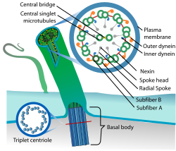
Ciliopathy (eukaryotic cilium diagram) Eukaryotic cilium diagram en.svg
Ciliopathy (eukaryotic cilium diagram)

Mechanism of nephronophthisis indicates that all proteins mutated in cystic kidney diseases express themselves in primary cilia. NPHP gene mutations cause defects in signaling resulting in flaws of planar cell polarity. The ciliary theory indicates that multiple organs are involved in NPHP (retinal degeneration, cerebellar hypoplasia, liver fibrosis, and intellectual disability). [9]

Nephronophthisis is a ciliopathy. Other known ciliopathies include primary ciliary dyskinesia, Bardet–Biedl syndrome, polycystic kidney and liver disease, Alström syndrome, Meckel–Gruber syndrome and some forms of retinal degeneration. [10]

NPHP2 is infantile type of nephropthisis[ check spelling ] and sometimes associated with situs inversus this can be explained by its relation with inversin gene. NPHP1, NPHP3, NPHP4, NPHP5, and NPHP6 are sometimes seen with retinitis pigmentosa, this particular association has a name, Senior-Loken syndrome. [11]

Diagnosis

Ultrasound Ultraschallgrat.jpg
Ultrasound

The diagnosis of nephronophthisis can be obtained via a kidney ultrasound, family history and clinical history of the affected individual according to Stockman, et al. [2]

Types

Management

The management of this condition can be done via-improvement of any electrolyte imbalance, as well as, high blood pressure and low red blood cell counts (anemia) treatment as the individual's condition warrants. [2]

Epidemiology

Nephronophthisis occurs equally in both sexes and has an estimate 9 in about 8 million rate in individuals. Nephronophthisis is the leading monogenic cause of end-stage kidney disease. [12]

Related Research Articles

<span class="mw-page-title-main">Autosomal dominant polycystic kidney disease</span> Medical condition

Autosomal dominant polycystic kidney disease (ADPKD) is one of the most common, life-threatening inherited human disorders and the most common hereditary kidney disease. It is associated with large interfamilial and intrafamilial variability, which can be explained to a large extent by its genetic heterogeneity and modifier genes. It is also the most common of the inherited cystic kidney diseases — a group of disorders with related but distinct pathogenesis, characterized by the development of renal cysts and various extrarenal manifestations, which in case of ADPKD include cysts in other organs, such as the liver, seminal vesicles, pancreas, and arachnoid membrane, as well as other abnormalities, such as intracranial aneurysms and dolichoectasias, aortic root dilatation and aneurysms, mitral valve prolapse, and abdominal wall hernias. Over 50% of patients with ADPKD eventually develop end stage kidney disease and require dialysis or kidney transplantation. ADPKD is estimated to affect at least one in every 1000 individuals worldwide, making this disease the most common inherited kidney disorder with a diagnosed prevalence of 1:2000 and incidence of 1:3000-1:8000 in a global scale.

<span class="mw-page-title-main">Cilium</span> Organelle found on eukaryotic cells

The cilium is a short hair-like membrane protrusion from many types of eukaryotic cell. The cilium has the shape of a slender threadlike projection that extends from the surface of the much larger cell body. Eukaryotic flagella found on sperm cells and many protozoans have a similar structure to motile cilia that enables swimming through liquids; they are longer than cilia and have a different undulating motion.

<span class="mw-page-title-main">Joubert syndrome</span> Medical condition

Joubert syndrome is a rare autosomal recessive genetic disorder that affects the cerebellum, an area of the brain that controls balance and coordination.

Exencephaly is a type of cephalic disorder wherein the brain is located outside of the skull. This condition is usually found in embryos as an early stage of anencephaly. As an exencephalic pregnancy progresses, the neural tissue gradually degenerates.

<span class="mw-page-title-main">Bardet–Biedl syndrome</span> Ciliopathic recessive genetic disorder

Bardet–Biedl syndrome (BBS) is a ciliopathic human genetic disorder that produces many effects and affects many body systems. It is characterized by rod/cone dystrophy, polydactyly, central obesity, hypogonadism, and kidney dysfunction in some cases. Historically, slower mental processing has also been considered a principal symptom but is now not regarded as such.

<span class="mw-page-title-main">Gitelman syndrome</span> Genetic kidney disorder

Gitelman syndrome (GS) is an autosomal recessive kidney tubule disorder characterized by low blood levels of potassium and magnesium, decreased excretion of calcium in the urine, and elevated blood pH. It is the most frequent hereditary salt-losing tubulopathy. Gitelman syndrome is caused by disease-causing variants on both alleles of the SLC12A3 gene. The SLC12A3 gene encodes the thiazide-sensitive sodium-chloride cotransporter, which can be found in the distal convoluted tubule of the kidney.

Agenesis of the corpus callosum (ACC) is a rare birth defect in which there is a complete or partial absence of the corpus callosum. It occurs when the development of the corpus callosum, the band of white matter connecting the two hemispheres in the brain, in the embryo is disrupted. The result of this is that the fibers that would otherwise form the corpus callosum are instead longitudinally oriented along the ipsilateral ventricular wall and form structures called Probst bundles.

<span class="mw-page-title-main">Medullary cystic kidney disease</span> Medical condition

Medullary cystic kidney disease (MCKD) is an autosomal dominant kidney disorder characterized by tubulointerstitial sclerosis leading to end-stage renal disease. Because the presence of cysts is neither an early nor a typical diagnostic feature of the disease, and because at least four different gene mutations may give rise to the condition, the name autosomal dominant tubulointerstitial kidney disease (ADTKD) has been proposed, to be appended with the underlying genetic variant for a particular individual. Importantly, if cysts are found in the medullary collecting ducts they can result in a shrunken kidney, unlike that of polycystic kidney disease. There are two known forms of medullary cystic kidney disease, mucin-1 kidney disease 1 (MKD1) and mucin-2 kidney disease/uromodulin kidney disease (MKD2). A third form of the disease occurs due to mutations in the gene encoding renin (ADTKD-REN), and has formerly been known as familial juvenile hyperuricemic nephropathy type 2.

<span class="mw-page-title-main">Meckel–Gruber syndrome</span> Medical condition

Meckel-Gruber syndrome is a rare, lethal ciliopathic genetic disorder, characterized by renal cystic dysplasia, central nervous system malformations, polydactyly (postaxial), hepatic developmental defects, and pulmonary hypoplasia due to oligohydramnios. Meckel–Gruber syndrome is named for Johann Meckel and Georg Gruber. This is called so because, of the following reasons.

<span class="mw-page-title-main">Senior–Løken syndrome</span> Congenital eye disorder

Senior–Løken syndrome is a congenital eye disorder, first characterized in 1961. It is a rare, ciliopathic, autosomal recessive disorder characterized by juvenile nephronophthis and progressive eye disease.

<span class="mw-page-title-main">NPHP1</span>

Nephrocystin-1 is a protein that in humans is encoded by the NPHP1 gene.

Conorenal syndrome, is a collection of medical conditions that seem to have a common genetic cause.

<span class="mw-page-title-main">Renal–hepatic–pancreatic dysplasia</span> Medical condition

Renal–hepatic–pancreatic dysplasia is an autosomal recessive congenital disorder characterized by pancreatic fibrosis, renal dysplasia and hepatic dysgenesis. An association with NPHP3 has been described. It was characterized in 1959.

<span class="mw-page-title-main">Ciliopathy</span> Genetic disease resulting in abnormal formation or function of cilia

A ciliopathy is any genetic disorder that affects the cellular cilia or the cilia anchoring structures, the basal bodies, or ciliary function. Primary cilia are important in guiding the process of development, so abnormal ciliary function while an embryo is developing can lead to a set of malformations that can occur regardless of the particular genetic problem. The similarity of the clinical features of these developmental disorders means that they form a recognizable cluster of syndromes, loosely attributed to abnormal ciliary function and hence called ciliopathies. Regardless of the actual genetic cause, it is clustering of a set of characteristic physiological features which define whether a syndrome is a ciliopathy.

<span class="mw-page-title-main">Orofaciodigital syndrome 1</span> Medical condition

Orofaciodigital syndrome 1 (OFD1), also called Papillon-Léage and Psaume syndrome, is an X-linked congenital disorder characterized by malformations of the face, oral cavity, and digits with polycystic kidney disease and variable involvement of the central nervous system.

Xaa-Pro aminopeptidase 3, also known as aminopeptidase P3, is an enzyme that in humans is encoded by the XPNPEP3 gene. XPNPEP3 localizes to mitochondria in renal cells and to kidney tubules in a cell type-specific pattern. Mutations in XPNPEP3 gene have been identified as a cause of a nephronophthisis-like disease.

Glomerulocystic kidney disease (GCKD) is a cystic disorder of the kidneys. GCKD involves cystic dilation of Bowman's capsule. It can occur with or without congenital abnormality.

<span class="mw-page-title-main">Juvenile nephronophthisis</span> Medical condition

Juvenile nephronophthisis is the juvenile form of nephronophthisis that causes end stage kidney disease around the age of 13; infantile nephronophthisis and adolescent nephronophthisis cause ESKD around the ages of 1 and 19, respectively.

<span class="mw-page-title-main">Martin Barratt</span> British paediatrician and professor

Thomas Martin Barratt was a British paediatrician and professor of paediatric nephrology. Barratt was most notable for developing a specialist service for children with kidney diseases in Britain, bringing peritoneal dialysis, haemodialysis, and later renal transplantation to ever younger children. Barratt was an early advocate for multidisciplinary care and developed a model that was later taken up by many other specialist centres across the world. His research led to a new treatments for many types of childhood kidney diseases., and for research into childhood Nephrotic syndrome and Hemolytic-uremic syndrome.

Friedhelm Hildebrandt is the William E. Harmon Professor of Pediatrics at Harvard Medical School and Chief of the Division of Nephrology at Boston Children's Hospital. He was formerly an Investigator of the Howard Hughes Medical Institute (HHMI) and the Frederick G.L. Huetwell Professor of Pediatrics at the University of Michigan.

References

  1. 1 2 Hildebrandt, Friedhelm; Zhou, Weibin (2007). "Nephronophthisis-Associated Ciliopathies". Journal of the American Society of Nephrology. 18 (6): 1855–71. doi: 10.1681/ASN.2006121344 . PMID   17513324.
  2. 1 2 3 4 5 6 7 8 Stokman, Marijn; Lilien, Marc; Knoers, Nine (1 January 1993). "Nephronophthisis-Related Ciliopathies". Nephronophthisis. University of Washington, Seattle. PMID   27336129 . Retrieved 1 August 2016.{{cite book}}: |work= ignored (help)update 2016
  3. "Nephronophthisis". Genetics Home Reference. Retrieved 2015-08-08.
  4. Hurd TW, Hildebrandt F (2011). "Mechanisms of nephronophthisis and related ciliopathies". Nephron Exp. Nephrol. 118 (1): e9–e14. doi:10.1159/000320888. PMC   2992643 . PMID   21071979.
  5. page 831, Chapter 35, in: Avner, Ellis D.; Harmon, William; Niaudet, Patrick; Yoshikawa, Norishige (2009-08-20). Pediatric Nephrology (Avner, Pediatric Nephrology). Springer. ISBN   978-3-540-76327-7. (stating the incidence in the United States as 9 per 8.3 million people.
  6. Kanwal, Kher (2007). Clinical Pediatric Nephrology, Second Edition (2nd ed.). McGraw-Hill. p. 205. ISBN   978-1-84184-447-3 . Retrieved 9 August 2015.
  7. Medullary Cystic Disease~clinical at eMedicine
  8. Salomon, Rémi; Saunier, Sophie; Niaudet, Patrick (2009). "Nephronophthisis". Pediatric Nephrology. 24 (12): 2333–44. doi:10.1007/s00467-008-0840-z. PMC   2770134 . PMID   18607645.
  9. Hildebrandt, Friedhelm; Attanasio, Massimo; Otto, Edgar (2009). "Nephronophthisis: Disease Mechanisms of a Ciliopathy". Journal of the American Society of Nephrology. 20 (1): 23–35. doi:10.1681/ASN.2008050456. PMC   2807379 . PMID   19118152.
  10. McCormack, Francis X.; Panos, Ralph J.; Trapnell, Bruce C. (2010-03-10). Molecular Basis of Pulmonary Disease: Insights from Rare Lung Disorders. Springer Science & Business Media. ISBN   9781597453844.
  11. Badano, Jose L.; Mitsuma, Norimasa; Beales, Phil L.; Katsanis, Nicholas (2006). "The Ciliopathies: An Emerging Class of Human Genetic Disorders". Annual Review of Genomics and Human Genetics. 7: 125–48. doi:10.1146/annurev.genom.7.080505.115610. PMID   16722803.
  12. Hildebrandt, Friedhelm (2009). "Nephronophthisis". In Lifton, Richard P.; Somlo, Stefan; Giebisch, Gerhard H.; et al. (eds.). Genetic Diseases of the Kidney. Academic Press. pp. 425–46. ISBN   978-0-08-092427-4.

Further reading