Odontogenic keratocyst

Last updated
Odontogenic keratocyst
Other namesKeratocystic odontogenic tumour (KCOT)
Keratocystic odontogenic tumour - 2 - very high mag.jpg
Micrograph of an odontogenic keratocyst. H&E stain.

An odontogenic keratocyst is a rare and benign but locally aggressive developmental cyst. It most often affects the posterior mandible and most commonly presents in the third decade of life. [1] Odontogenic keratocysts make up around 19% of jaw cysts. [2] . Despite its more common appearance in the bone region, it can affect soft tissue. [3]

Contents

In the WHO/IARC classification of head and neck pathology, this clinical entity had been known for years as the odontogenic keratocyst; it was reclassified as keratocystic odontogenic tumour (KCOT) from 2005 to 2017. [4] [5] In 2017 it reverted to the earlier name, as the new WHO/IARC classification reclassified OKC back into the cystic category. [6] Under The WHO/IARC classification, Odontogenic Keratocyst underwent the reclassification as it is no longer considered a neoplasm due to a lack of quality evidence regarding this hypothesis, especially with respect to clonality. Within the Head and Neck pathology community there is still controversy surrounding the reclassification, with some pathologists still considering Odontogenic Keratocyst as a neoplasm in line with the previous classification. [7]

Signs and symptoms

Odontogenic keratocysts can occur at any age, however they are more common in the third to sixth decades. The male to female ratio is approximately 2:1. The majority are found in the mandible, with half occurring at the angle of the mandible.

Early odontogenic keratocysts usually do not display symptoms. Typically, clinical signs and symptoms present with bony expansion, or infection. However, bony expansion is uncommon as odontogenic keratocysts grow due to increased epithelial turnover rather than osmotic pressure. When symptoms are present they usually take the form of pain, swelling and discharge due to secondary infection. Odontogenic keratocysts are usually noted as incidental radiographic findings. Radiographically they can be seen as unilocular or multilocular radiolucencies. They can be mistaken for other cysts such as residual cysts or a dentigerous cyst if they occur over an unerupted tooth. [8]

Relative incidence of odontogenic cysts. Odontogenic keratocyst is labeled at bottom right. Relative incidence of odontogenic cysts.jpg
Relative incidence of odontogenic cysts. Odontogenic keratocyst is labeled at bottom right.

Pathogenesis

Odontogenic keratocysts originate from the odontogenic epithelium (dental lamina) in the alveolus left from tooth development stages. They are mainly thought to arise from rests of Serres. [10]

Genetics

Sporadic (non-syndromic) and syndromic OKCs are associated with mutations in the gene PTCH found on chromosome 9q, which is part of the Hedgehog signaling pathway. [5] [11] [12] PTCH is a tumour suppressor gene. Loss of PTCH activity leads to a brake in the cell cycle. A third of OKCs show mutations in PTCH, resulting in the cyst epithelium undergoing highly proliferative activity. This leads to growth of the cyst wall and when removed favours recurrence if following incomplete removal of the epithelium. [10]

Nevoid basal-cell carcinoma syndrome

Multiple odontogenic keratocysts are a feature, and major diagnostic criteria, of nevoid basal cell carcinoma syndrome (NBCCS, also known as Gorlin-Goltz Syndrome). Almost all individuals with NBCCS have odontogenic keratocysts which require numerous treatments. Consideration of the syndrome needs to be taken into account if found in children or if multiple OKCs are present; diagnosis of multiple OKCs in a child necessitates referral for genetic evaluation. Histologically, the cysts are indistinguishable to non-syndromic cysts and over 80% will have PTCH mutations. [10]

Diagnosis

Classic look of an odontogenic keratocyst of the right mandible in the place of a former wisdom tooth. Well defined, unilocular, radiolucent lesion within the bone. Classic keratocystic odontogenic tumour.jpg
Classic look of an odontogenic keratocyst of the right mandible in the place of a former wisdom tooth. Well defined, unilocular, radiolucent lesion within the bone.

Diagnosis is usually radiological. However, definitive diagnosis is through biopsy. Aspirational biopsy of odontogenic keratocysts contains a greasy fluid which is pale in colour and contains keratotic squames. [13] [2] Protein content of cyst fluid below 4g% is diagnostic of odontogenic keratocysts. [2] Smaller and unilocular lesions resembling other types of cysts may require a biopsy to confirm the diagnosis. [10] On a CT scan, the radiodensity of a keratocystic odontogenic tumour is about 30 Hounsfield units, which is about the same as ameloblastomas. However, ameloblastomas show more bone expansion and seldom show high density areas. [14]

Radiographs of odontogenic keratocysts show well-defined radiolucent areas with rounded or scalloped margins which are well demarcated. [13] These areas can be multilocular or unilocular. The growth pattern of the lesion is very characteristic from which a diagnosis can be made as there is growth and spread both forward and backward along the medullary cavity with little expansion. No resorption of teeth or inferior dental canal and minimal displacement of teeth is seen. Due to lack of expansion of the odontogenic keratocyst, the lesion can be very large when radiographically discovered. [10]

Differential diagnosis

Radiologically

Histologically

Histology

Odontogenic keratocysts have a diagnostic histological appearance. Under the microscope, OKCs vaguely resemble keratinized squamous epithelium; [15] however, they lack rete ridges and often have an artifactual separation from their basement membrane. [2]

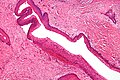
The fibrous wall of the cyst is usually thin and uninflamed. The epithelial lining is thin with even thickness and parakeratinised with columnar cells in the basal layer which have focal reverse polarisation (nuclei are on the opposite pole of the cell). [13] The basal cells are an indication of the odontogenic origin as they resemble pre-ameloblasts. The epithelium can separate from the wall, resulting in islands of epithelium. These can go on to form 'satellite' or 'daughter' cysts, leading to an overall multilocular cyst. [10] Presence of daughter cysts is particularly seen in those with NBCCS. [13] Inflamed cysts show hyperplastic epithelium which is no longer characteristic of OKCs and can have resemblance to radicular cysts instead. Due to areas of focal inflammation, a larger biopsy is required for correct diagnosis of odontogenic keratocysts. [10]

Treatment

Large odontogenic keratocyst with impacted wisdom teeth superficial to lesion Massive keratocystic odontogenic tumour.jpg
Large odontogenic keratocyst with impacted wisdom teeth superficial to lesion

As the condition is quite rare, opinions among experts about how to treat OKCs differ. A 2015 Cochrane review found that there is currently no high quality evidence to suggest the effectiveness of specific treatments for the treatment of odontogenic keratocysts. [8] Treatment depends on extent of multilocularity and cyst. Small multilocular and unilocular cysts can be treated more conservatively through enucleation and curretage. Treatment options for KTOC may vary according to its size, extent, site, and adjacent structures.

Treatment options: [5] [10] [8]

Follow-up

Annual radiographic review has been recommended. [13] Long-term clinical follow-up is also recommended due to recurrences occurring many years after treatment. [2]

Recurrence and neoplastic nature

Malignant transformation to squamous cell carcinoma may occur, but is unusual. [19]

Recurrence is likely when treated by simple enucleation. Contributing causes include thin and fragile epithelium leading to incomplete removal, cyst extensions extending into cancellous bone, satellite cysts found in the wall, experience of the surgeon, formation of further new cysts from other remnants of the dental epithelium. With current treatment techniques the recurrence rate is around 2-3% but can be as high as 50%. Recurrence can occur as early as 5 years and as late as 40 years after removal. [10] Recurrence is usually seen within 5 years of treatment. Early findings of recurrence can be easily treated with minor surgery and curretage. [10] Any fragment of the cyst that is left behind has the potential to survive and grow. Therefore, the success of enucleation depends on how well the cyst is removed. Larger cysts have a higher rate of recurrence after enucleation as they are more difficult to remove.

Pronto genie keratocysts are well known to recur in the posterior mandible. A substantial amount of odontogenic keratocysts also recur in the tooth-bearing area of the jaws, requiring attention from clinicians. [20]

The neoplastic nature of odontogenic keratocysts has been debated. Due to high recurrence rate, late detection when the cyst has grown very large and causation by tumour suppressor gene inactivation, some have classified OKCs as benign neoplasms. The best evidence to suggest that this type of cyst is not a neoplasm is that it responds very well to marsupialisation. [10]

See also

Related Research Articles

<span class="mw-page-title-main">Ameloblastoma</span> Medical condition

Ameloblastoma is a rare, benign or cancerous tumor of odontogenic epithelium much more commonly appearing in the lower jaw than the upper jaw. It was recognized in 1827 by Cusack. This type of odontogenic neoplasm was designated as an adamantinoma in 1885 by the French physician Louis-Charles Malassez. It was finally renamed to the modern name ameloblastoma in 1930 by Ivey and Churchill.

<span class="mw-page-title-main">Dental follicle</span> Anatomical entity

The dental follicle, also known as dental sac, is made up of mesenchymal cells and fibres surrounding the enamel organ and dental papilla of a developing tooth. It is a vascular fibrous sac containing the developing tooth and its odontogenic organ. The dental follicle (DF) differentiates into the periodontal ligament. In addition, it may be the precursor of other cells of the periodontium, including osteoblasts, cementoblasts and fibroblasts. They develop into the alveolar bone, the cementum with Sharpey's fibers and the periodontal ligament fibers respectively. Similar to dental papilla, the dental follicle provides nutrition to the enamel organ and dental papilla and also have an extremely rich blood supply.

<span class="mw-page-title-main">Dentigerous cyst</span> Medical condition

A dentigerous cyst, also known as a follicular cyst, is an epithelial-lined developmental cyst formed by accumulation of fluid between the reduced enamel epithelium and the crown of an unerupted tooth. It is formed when there is an alteration in the reduced enamel epithelium and encloses the crown of an unerupted tooth at the cemento-enamel junction. Fluid is accumulated between reduced enamel epithelium and the crown of an unerupted tooth.

Giant-cell fibroma is a benign localized fibrous mass. It often mimics other fibroepithelial growths and can be distinguished by its histopathology. The exact cause of giant-cell fibromas is unknown however there is no evidence to show that it can be caused by irritation. Giant-cell fibromas can be removed by surgical incision, electrosurgery, or laser excision.

<span class="mw-page-title-main">Central giant-cell granuloma</span> Medical condition

Central giant-cell granuloma (CGCG) is a localised benign condition of the jaws. It is twice as common in females and is more likely to occur before age 30. Central giant-cell granulomas are more common in the anterior mandible, often crossing the midline and causing painless swellings.

<span class="mw-page-title-main">Periapical cyst</span> Medical condition

Commonly known as a dental cyst, the periapical cyst is the most common odontogenic cyst. It may develop rapidly from a periapical granuloma, as a consequence of untreated chronic periapical periodontitis.

Lateral periodontal cysts (LPCs) are defined as non-keratinised and non-inflammatory developmental cysts located adjacent or lateral to the root of a vital tooth.” LPCs are a rare form of jaw cysts, with the same histopathological characteristics as gingival cysts of adults (GCA). Hence LPCs are regarded as the intraosseous form of the extraosseous GCA. They are commonly found along the lateral periodontium or within the bone between the roots of vital teeth, around mandibular canines and premolars. Standish and Shafer reported the first well-documented case of LPCs in 1958, followed by Holder and Kunkel in the same year although it was called a periodontal cyst. Since then, there has been more than 270 well-documented cases of LPCs in literature.

Botryoid odontogenic cyst (BOC) is a type of developmental odontogenic cyst that is extremely rare. It is thought to be a lateral periodontal cyst (LPC) variant with a higher risk of recurrence. Weathers and Waldron coined the term BOC in 1973. Adults over the age of 50 are the most affected. BOC appears as a slow-growing lesion that is symptomatic in approximately 70% of cases.

<span class="mw-page-title-main">Calcifying odontogenic cyst</span> Medical condition

Calcifying odontogenic cyst (COC) is a rare developmental lesion that comes from odontogenic epithelium. It is also known as a calcifying cystic odontogenic tumor, which is a proliferation of odontogenic epithelium and scattered nest of ghost cells and calcifications that may form the lining of a cyst, or present as a solid mass.

<span class="mw-page-title-main">Glandular odontogenic cyst</span> Human jaw cyst

A glandular odontogenic cyst (GOC) is a rare and usually benign odontogenic cyst developed at the odontogenic epithelium of the mandible or maxilla. Originally, the cyst was labeled as "sialo-odontogenic cyst" in 1987. However, the World Health Organization (WHO) decided to adopt the medical expression "glandular odontogenic cyst". Following the initial classification, only 60 medically documented cases were present in the population by 2003. GOC was established as its own biological growth after differentiation from other jaw cysts such as the "central mucoepidermoid carcinoma (MEC)", a popular type of neoplasm at the salivary glands. GOC is usually misdiagnosed with other lesions developed at the glandular and salivary gland due to the shared clinical signs. The presence of osteodentin supports the concept of an odontogenic pathway. This odontogenic cyst is commonly described to be a slow and aggressive development. The inclination of GOC to be large and multilocular is associated with a greater chance of remission. GOC is an infrequent manifestation with a 0.2% diagnosis in jaw lesion cases. Reported cases show that GOC mainly impacts the mandible and male individuals. The presentation of GOC at the maxilla has a very low rate of incidence. The GOC development is more common in adults in their fifth and sixth decades.

Squamous odontogenic tumors (SOTs) are very rare benign locally infiltrative odontogenic neoplasms of epithelial origin. Only some 50 cases have been documented. They occur mostly from 20-40 and are more common in males. Treatment is by simple enucleation and local curettage, and recurrence is rare.

<span class="mw-page-title-main">Ameloblastic fibroma</span> Medical condition

An ameloblastic fibroma is a fibroma of the ameloblastic tissue, that is, an odontogenic tumor arising from the enamel organ or dental lamina. It may be either truly neoplastic or merely hamartomatous. In neoplastic cases, it may be labeled an ameloblastic fibrosarcoma in accord with the terminological distinction that reserves the word fibroma for benign tumors and assigns the word fibrosarcoma to malignant ones. It is more common in the first and second decades of life, when odontogenesis is ongoing, than in later decades. In 50% of cases an unerupted tooth is involved.

<span class="mw-page-title-main">Odontoma</span> Benign tumour of dental tissue

An odontoma, also known as an odontome, is a benign tumour linked to tooth development. Specifically, it is a dental hamartoma, meaning that it is composed of normal dental tissue that has grown in an irregular way. It includes both odontogenic hard and soft tissues. As with normal tooth development, odontomas stop growing once mature which makes them benign.

The odontogenic myxoma is an uncommon benign odontogenic tumor arising from embryonic connective tissue associated with tooth formation. As a myxoma, this tumor consists mainly of spindle shaped cells and scattered collagen fibers distributed through a loose, mucoid material.

The calcifying epithelial odontogenic tumor (CEOT), also known as a Pindborg tumor, is an odontogenic tumor first recognized by the Danish pathologist Jens Jørgen Pindborg in 1955. It was previously described as an adenoid adamantoblastoma, unusual ameloblastoma and a cystic odontoma. Like other odontogenic neoplasms, it is thought to arise from the epithelial element of the enamel origin. It is a typically benign and slow growing, but invasive neoplasm.

Carnoy's solution is a fixative composed of 60% ethanol, 30% chloroform and 10% glacial acetic acid, 1 gram of ferric chloride.

Odontogenic cyst are a group of jaw cysts that are formed from tissues involved in odontogenesis. Odontogenic cysts are closed sacs, and have a distinct membrane derived from rests of odontogenic epithelium. It may contain air, fluids, or semi-solid material. Intra-bony cysts are most common in the jaws, because the mandible and maxilla are the only bones with epithelial components. That odontogenic epithelium is critical in normal tooth development. However, epithelial rests may be the origin for the cyst lining later. Not all oral cysts are odontogenic cysts. For example, mucous cyst of the oral mucosa and nasolabial duct cyst are not of odontogenic origin.

A cyst is a pathological epithelial lined cavity that fills with fluid or soft material and usually grows from internal pressure generated by fluid being drawn into the cavity from osmosis. The bones of the jaws, the mandible and maxilla, are the bones with the highest prevalence of cysts in the human body. This is due to the abundant amount of epithelial remnants that can be left in the bones of the jaws. The enamel of teeth is formed from ectoderm, and so remnants of epithelium can be left in the bone during odontogenesis. The bones of the jaws develop from embryologic processes which fuse, and ectodermal tissue may be trapped along the lines of this fusion. This "resting" epithelium is usually dormant or undergoes atrophy, but, when stimulated, may form a cyst. The reasons why resting epithelium may proliferate and undergo cystic transformation are generally unknown, but inflammation is thought to be a major factor. The high prevalence of tooth impactions and dental infections that occur in the bones of the jaws is also significant to explain why cysts are more common at these sites.

Ameloblastic carcinoma is a rare form of malignant odontogenic tumor, that develops in the jawbones from the epithelial cells that generate the tooth enamel. It is usually treated with surgery; chemotherapy has not been proven to be effective.

The ameloblastic fibro-odontoma (AFO) is essentially a benign tumor with the features characteristic of ameloblastic fibroma along with enamel and dentin. Though it is generally regarded as benign, there have been cases of its malignant transformation into ameloblastic fibrosarcoma and odontogenic sarcoma. Cahn LR and Blum T, believed in "maturation theory", which suggested that AFO was an intermediate stage and eventually developed during the period of tooth formation to a complex odontoma thus, being a hamartoma.

References

  1. MacDonald-Jankowski DS (January 2011). "Keratocystic odontogenic tumour: systematic review". Dento Maxillo Facial Radiology. 40 (1): 1–23. doi:10.1259/dmfr/29949053. PMC   3611466 . PMID   21159911.
  2. 1 2 3 4 5 Crispian S (2008). Oral and maxillofacial medicine : the basis of diagnosis and treatment (2nd ed.). Edinburgh: Churchill Livingstone. ISBN   9780443068188. OCLC   123962943.
  3. Stoelinga, P, et al. (January 2022). "The extra-osseous odontogenic keratocyst: An anachronism?". Journal of Stomatology Oral and Maxillofacial Surgery. 123 (6): e790–e793. doi:10.1016/j.jormas.2022.07.001. PMID   35798194.
  4. Barnes L, Eveson JW, Reichart P, Sidransky D, eds. (2005). World Health Organization Classification of Tumours: Pathology and Genetics of Head and Neck Tumours (PDF) (2005 ed.). Lyon, France: IARC Press. ISBN   978-92-832-2417-4. Archived from the original (PDF) on 2015-09-24.
  5. 1 2 3 Madras J, Lapointe H (March 2008). "Keratocystic odontogenic tumour: reclassification of the odontogenic keratocyst from cyst to tumour". Journal. 74 (2): 165–165h. PMID   18353202.
  6. El-Naggar AK, Chan JK, Grandis JR, Takata T, Slootweg PJ, eds. (2017). WHO Classification of Head and Neck Tumours. WHO/IARC Classification of Tumours. Vol. 9 (4th ed.). Lyon, France: IARC Press. ISBN   978-92-832-2438-9.
  7. Wright JM, Vered M (March 2017). "Update from the 4th Edition of the World Health Organization Classification of Head and Neck Tumours: Odontogenic and Maxillofacial Bone Tumors". Head and Neck Pathology. 11 (1): 68–77. doi:10.1007/s12105-017-0794-1. PMC   5340735 . PMID   28247226.
  8. 1 2 3 Sharif FN, Oliver R, Sweet C, Sharif MO (November 2015). "Interventions for the treatment of keratocystic odontogenic tumours". The Cochrane Database of Systematic Reviews. 2016 (11): CD008464. doi:10.1002/14651858.cd008464.pub3. PMC   7173719 . PMID   26545201.
  9. Borges LB, Fechine FV, Mota MR, Sousa FB, Alves AP (2012). "Odontogenic lesions of the jaw: a clinical-pathological study of 461 cases". Revista Gaúcha de Odontologia. 60 (1).
  10. 1 2 3 4 5 6 7 8 9 10 11 Odell EW, Cawson RA (2017). Cawson's Essentials of Oral Pathology and Oral Medicine (Ninth ed.). Edinburgh. ISBN   978-0-7020-4982-8. OCLC   960030340.{{cite book}}: CS1 maint: location missing publisher (link)
  11. "PATCHED, DROSOPHILA, HOMOLOG OF, 1; PTCH1". OMIM .
  12. Ren C, Amm HM, DeVilliers P, Wu Y, Deatherage JR, Liu Z, MacDougall M (August 2012). "Targeting the sonic hedgehog pathway in keratocystic odontogenic tumor". The Journal of Biological Chemistry. 287 (32): 27117–27125. doi: 10.1074/jbc.M112.367680 . PMC   3411054 . PMID   22679015.
  13. 1 2 3 4 5 Coulthard P, Heasman PA, Horner K, Sloan P, Theaker ED (2013-05-17). Master dentistry. Volume 1, Oral and maxillofacial surgery, radiology, pathology and oral medicine (Third ed.). Edinburgh. ISBN   9780702046001. OCLC   826658944.{{cite book}}: CS1 maint: location missing publisher (link)
  14. Ariji Y, Morita M, Katsumata A, Sugita Y, Naitoh M, Goto M, et al. (March 2011). "Imaging features contributing to the diagnosis of ameloblastomas and keratocystic odontogenic tumours: logistic regression analysis". Dento Maxillo Facial Radiology. 40 (3): 133–140. doi:10.1259/dmfr/24726112. PMC   3611454 . PMID   21346078.
  15. Thompson LD (2006). Goldblum JR (ed.). Head and Neck Pathology. Foundations in Diagnostic Pathology. Churchill Livingstone. ISBN   0-443-06960-3.
  16. Schmidt BL, Pogrel MA (July 2001). "The use of enucleation and liquid nitrogen cryotherapy in the management of odontogenic keratocysts". Journal of Oral and Maxillofacial Surgery. 59 (7): 720–5, discussion 726–7. doi:10.1053/joms.2001.24278. PMID   11429726.
  17. de Castro MS, Caixeta CA, de Carli ML, Ribeiro Júnior NV, Miyazawa M, Pereira AA, et al. (June 2018). "Conservative surgical treatments for nonsyndromic odontogenic keratocysts: a systematic review and meta-analysis". Clinical Oral Investigations. 22 (5): 2089–2101. doi:10.1007/s00784-017-2315-8. PMID   29264656. S2CID   31083453.
  18. Ledderhof NJ, Caminiti MF, Bradley G, Lam DK (March 2017). "Topical 5-Fluorouracil is a Novel Targeted Therapy for the Keratocystic Odontogenic Tumor". Journal of Oral and Maxillofacial Surgery. 75 (3): 514–524. doi: 10.1016/j.joms.2016.09.039 . PMID   27789270.
  19. Piloni MJ, Keszler A, Itoiz ME (2005). "Agnor as a marker of malignant transformation in odontogenic keratocysts". Acta Odontologica Latinoamericana. 18 (1): 37–42. PMID   16302459.
  20. Slusarenko da Silva Y, Stoelinga PJ, Naclério-Homem MD (June 2019). "The presentation of odontogenic keratocysts in the jaws with an emphasis on the tooth-bearing area: a systematic review and meta-analysis". Oral and Maxillofacial Surgery. 23 (2): 133–147. doi:10.1007/s10006-019-00754-5. PMID   30825057. S2CID   71145159.