Hamartoma

Last updated
Hamartoma
Spleen hamartom.jpg
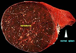
A large hamartoma of the spleen. The hamartoma is the dark circular object on the left that dominates the image. This is a cross-section; the growth is about 9 cm in diameter, while the entire spleen is about 11 cm in diameter. [1]
Pronunciation
  • hăm-ăr-tō′mă
Specialty Medical genetics, pathology   OOjs UI icon edit-ltr-progressive.svg
Diagnostic method Chest x-ray, CT scan, MRI, ultrasound, and bronchoscopy. [2]

A hamartoma is a mostly benign, [3] local malformation of cells that resembles a neoplasm of local tissue but is usually due to an overgrowth of multiple aberrant cells, with a basis in a systemic genetic condition, rather than a growth descended from a single mutated cell (monoclonality), as would typically define a benign neoplasm/tumor. [4] Despite this, many hamartomas are found to have clonal chromosomal aberrations that are acquired through somatic mutations, and on this basis the term hamartoma is sometimes considered synonymous with neoplasm. Hamartomas are by definition benign, slow-growing or self-limiting, [3] [4] though the underlying condition may still predispose the individual towards malignancies.

Contents

Hamartomas are usually caused by a genetic syndrome that affects the development cycle of all or at least multiple cells. [4] Many of these conditions are classified as overgrowth syndromes or cancer syndromes. Hamartomas occur in many different parts of the body and are most often asymptomatic incidentalomas (undetected until they are found incidentally on an imaging study obtained for another reason). Additionally, the definition of hamartoma versus benign neoplasm is often unclear, since both lesions can be clonal. Lesions such as adenomas, developmental cysts, hemangiomas, lymphangiomas and rhabdomyomas within the kidneys, lungs or pancreas are interpreted by some experts as hamartomas while others consider them true neoplasms. Moreover, even though hamartomas show a benign histology, there is a risk of some rare but life-threatening complications such as those found in neurofibromatosis type I and tuberous sclerosis. [5]

It is different from choristoma, a closely related form of heterotopia. [6] [7] The two can be differentiated as follows: a hamartoma is an excess of normal tissue in a normal situation (e.g., a birthmark on the skin), while a choristoma is an excess of tissue in an abnormal situation (e.g., pancreatic tissue in the duodenum). [8] [9] The term hamartoma is from the Greek ἁμαρτία, hamartia ("error"), and was introduced by D.P.G. Albrecht in 1904. [10]

Causes

Hamartomas are caused by abnormal formation in normal tissue and can occur spontaneously or as a result of an underlying disorder. Hamartomas are most likely the result of developmental error and can manifest itself in multiple locations. The development of hamartomas has also been linked to certain genes such as SMAD4, PTEN, STK1, and BMPR1A. [2]

Disorders associated with hamartomas include tuberous sclerosis, cowden syndrome, PTEN hamartoma tumour syndrome, and Peutz–Jeghers syndrome. [2]

Diagnosis

Classification

Lung

Parenchymal hamartoma of the lung. The surrounding lung falls away from the well-circumscribed mass, a typical feature of these lesions. The hamartoma shows a variegated yellow and white appearance, which corresponds respectively to fat and cartilage. Hamartoma of the lung.jpg
Parenchymal hamartoma of the lung. The surrounding lung falls away from the well-circumscribed mass, a typical feature of these lesions. The hamartoma shows a variegated yellow and white appearance, which corresponds respectively to fat and cartilage.

About 5–8% of all solitary lung nodules and about 75% of all benign lung tumors, are hamartomas. [11] Ten percent of hamartomas are endobronchial lesions, with the majority occurring in the peripheral lung parenchyma. [12] Peripheral pulmonary hamartomas typically do not cause any symptoms. [13] Patients may experience hemoptysis, obstructive pneumonia, dyspnea, persistent cough, and chest pain, depending on the size and location. [14] Typically, lung hamartomas appear as solitary nodules on thoracic computed tomography (CT) scans, with a diameter of less than 4 cm. [15]

Lung hamartomas are more common in men than in women, and may present additional difficulties in smokers. [16]

Heart

Cardiac rhabdomyomas is an uncommon, benign mesenchymal tumor that originated from striated muscle. Usually, it affects the head and neck. [17] It has been found that tuberous sclerosis is linked to 80–90% of cardiac rhabdomyomas. [18] The symptoms may manifest as pericardial effusion, hydrops fetalis, or heart blocks. [19]

Nerves

Sometimes nerves can also be affected. The most common nerve to be affected by hamartoma is reported to be median nerve. [20]

Hypothalamus

One of the most troublesome hamartomas occurs on the hypothalamus. Unlike most such growths, a hypothalamic hamartoma is symptomatic; it most often causes gelastic seizures, and can cause visual problems, other seizures, rage disorders associated with hypothalamic diseases, and early onset of puberty. The symptoms typically begin in early infancy and are progressive, often into general cognitive and/or functional disability. Moreover, resection is usually difficult, as the growths are generally adjacent to, or even intertwined with, the optic nerve. Symptoms tend to be resistant to medical control; however, surgical techniques are improving and can result in immense improvement of prognosis. [21]

Kidneys, stomach, spleen and other vascular organs

Hamartoma in breast with ultrasound Hamartoma in breast.jpg
Hamartoma in breast with ultrasound

Renal hamartomas are benign tumors that most likely developed from birth defects in the organ. They are frequently abundant in blood vessels and contain varying amounts of fat and smooth muscle components. [22]

A myoepithelial hamartoma, also known as a pancreatic rest, is ectopic pancreatic tissue found in the stomach, duodenum, or proximal jejunum. When seen on upper gastrointestinal series, a pancreatic rest may appear to be a submucosal mass or gastric neoplasm. Most are asymptomatic, but they can cause dyspepsia or upper gastrointestinal bleeding. [23]

A hamartoma has been identified as a cause of partial outflow obstruction in the abomasum (true gastric stomach) of a dairy goat. [24]

Splenic hamartoma is an uncommon benign vascular proliferative tumor that is identified by the vascular endothelial lining cells' CD8 immunopositivity. It is made up of an unusual combination of typical splenic components, like red and white pulp. [25]

Cowden syndrome

Cowden syndrome is an uncommon hereditary disorder marked by numerous hamartomas in a range of tissues from all three layers of the embryo. This is a syndrome that predisposes people to cancer and increases the risk of developing cancer in many different tissues, but particularly in the endometrium, thyroid, and breast. It is inherited autosomally dominantly, with a germ-line mutation of the PTEN tumor suppressor gene present in about 80% of patients. [26]

Prognosis

Hamartomas, while generally benign, can cause problems due to their location. For example, when located on the skin, especially on the face or neck, they can be very disfiguring. Cases have been reported of hamartomas the size of a small orange. [27] They may obstruct practically any organ in the body, such as the colon, eye, etc. They are particularly likely to cause major health issues when located in the hypothalamus, kidneys, lips, or spleen. They can be removed surgically if necessary, and are not likely to recur. Prognosis will depend upon the location and size of the lesion, as well as the overall health of the patient.[ citation needed ]

See also

Related Research Articles

<span class="mw-page-title-main">Proteus syndrome</span> Human genetic disorder

Proteus syndrome is a rare disorder with a genetic background that can cause tissue overgrowth involving all three embryonic lineages. Patients with Proteus syndrome tend to have an increased risk of embryonic tumor development. The clinical and radiographic symptoms of Proteus syndrome are highly variable, as are its orthopedic manifestations.

<span class="mw-page-title-main">Tuberous sclerosis</span> Genetic condition causing non-cancerous tumours

Tuberous sclerosis complex (TSC) is a rare multisystem autosomal dominant genetic disease that causes non-cancerous tumours to grow in the brain and on other vital organs such as the kidneys, heart, liver, eyes, lungs and skin. A combination of symptoms may include seizures, intellectual disability, developmental delay, behavioral problems, skin abnormalities, lung disease, and kidney disease.

<span class="mw-page-title-main">Rhabdomyoma</span> Medical condition

A rhabdomyoma is a benign tumor of striated muscle. Rhabdomyomas may be either cardiac or extracardiac. Extracardiac forms of rhabdomyoma are sub-classified into three distinct types: adult type, fetal type, and genital type.

<span class="mw-page-title-main">Benign tumor</span> Mass of cells which cannot spread throughout the body

A benign tumor is a mass of cells (tumor) that does not invade neighboring tissue or metastasize. Compared to malignant (cancerous) tumors, benign tumors generally have a slower growth rate. Benign tumors have relatively well differentiated cells. They are often surrounded by an outer surface or stay contained within the epithelium. Common examples of benign tumors include moles and uterine fibroids.

<span class="mw-page-title-main">Birt–Hogg–Dubé syndrome</span> Rare autosomal dominant cancer syndrome

Birt–Hogg–Dubé syndrome (BHD), also Hornstein–Birt–Hogg–Dubé syndrome, Hornstein–Knickenberg syndrome, and fibrofolliculomas with trichodiscomas and acrochordons is a human, adult onset, autosomal dominant genetic disorder caused by the FLCN gene. It can cause susceptibility to kidney cancer, renal and pulmonary cysts, and noncancerous tumors of the hair follicles, called fibrofolliculomas. The symptoms seen in each family are unique, and can include any combination of the three symptoms. Fibrofolliculomas are the most common manifestation, found on the face and upper trunk in over 80% of people with BHD over the age of 40. Pulmonary cysts are equally common (84%) and 24% of people with BHD eventually experience a collapsed lung. Kidney tumors, both cancerous and benign, occur in 14–34% of people with BHD; the associated kidney cancers are often rare hybrid tumors.

<span class="mw-page-title-main">Cowden syndrome</span> Medical condition

Cowden syndrome is an autosomal dominant inherited condition characterized by benign overgrowths called hamartomas as well as an increased lifetime risk of breast, thyroid, uterine, and other cancers. It is often underdiagnosed due to variability in disease presentation, but 99% of patients report mucocutaneous symptoms by age 20–29. Despite some considering it a primarily dermatologic condition, Cowden's syndrome is a multi-system disorder that also includes neurodevelopmental disorders such as macrocephaly.

Phakomatoses, also known as neurocutaneous syndromes, are a group of multisystemic diseases that most prominently affect structures primarily derived from the ectoderm such as the central nervous system, skin and eyes. The majority of phakomatoses are single-gene disorders that may be inherited in an autosomal dominant, autosomal recessive or X-linked pattern. Presentations may vary dramatically between patients with the same particular syndrome due to mosaicism, variable expressivity, and penetrance.

<i>PTEN</i> (gene) Tumor suppressor gene

Phosphatase and tensin homolog (PTEN) is a phosphatase in humans and is encoded by the PTEN gene. Mutations of this gene are a step in the development of many cancers, specifically glioblastoma, lung cancer, breast cancer, and prostate cancer. Genes corresponding to PTEN (orthologs) have been identified in most mammals for which complete genome data are available.

<span class="mw-page-title-main">Bannayan–Riley–Ruvalcaba syndrome</span> Medical condition

Bannayan–Riley–Ruvalcaba syndrome (BRRS) is a rare overgrowth syndrome and hamartomatous disorder with occurrence of multiple subcutaneous lipomas, macrocephaly and hemangiomas. The disease is inherited in an autosomal dominant manner. The disease belongs to a family of hamartomatous polyposis syndromes, which also includes Peutz–Jeghers syndrome, juvenile polyposis and Cowden syndrome. Mutation of the PTEN gene underlies this syndrome, as well as Cowden syndrome, Proteus syndrome, and Proteus-like syndrome, these four syndromes are referred to as PTEN Hamartoma-Tumor Syndromes.

<span class="mw-page-title-main">Angiomyolipoma</span> Medical condition

Angiomyolipomas are the most common benign tumour of the kidney. Although regarded as benign, angiomyolipomas may grow such that kidney function is impaired or the blood vessels may dilate and burst, leading to bleeding.

<span class="mw-page-title-main">Neuroendocrine tumor</span> Medical condition

Neuroendocrine tumors (NETs) are neoplasms that arise from cells of the endocrine (hormonal) and nervous systems. They most commonly occur in the intestine, where they are often called carcinoid tumors, but they are also found in the pancreas, lung, and the rest of the body.

A gelastic seizure, also known as "gelastic epilepsy", is a rare type of seizure that involves a sudden burst of energy, usually in the form of laughing. This syndrome usually occurs for no obvious reason and is uncontrollable. It is slightly more common in males than females.

Choristomas, a form of heterotopia, are masses of normal tissues found in abnormal locations. In contrast to a neoplasm or tumor, the growth of a choristoma is normally regulated.

<span class="mw-page-title-main">Lhermitte–Duclos disease</span> Medical condition

Lhermitte–Duclos disease (LDD), also called dysplastic gangliocytoma of the cerebellum (DGC), is a rare, slowly growing tumor of the cerebellum, a gangliocytoma sometimes considered to be a hamartoma, characterized by diffuse hypertrophy of the granular layer of the cerebellum. It is often associated with Cowden syndrome. It was described by Jacques Jean Lhermitte and P. Duclos in 1920.

<span class="mw-page-title-main">Lung nodule</span> Medical condition

A lung nodule or pulmonary nodule is a relatively small focal density in the lung. A solitary pulmonary nodule (SPN) or coin lesion, is a mass in the lung smaller than three centimeters in diameter. A pulmonary micronodule has a diameter of less than three millimetres. There may also be multiple nodules.

<span class="mw-page-title-main">Angiofibroma</span> Medical condition

Angiofibroma (AGF) is a descriptive term for a wide range of benign skin or mucous membrane lesions in which individuals have:

  1. benign papules, i.e. pinhead-sized elevations that lack visible evidence of containing fluid;
  2. nodules, i.e. small firm lumps usually >0.1 cm in diameter; and/or
  3. tumors, i.e. masses often regarded as ~0.8 cm or larger.
<span class="mw-page-title-main">Koenen's tumor</span> Medical condition

Koenen's tumor (KT), also commonly termed periungual angiofibroma, is a subtype of the angiofibromas. Angiofibromas are benign papule, nodule, and/or tumor lesions that are separated into various subtypes based primarily on the characteristic locations of their lesions. KTs are angiofibromas that develop in and under the toenails and/or fingernails. KTs were once considered as the same as another subtype of the angiofibromas viz., acral angiofibromas. While the literature may still sometimes regard KTs as acral angiofibromas, acral angiofibromas are characteristically located in areas close to but not in the toenails and fingernails as well as in the soles of the feet and palms of the hands. KTs are here regarded as distinct from acral angiofibromas.

<span class="mw-page-title-main">Sebaceous adenoma</span> Medical condition

A sebaceous adenoma, a type of adenoma, a cutaneous condition characterized by a slow-growing tumor usually presenting as a pink, flesh-coloured, or yellow papule or nodule.

Plasma cell granulomas (PCGs) are uncommon, non-neoplastic lesions of unknown etiology and are considered an entity of IgG4-related diseases.

<span class="mw-page-title-main">Multifocal micronodular pneumocyte hyperplasia</span>

Multifocal micronodular pneumocyte hyperplasia (MMPH) is a subtype of pneumocytic hyperplasia.

References

  1. Uthman E (2 January 1999). "Hamartoma of the spleen". [Personal website]. Archived from the original on 15 June 2014. Retrieved 30 July 2014.
  2. 1 2 3 Ali, Syed Awab; Mulita, Francesk (March 14, 2023). "Hamartoma". StatPearls Publishing. PMID   32965969 . Retrieved October 10, 2023.
  3. 1 2 "Hamartoma definition". Taber's Medical Dictionary. Archived from the original on 7 December 2008. Retrieved 2008-09-25.
  4. 1 2 3 Batsakis JG (1984-01-01). "Pathology consultation. Nomenclature of developmental tumors". The Annals of Otology, Rhinology, and Laryngology. 93 (1 Pt 1): 98–99. doi:10.1177/000348948409300122. PMID   6703601. S2CID   75206651.
  5. Kumar V, Abbas AK, Fausto N, Aster JC (27 August 2014). Robbin's Pathologic Basis of Disease (9th ed.). Elsevier Health Sciences. p. 481. ISBN   978-0-323-29639-7.
  6. "Choristoma" at Dorland's Medical Dictionary
  7. Lee KH, Roland PS (2013). "Heterotopias, Teratoma, and Choristoma". Encyclopedia of Otolaryngology, Head and Neck Surgery. pp. 1179–1183. doi:10.1007/978-3-642-23499-6_642. ISBN   978-3-642-23498-9.
  8. Jorquera JP, Rubio-Palau J, Cazalla AA, Rodríguez-Carunchio L (2016). "Choristoma: A rare congenital tumor of the tongue". Annals of Maxillofacial Surgery. 6 (2): 311–313. doi: 10.4103/2231-0746.200342 . PMC   5343649 . PMID   28299279.
  9. Goswamy M, Tabasum S, Kudva P, Gupta S (January 2012). "Osseous choristoma of the periodontium". Journal of Indian Society of Periodontology. 16 (1): 120–122. doi: 10.4103/0972-124X.94619 . PMC   3357020 . PMID   22628977.
  10. Patil, Shankargouda; Rao, RoopaS; Majumdar, Barnali (2015). "Hamartomas of the oral cavity". Journal of International Society of Preventive and Community Dentistry. 5 (5). Medknow: 347–353. doi: 10.4103/2231-0762.164789 . ISSN   2231-0762. PMC   4606596 . PMID   26539384.
  11. De Cicco C, Bellomi M, Bartolomei M, Carbone G, Pelosi G, Veronesi G, et al. (December 2008). "Imaging of lung hamartomas by multidetector computed tomography and positron emission tomography". The Annals of Thoracic Surgery. 86 (6): 1769–1772. doi:10.1016/j.athoracsur.2008.08.033. PMID   19021972.
  12. Wu, Lei; Chen, Wei; Li, PengCheng; Li, Shuxian; Chen, Zhimin (April 27, 2021). "Case Report: Resection of Giant Endotracheal Hamartoma by Electrosurgical Snaring via Fiberoptic Bronchoscopy in a 9-Year-Old Boy". Frontiers in Pediatrics. 9. Frontiers Media SA. doi: 10.3389/fped.2021.528966 . ISSN   2296-2360. PMC   8111287 . PMID   33987147.
  13. SINGH, HARIQBAL; KHANNA, SK; CHANDRAN, VIJAY; Jetley, RK (1999). "Pulmonary Hamartoma". Medical Journal Armed Forces India. 55 (1). Elsevier BV: 79–80. doi:10.1016/s0377-1237(17)30328-3. ISSN   0377-1237. PMC   5531795 . PMID   28775580.
  14. Grigoraş, Adriana; Amălinei, Cornelia; Lovin, Ciprian Sebastian; Grigoraş, Constantin Cristian; Pricope, Diana Lavinia; Costin, Constantin Aleodor; Chiseliţă, Irina Rodica; Crişan-Dabija, Radu Adrian (February 18, 2023). "The clinicopathological challenges of symptomatic and incidental pulmonary hamartomas diagnosis". Romanian Journal of Morphology and Embryology. 63 (4). Societatea Romana de Morfologie: 607–613. doi:10.47162/rjme.63.4.02. ISSN   1220-0522. PMC   10028331 . PMID   36808195.
  15. Saadi, Muslim M.; Barakeh, Duna H.; Husain, Sufia; Hajjar, Waseem M. (2015). "Large multicystic pulmonary chondroid hamartoma in a child presenting as pneumothorax". Saudi Medical Journal. 36 (4): 487–489. doi:10.15537/smj.2015.4.10210. ISSN   0379-5284. PMC   4404485 . PMID   25828288.
  16. Vuckovic, Dejan C; Koledin, Milos P; Vuckovic, Nada M; Koledin, Bojan M (March 25, 2020). "Mediastinal Cartilaginous Hamartoma". Cureus. 12 (3). Cureus, Inc.: e7411. doi: 10.7759/cureus.7411 . ISSN   2168-8184. PMC   7182163 . PMID   32337135.
  17. Hansen, Torsten; Katenkamp, Detlef (August 18, 2005). "Rhabdomyoma of the head and neck: morphology and differential diagnosis". Virchows Archiv. 447 (5). Springer Science and Business Media LLC: 849–854. doi:10.1007/s00428-005-0038-8. ISSN   0945-6317. PMID   16133368. S2CID   8031365.
  18. Sarkar, Sharmila; Siddiqui, Waqas J. (November 14, 2022). "Cardiac Rhabdomyoma". StatPearls Publishing. PMID   32809444 . Retrieved January 28, 2024.
  19. Hinton, Robert B.; Prakash, Ashwin; Romp, Robb L.; Krueger, Darcy A.; Knilans, Timothy K. (December 17, 2014). "Cardiovascular Manifestations of Tuberous Sclerosis Complex and Summary of the Revised Diagnostic Criteria and Surveillance and Management Recommendations From the International Tuberous Sclerosis Consensus Group". Journal of the American Heart Association. 3 (6). Ovid Technologies (Wolters Kluwer Health): e001493. doi:10.1161/jaha.114.001493. ISSN   2047-9980. PMC   4338742 . PMID   25424575.
  20. Ranjan, Rajni; Kumar, Rakesh; Jeyaraman, Madhan; Kumar, Sudhir (June 1, 2020). "Fibrolipomatous Hamartoma (FLH) of Median Nerve: A Rare Case Report and Review". Indian Journal of Orthopaedics. 55 (S1). Springer Science and Business Media LLC: 267–272. doi:10.1007/s43465-020-00149-9. ISSN   0019-5413. PMC   8149561 . PMID   34113430.
  21. "Hypothalmic Hamartoma". Barrow Neurological Institute. Archived from the original on 2015-09-21. Retrieved 2013-01-09.
  22. Palmisano, Peter J. (1967). "Renal Hamartoma (Angiomyolipoma): Its Angiographic Appearance and Response to Intra-Arterial Epinephrine". Radiology. 88 (2): 249–252. doi:10.1148/88.2.249. ISSN   0033-8419. PMID   6016922.
  23. Shah, A.; Gordon, A.R.; Ginsberg, G.G.; Furth, E.E.; Levine, M.S. (2007). "Ectopic pancreatic rest in the proximal stomach mimicking gastric neoplasms". Clinical Radiology. 62 (6). Elsevier BV: 600–602. doi:10.1016/j.crad.2007.01.001. ISSN   0009-9260. PMID   17467399.
  24. Smith J, Klostermann C, Harm T, Breuer R, Kovalik DA, Bornkamp J, Yaeger M (August 2017). "Abomasal hamartoma in a La Mancha wether". Veterinary Record Case Reports. 5 (3): e000515. doi:10.1136/vetreccr-2017-000515. S2CID   80240747.
  25. Sim, Jongmin; Ahn, Hye In; Han, Hulin; Jun, Young Jin; Rehman, Abdul; Jang, Se Min; Jang, Kiseok; Paik, Seung Sam (2013). "Splenic hamartoma: A case report and review of the literature". World Journal of Clinical Cases. 1 (7). Baishideng Publishing Group Inc.: 217. doi: 10.12998/wjcc.v1.i7.217 . ISSN   2307-8960. PMC   3856295 .
  26. Farooq, A.; Walker, L.J.; Bowling, J.; Audisio, R.A. (2010). "Cowden syndrome". Cancer Treatment Reviews. 36 (8). Elsevier BV: 577–583. doi:10.1016/j.ctrv.2010.04.002. ISSN   0305-7372. PMID   20580873.
  27. "Dermatology Images". Dermatology Image Atlas. Archived from the original on 2006-05-15.